Esthesioneuroblastoma

Last updated
Esthesioneuroblastoma
Other namesNeuroblastoma
Esthesioneuroblastoma.jpg
Esthesioneuroblastoma
Specialty Neuro-oncology

Esthesioneuroblastoma is a rare cancer of the nasal cavity. Arising from the upper nasal tract, esthesioneuroblastoma is believed to originate from sensory neuroepithelial cells, also known as neuroectodermal olfactory cells. [1]

Contents

Due to the location of the tumor and its proximity to the cranial cavity, esthesioneuroblastoma can be highly invasive and challenging to treat. There is no consensus on an appropriate treatment approach of esthesioneuroblastoma because of the rarity of the disease. Most studies reported cranial surgical resection with radiotherapy or chemotherapy to target the tumor. [2]

Signs and symptoms

Esthesioneuroblastoma frequently first presents as a nasal mass. [3] The most common signs and symptoms of esthesioneuroblastoma are nasal obstruction (70%) and epistaxis (50%). [4] [1] Less common symptoms include hyposmia (loss of smell), headache, rhinorrhea, vision loss, proptosis, facial pain, diplopia (double vision), masses in the neck and changes in mental status. [4] Esthesioneuroblastoma occurs in the upper nasal cavity, near the optic nerves and optic chiasm. Thus, tumor growth can impinge nerve function and result in vision loss and diplopia. [5] As the tumor metastasizes to the oral cavity, there can be tooth pain and tooth mobility. [6]

Genetics

There is limited research on the genetic role in esthesioneuroblastoma development. Of the research to date, the sonic hedgehog pathway, MYC and KDR genes are implicated for esthesioneuroblastoma. [7] [8]

Mutations in dystrophin and Laminin, alpha 2 (LAMA2) have also been implicated in this disease. [9]

Pathophysiology

Esthesioneuroblastoma is of neurocrest origin, arising from olfactory sensory cells in the olfactory epithelium. [1] The olfactory epithelium consists of olfactory sensory cells, sustentacular cells and basal cells. [1] Esthesioneuroblastoma consists of lobular sheets with neurofibrullar fibers and rosettes. [4] Hyam's classifications are an important way of determining prognosis. [10]

Hyam's histopathological grades for esthesioneuroblastoma [11]

GradeLobular architexture preservationMitotic indexNuclear polymorphismFibrillary matrixRosettesNecrosis
I+nonenoneprominent Homer Wright (HW) rosettes none
II+lowmoderatepresentHW rosettesnone
III+/-moderateprominentlow Flexner-Wintersteiner rosettes rare
IV+/-highmarkedabsentnonefrequent

Diagnosis

Esthesioneuroblastoma can resemble small blue cell tumors like squamous cell carcinoma, sinonasal undifferentiated carcinoma, extranodal NK/T cell lymphoma, nasal type, rhabdomyosarcoma, Ewing/PNET, mucosal malignant melanoma and neuroendocrine carcinomas (NEC) that occur in the intranasal tract. [1] Compared to other tumors in the region, esthesioneuroblastoma has the best prognosis, with an overall five-year survival rate of 60–80%. [1] Fewer than 700 cases have been documented in the United States alone. [12] Esthesioneuroblastoma is characterized by neurofibrillary stroma and neurosecretary granules that are not seen concurrently by any other pathologies in the region. [1] Histological tests such as keratin, CK5/6, S-100 protein or NSE can be run to further differentiate esthesioneuroblastoma from other tumors. [1]

Staging

The Kadish classification is used for clinical classification of sinonasal tumors including esthesioneuroblastoma. Subsequent research articles have been published to determine prognosis based on tumor grade.

Modified Kadish classification [13] [4]

StageDescription5 year survival
ATumor confined to nasal cavity 75–91
BNasal cavity and paranasal sinuses 68–71
CTumor extends beyond nasal cavity and paranasal sinuses, including skull base, orbit or cribiform plate41–47
DTumor metastasizes to cervical lymph nodes and beyond<40

Dulguerov classification [14]

StageCharacteristics
T1Tumour involving the nasal cavity and/or paranasal sinuses (excluding sphenoid), sparing the most superior ethmoidal cells
T2Tumour involving the nasal cavity and/or paranasal sinuses (including the sphenoid) with extension to or erosion of the cribriform plate
T3Tumour extending into the orbit or protruding into the anterior cranial fossa, without dural invasion
T4Tumour involving the brain
N0No cervical lymph-node metastasis
N1Any form of cervical lymph-node metastasis
M0No metastases
M1Distant metastasis

Treatment

The preferred treatment for esthesioneuroblastoma is surgery followed by radiotherapy to prevent recurrence of the tumor. [14]

Surgical approaches

Several surgical approaches have been described, [15] [16] [17] [18] [19] but post-excision recurrence rates have remained relatively high. Studies suggest better results with a bilateral approach. For cases with cribriform plate involvement, tumors are resected bilaterally using a transfacial and craniotomy approach. [20] In a research study, the craniofacial approach decreased recurrence of esthesioneuroblastoma by 20%. [20] Craniofacial resection can help preserve the optic nerves and brain while removing the cribriform plate, olfactory bulb, dura surrounding the bulb and even the orbital periosteum. [14]

Radiotherapy

Radiotherapy alone is reserved only for small lesions not appropriate for either surgery or chemotherapy. [4] Both photon and proton radiotherapy have been used effectively to treat esthesioneuroblastoma. [14] [21] Proton radiotherapy has recently been shown to be effective in a 10-person study with Kadish C tumors, while delivering less toxicity to the nervous system. [21]

Chemotherapy

Chemotherapy is used in a multimodality treatment plan generally for more advanced, unresectable or reoccurring tumors. [4] Cyclophosphamide, vincristine and doxorubicin have been used as neoadjuvant chemotherapy drugs for grade C esthesioneuroblastoma before surgical resection, producing fair outcomes. Cisplatin and etoposide are often used to treat esthesioneuroblastoma as neoadjuvants or adjuvants with radiotherapy or surgery. [22] [23] [24]

Study results are promising. In advanced stage esthesioneuroblastoma in pediatric patients, where surgery is no longer possible, aggressive chemotherapy and radiotherapy has resulted in some tumor control and long-term survival. [25]

Prognosis

Esthesioneuroblastoma is a slow developing but malignant tumor with high recurrence rates because of its anatomical position. [10] The tumor composition, location and metastatic characteristics as well as the treatment plan determine prognosis. Common clinical classification systems for esthesioneuroblastoma include the Kadish classification and the Dulguerov classification. Histopathological characteristics on top of Kadish classification can further determine cancer prognosis. In severe, Kadish class C tumors, Haym's grades of pathology are important for prognosis. Patients with low grade Kadish class C tumors have a 10-year survival rate of 86 percent compared to patients with high grade class C tumors who have a survival rate of 28 percent. [10] Surgically treated patients with high grade tumors are more likely to experience leptomeningeal metastases or involvement of the cerebral spinal fluid unlike patients with low grade tumors who usually only see local recurrence. [26]

Survival rates for treated esthesioneuroblastoma are best for surgery with radiotherapy (65%), then for radiotherapy and chemotherapy (51%), just surgery (48%), surgery, radiotherapy and chemotherapy (47%) and finally just radiotherapy (37%). [14] From the literature, radiotherapy and surgery seem to boast the best outcome for patients. However, it is important to understand that to some degree, prognosis is related to tumor severity. More progressed, higher grade tumors would result in chemotherapy or radiotherapy as the only treatment. It is no surprise that the prognosis would be worse in these cases.[ citation needed ]

Incidence

Esthesioneuroblastoma accounts for 2% of all intranasal tumors with an incidence of 0.4 cases per million people. [1] Fewer than 700 cases have been documented in the United States. [12] Fewer than 400 unique cases have been reported globally. [4] [1] Esthesioneuroblastoma can occur at any time, with peak occurrence reported in the second and sixth decades of life. [1]

History

Esthesioneuroblastoma was first characterized in 1924. [27] [28]

Notable cases

The disease was brought into prominence by the case of Chantal Sébire, who was suffering from the disease and ended her life after being denied euthanasia. [29]

Related Research Articles

<span class="mw-page-title-main">Brain tumor</span> Neoplasm in the brain

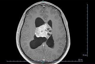
A brain tumor occurs when abnormal cells form within the brain. There are two main types of tumors: malignant tumors and benign (non-cancerous) tumors. These can be further classified as primary tumors, which start within the brain, and secondary tumors, which most commonly have spread from tumors located outside the brain, known as brain metastasis tumors. All types of brain tumors may produce symptoms that vary depending on the size of the tumor and the part of the brain that is involved. Where symptoms exist, they may include headaches, seizures, problems with vision, vomiting and mental changes. Other symptoms may include difficulty walking, speaking, with sensations, or unconsciousness.

<span class="mw-page-title-main">Osteosarcoma</span> Cancerous tumour in a bone

An osteosarcoma (OS) or osteogenic sarcoma (OGS) is a cancerous tumor in a bone. Specifically, it is an aggressive malignant neoplasm that arises from primitive transformed cells of mesenchymal origin and that exhibits osteoblastic differentiation and produces malignant osteoid.

<span class="mw-page-title-main">Thymoma</span> Medical condition

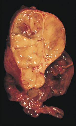
A thymoma is a tumor originating from the epithelial cells of the thymus that is considered a rare malignancy. Thymomas are frequently associated with neuromuscular disorders such as myasthenia gravis; thymoma is found in 20% of patients with myasthenia gravis. Once diagnosed, thymomas may be removed surgically. In the rare case of a malignant tumor, chemotherapy may be used.

Spinal tumors are neoplasms located in either the vertebral column or the spinal cord. There are three main types of spinal tumors classified based on their location: extradural and intradural. Extradural tumors are located outside the dura mater lining and are most commonly metastatic. Intradural tumors are located inside the dura mater lining and are further subdivided into intramedullary and extramedullary tumors. Intradural-intramedullary tumors are located within the dura and spinal cord parenchyma, while intradural-extramedullary tumors are located within the dura but outside the spinal cord parenchyma. The most common presenting symptom of spinal tumors is nocturnal back pain. Other common symptoms include muscle weakness, sensory loss, and difficulty walking. Loss of bowel and bladder control may occur during the later stages of the disease.

<span class="mw-page-title-main">Ependymoma</span> Medical condition

An ependymoma is a tumor that arises from the ependyma, a tissue of the central nervous system. Usually, in pediatric cases the location is intracranial, while in adults it is spinal. The common location of intracranial ependymomas is the fourth ventricle. Rarely, ependymomas can occur in the pelvic cavity.

<span class="mw-page-title-main">Chondrosarcoma</span> Medical condition

Chondrosarcoma is a bone sarcoma, a primary cancer composed of cells derived from transformed cells that produce cartilage. A chondrosarcoma is a member of a category of tumors of bone and soft tissue known as sarcomas. About 30% of bone sarcomas are chondrosarcomas. It is resistant to chemotherapy and radiotherapy. Unlike other primary bone sarcomas that mainly affect children and adolescents, a chondrosarcoma can present at any age. It more often affects the axial skeleton than the appendicular skeleton.

<span class="mw-page-title-main">Pseudomyxoma peritonei</span> Medical condition

Pseudomyxoma peritonei (PMP) is a clinical condition caused by cancerous cells that produce abundant mucin or gelatinous ascites. The tumors cause fibrosis of tissues and impede digestion or organ function, and if left untreated, the tumors and mucin they produce will fill the abdominal cavity. This will result in compression of organs and will destroy the function of the colon, small intestine, stomach, or other organs. Prognosis with treatment in many cases is optimistic, but the disease is lethal if untreated, with death occurring via cachexia, bowel obstruction, or other types of complications.

<span class="mw-page-title-main">Desmoplastic small-round-cell tumor</span> Aggressive and rare cancer

Desmoplastic small-round-cell tumor (DSRCT) is an aggressive and rare cancer that primarily occurs as masses in the abdomen. Other areas affected may include the lymph nodes, the lining of the abdomen, diaphragm, spleen, liver, chest wall, skull, spinal cord, large intestine, small intestine, bladder, brain, lungs, testicles, ovaries, and the pelvis. Reported sites of metastatic spread include the liver, lungs, lymph nodes, brain, skull, and bones. It is characterized by the EWS-WT1 fusion protein.

<span class="mw-page-title-main">Hemangiopericytoma</span> Medical condition

A hemangiopericytoma is a type of soft-tissue sarcoma that originates in the pericytes in the walls of capillaries. When inside the nervous system, although not strictly a meningioma tumor, it is a meningeal tumor with a special aggressive behavior. It was first characterized in 1942.

Adjuvant therapy, also known as adjunct therapy, adjuvant care, or augmentation therapy, is a therapy that is given in addition to the primary or initial therapy to maximize its effectiveness. The surgeries and complex treatment regimens used in cancer therapy have led the term to be used mainly to describe adjuvant cancer treatments. An example of such adjuvant therapy is the additional treatment usually given after surgery where all detectable disease has been removed, but where there remains a statistical risk of relapse due to the presence of undetected disease. If known disease is left behind following surgery, then further treatment is not technically adjuvant.

A blastoma is a type of cancer, more common in children, that is caused by malignancies in precursor cells, often called blasts. Examples are nephroblastoma, medulloblastoma, and retinoblastoma. The suffix -blastoma is used to imply a tumor of primitive, incompletely differentiated cells, e.g., chondroblastoma is composed of cells resembling the precursor of chondrocytes.

<span class="mw-page-title-main">Mucoepidermoid carcinoma</span> Medical condition

Mucoepidermoid carcinoma (MEC) is the most common type of minor salivary gland malignancy in adults. Mucoepidermoid carcinoma can also be found in other organs, such as bronchi, lacrimal sac, and thyroid gland.

<span class="mw-page-title-main">Undifferentiated pleomorphic sarcoma</span> Medical condition

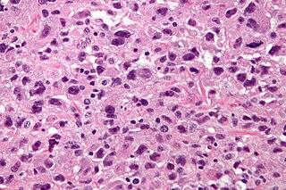
Undifferentiated pleomorphic sarcoma (UPS), also termed pleomorphic myofibrosarcoma, high-grade myofibroblastic sarcoma, and high-grade myofibrosarcoma, is characterized by the World Health Organization (WHO), 2020, as a rare, poorly differentiated neoplasm, i.e. an abnormal growth of cells that have an unclear identity and/or cell of origin. WHO classified it as one of the undifferentiated/unclassified sarcomas in the category of tumors of uncertain differentiation. Sarcomas are cancers known or thought to derive from mesenchymal stem cells that typically develop in bone, muscle, fat, blood vessels, lymphatic vessels, tendons, and ligaments. More than 70 sarcoma subtypes have been described. The UPS subtype of these sarcomas consists of tumor cells that are poorly differentiated and may appear as spindle-shaped cells, histiocytes, and giant cells. UPS is considered a diagnosis that defies formal sub-classification after thorough histologic, immunohistochemical, and ultrastructural examinations fail to identify the type of cells involved.

<span class="mw-page-title-main">Thymic carcinoma</span> Medical condition

Thymic carcinoma, or type C thymoma, is a malignancy of the thymus. It is a rare cancer that is often diagnosed at advanced stages. Recurrence following treatment is common, and thymic carcinoma is associated with a poor prognosis.

<span class="mw-page-title-main">Inverted papilloma</span> Medical condition

An inverted papilloma, also known as Ringertz tumour, is a type of tumor in which surface epithelial cells grow downward into the underlying supportive tissue. It may occur in the nose and/or sinuses or in the urinary tract. When it occurs in the nose or sinuses, it may cause symptoms similar to those caused by sinusitis, such as nasal congestion. When it occurs in the urinary tract, it may cause blood in the urine.

<span class="mw-page-title-main">Choroid plexus carcinoma</span> Medical condition

A choroid plexus carcinoma is a type of choroid plexus tumor that affects the choroid plexus of the brain. It is considered the worst of the three grades of chord plexus tumors, having a much poorer prognosis than choroid atypical plexus papilloma and choroid plexus papilloma. The disease creates lesions in the brain and increases cerebrospinal fluid volume, resulting in hydrocephalus.

<span class="mw-page-title-main">Central neurocytoma</span> Medical condition

Central neurocytoma (CNC) is an extremely rare, ordinarily benign intraventricular brain tumour that typically forms from the neuronal cells of the septum pellucidum. The majority of central neurocytomas grow inwards into the ventricular system forming interventricular neurocytomas. This leads to two primary symptoms of CNCs, blurred vision and increased intracranial pressure. Treatment for a central neurocytoma typically involves surgical removal, with an approximate 1 in 5 chance of recurrence. Central neurocytomas are classified as a grade II tumor under the World Health Organization's classification of tumors of the nervous system.

<span class="mw-page-title-main">Paranasal sinus and nasal cavity cancer</span> Medical condition

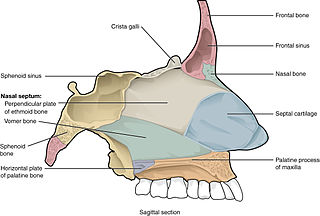
Paranasal sinus and nasal cavity cancer is a type of cancer that is caused by the appearance and spread of malignant cells into the paranasal sinus and nasal cavity. The cancer most commonly occurs in people between 50 and 70 years old, and occurs twice as often in males as in females. During early phases of the cancer, symptoms may include nasal obstruction and hyposmia, as well as other symptoms. More symptoms may develop as malignant cells further grow and spread into other nearby tissue such as the palate or orbital floor. X-rays of the head and MRI can aid in diagnosis of the cancer while tumor resection surgery, radiation therapy and chemotherapy can be used for treatment of the cancer.

Myxofibrosarcoma (MFS), although a rare type of tumor, is one of the most common soft tissue sarcomas, i.e. cancerous tumors, that develop in the soft tissues of elderly individuals. Initially considered to be a type of histiocytoma termed fibrous histiocytoma or myxoid variant of malignant fibrous histiocytoma, Angervall et al. termed this tumor myxofibrosarcoma in 1977. In 2020, the World Health Organization reclassified MFS as a separate and distinct tumor in the category of malignant fibroblastic and myofibroblastic tumors.

Sclerosing epithelioid fibrosarcoma (SEF) is a very rare malignant tumor of soft tissues that on microscopic examination consists of small round or ovoid neoplastic epithelioid fibroblast-like cells, i.e. cells that have features resembling both epithelioid cells and fibroblasts. In 2020, the World Health Organization classified SEF as a distinct tumor type in the category of malignant fibroblastic and myofibroblastic tumors. However, current studies have reported that low-grade fibromyxoid sarcoma (LGFMS) has many clinically and pathologically important features characteristic of SEF; these studies suggest that LGSFMS may be an early form of, and over time progress to become, a SEF. Since the World Health Organization has classified LGFMS as one of the malignant fibroblastic and myofibroblastic tumors that is distinctly different than SEF, SEF and LGFMS are here regarded as different tumor forms.

References

  1. 1 2 3 4 5 6 7 8 9 10 11 Thompson LD (2009). Olfactory neuroblastoma. Head and neck pathology. 3: 252–259
  2. Jiang GY, Li FC, Chen WK, Liu AM, Cai WQ (December 2011). "Therapy and prognosis of intracranial invasive olfactory neuroblastoma". Otolaryngology–Head and Neck Surgery. 145 (6): 951–955. doi: 10.1177/0194599811416752 . PMID   21825099. S2CID   9860811.
  3. Ghaffar S, Salahuddin I (March 2005). "Olfactory neuroblastoma: a case report and review of the literature". Ear, Nose, & Throat Journal. 84 (3): 150–152. doi:10.1177/014556130508400311. PMID   15871582. S2CID   20079359.
  4. 1 2 3 4 5 6 7 Sheehan JM (2011). "Esthesioneuroblastoma". In Jane JA (ed.). Youmans Neurological Surgery (6th ed.). Retrieved 7 December 2016.
  5. Oskouian RJ, Jane JA, Dumont AS, Sheehan JM, Laurent JJ, Levine PA (May 2002). "Esthesioneuroblastoma: clinical presentation, radiological, and pathological features, treatment, review of the literature, and the University of Virginia experience". Neurosurgical Focus. 12 (5): e4. doi: 10.3171/foc.2002.12.5.5 . PMID   16119902.
  6. von Zeidler SV, Guidi R, Alencar R, Aguiar R, Mendonça EF, Batista AC, Ribeiro-Rotta RF (January 2014). "Atypical esthesioneuroblastoma invading oral cavity: a case report and review of the literature". Diagnostic Pathology. 9: 10. doi: 10.1186/1746-1596-9-10 . PMC   3943513 . PMID   24443792.
  7. Mao L, Xia YP, Zhou YN, Dai RL, Yang X, Wang YJ, et al. (2009). "Activation of sonic hedgehog signaling pathway in olfactory neuroblastoma". Oncology. 77 (3–4): 231–243. doi:10.1159/000236047. PMID   19738389. S2CID   33078469.
  8. Weiss GJ, Liang WS, Izatt T, Arora S, Cherni I, Raju RN, et al. (2012). "Paired tumor and normal whole genome sequencing of metastatic olfactory neuroblastoma". PLOS ONE. 7 (5): e37029. Bibcode:2012PLoSO...737029W. doi: 10.1371/journal.pone.0037029 . PMC   3359355 . PMID   22649506.
  9. Gallia GL, Zhang M, Ning Y, Haffner MC, Batista D, Binder ZA, et al. (December 2018). "Genomic analysis identifies frequent deletions of Dystrophin in olfactory neuroblastoma". Nature Communications. 9 (1): 5410. Bibcode:2018NatCo...9.5410G. doi:10.1038/s41467-018-07578-z. PMC   6303314 . PMID   30575736.
  10. 1 2 3 Kaur G, Kane AJ, Sughrue ME, Madden M, Oh MC, Sun MZ, et al. (February 2013). "The prognostic implications of Hyam's subtype for patients with Kadish stage C esthesioneuroblastoma". Journal of Clinical Neuroscience. 20 (2): 281–286. doi:10.1016/j.jocn.2012.05.029. PMC   3795510 . PMID   23266076.
  11. Hyams VG, Baksakis JG, Michaels L, eds. (1988). Tumors of the upper respiratory tract and ear. Washington DC: Armed Forces Institute of Pathology. pp. 240–248.
  12. 1 2 Stokes WA, Camilon PR, Banglawala SM, Nguyen SA, Harvey R, Vandergrift WA, Schlosser RJ (2015). "Is sex an independent prognostic factor in esthesioneuroblastoma?". American Journal of Rhinology & Allergy. 29 (5): 369–372. doi:10.2500/ajra.2015.29.4204. PMID   26358349. S2CID   41961725.
  13. Kadish S, Goodman M, Wang CC (March 1976). "Olfactory neuroblastoma. A clinical analysis of 17 cases". Cancer. 37 (3): 1571–1576. doi: 10.1002/1097-0142(197603)37:3<1571::aid-cncr2820370347>3.0.co;2-l . PMID   1260676.
  14. 1 2 3 4 5 Dulguerov P, Allal AS, Calcaterra TC (November 2001). "Esthesioneuroblastoma: a meta-analysis and review". The Lancet. Oncology. 2 (11): 683–90. doi:10.1016/S1470-2045(01)00558-7. PMID   11902539.
  15. Roxbury CR, Ishii M, Gallia GL, Reh DD (February 2016). "Endoscopic Management of Esthesioneuroblastoma". Otolaryngologic Clinics of North America. 49 (1): 153–165. doi:10.1016/j.otc.2015.09.010. PMID   26614835.
  16. Vergani F, Pirola E, Fiori L, Pagni F, Parmigiani F, Sganzerla EP (June 2007). "Combined transcranial and endoscopic nasal resection for esthesioneuroblastoma. Technical note". Journal of Neurosurgical Sciences. 51 (2): 99–102. PMID   17571044.
  17. Unger F, Walch C, Stammberger H, Papaefthymiou G, Haselsberger K, Pendl G (June 2001). "Olfactory neuroblastoma (esthesioneuroblastoma): report of six cases treated by a novel combination of endoscopic surgery and radiosurgery". Minimally Invasive Neurosurgery. 44 (2): 79–84. doi:10.1055/s-2001-16000. PMID   11487789. S2CID   13035634.
  18. Cakmak O, Ergin NT, Yilmazer C, Kayaselçuk F, Barutcu O (July 2002). "Endoscopic removal of esthesioneuroblastoma". International Journal of Pediatric Otorhinolaryngology. 64 (3): 233–238. doi:10.1016/S0165-5876(02)00036-8. PMID   12090952.
  19. Prasad KC, Kumar A, Prasad SC, Jain D (September 2007). "Endoscopic-assisted excision of esthesioneuroblastoma". The Journal of Craniofacial Surgery. 18 (5): 1034–1038. doi:10.1097/scs.0b013e318157264c. PMID   17912077. S2CID   27445039.
  20. 1 2 Spaulding CA, Kranyak MS, Constable WC, Stewart FM (September 1988). "Esthesioneuroblastoma: a comparison of two treatment eras". International Journal of Radiation Oncology, Biology, Physics. 15 (3): 581–590. doi:10.1016/0360-3016(88)90298-2. PMID   3138210.
  21. 1 2 Nichols AC, Chan AW, Curry WT, Barker FG, Deschler DG, Lin DT (September 2008). "Esthesioneuroblastoma: the massachusetts eye and ear infirmary and massachusetts general hospital experience with craniofacial resection, proton beam radiation, and chemotherapy". Skull Base. 18 (5): 327–337. doi:10.1055/s-2008-1076098. PMC   2637063 . PMID   19240832.
  22. Porter AB, Bernold DM, Giannini C, Foote RL, Link MJ, Olsen KD, et al. (November 2008). "Retrospective review of adjuvant chemotherapy for esthesioneuroblastoma". Journal of Neuro-Oncology. 90 (2): 201–204. doi:10.1007/s11060-008-9645-y. PMID   18633576. S2CID   25788460.
  23. Bhattacharyya N, Thornton AF, Joseph MP, Goodman ML, Amrein PC (January 1997). "Successful treatment of esthesioneuroblastoma and neuroendocrine carcinoma with combined chemotherapy and proton radiation. Results in 9 cases". Archives of Otolaryngology–Head & Neck Surgery. 123 (1): 34–40. doi:10.1001/archotol.1997.01900010038005. PMID   9006501.
  24. Kim DW, Jo YH, Kim JH, Wu HG, Rhee CS, Lee CH, et al. (November 2004). "Neoadjuvant etoposide, ifosfamide, and cisplatin for the treatment of olfactory neuroblastoma". Cancer. 101 (10): 2257–2260. doi: 10.1002/cncr.20648 . PMID   15484215. S2CID   44518377.
  25. Bisogno G, Soloni P, Conte M, Podda M, Ferrari A, Garaventa A, et al. (March 2012). "Esthesioneuroblastoma in pediatric and adolescent age. A report from the TREP project in cooperation with the Italian Neuroblastoma and Soft Tissue Sarcoma Committees". BMC Cancer. 12: 117. doi: 10.1186/1471-2407-12-117 . PMC   3368746 . PMID   22443159.
  26. Malouf GG, Casiraghi O, Deutsch E, Guigay J, Temam S, Bourhis J (April 2013). "Low- and high-grade esthesioneuroblastomas display a distinct natural history and outcome". European Journal of Cancer. 49 (6): 1324–1334. doi:10.1016/j.ejca.2012.12.008. PMID   23312882.
  27. Fordice JO (3 March 1994). "Esthesioneuroblastoma". Baylor College of Medicine. Archived from the original on April 9, 2007. Retrieved 2008-03-22.
  28. Berger L, Luc G, Richard D (1924). "L'esthésioneuroépithéliome olfactif". Bull Assoc Franç Étude Cancer. 13: 410–421.
  29. "BBC NEWS - Europe - Tumour woman's death not natural". BBC. 21 March 2008. Retrieved 1 November 2016.