Follicular B helper T cells

Last updated

Follicular helper T cells (also known as follicular B helper T cells and abbreviated as TFH), are antigen-experienced CD4 + T cells found in the periphery within B cell follicles of secondary lymphoid organs such as lymph nodes, spleen and Peyer's patches, and are identified by their constitutive expression of the B cell follicle homing receptor CXCR5. [1] Upon cellular interaction and cross-signaling with their cognate follicular (Fo B) B cells, TFH cells trigger the formation and maintenance of germinal centers through the expression of CD40 ligand (CD40L) and the secretion of IL-21 [2] and IL-4. [3] TFH cells also migrate from T cell zones into these seeded germinal centers, predominantly composed of rapidly dividing B cells mutating their Ig genes. Within germinal centers, TFH cells play a critical role in mediating the selection and survival of B cells that go on to differentiate either into long-lived plasma cells capable of producing high affinity antibodies against foreign antigen, or germinal center-dependent memory B cells capable of quick immune re-activation in the future if ever the same antigen is re-encountered. [4] TFH cells are also thought to facilitate negative selection of potentially autoimmune-causing mutated B cells in the germinal center. However, the biomechanisms by which TFH cells mediate germinal center tolerance are yet to be fully understood.

Contents

It is possible that TFH cells might arise as branches in the Th1 and Th2 differentiation pathways but their precise lineage relationship to the other effector CD4+ T cell subsets is still uncertain. Studies have however shown that TFH have distinct gene expression profiles, supporting the theory that TFH are a subset of CD4+ T cells distinct from Th-1, Th-2, Th-17 or Tregs. [5] [6]

The function of TFH cells.
A subset of naive T cells in the T cell zone are activated by antigen and migrate to the follicles where they differentiate into TFH cells that interact with and instruct Follicular B (Fo B) cells to undergo isotype switching, somatic hypermutation, and rapid cellular division to seed germinal centers (GC). Within these germinal centers, TFH cells continue to provide help to GC B cells to facilitate their production of high affinity antibody producing plasma cells (PC) and long-lived memory (Mem) B cells. Tfh.pdf
The function of TFH cells.
A subset of naive T cells in the T cell zone are activated by antigen and migrate to the follicles where they differentiate into TFH cells that interact with and instruct Follicular B (Fo B) cells to undergo isotype switching, somatic hypermutation, and rapid cellular division to seed germinal centers (GC). Within these germinal centers, TFH cells continue to provide help to GC B cells to facilitate their production of high affinity antibody producing plasma cells (PC) and long-lived memory (Mem) B cells.

Biomolecular characterization

The inducible T-cell co-stimulator (CD278 or ICOS) is proven to provide a particularly critical signal for TFH cells since experimental mice deficient in ICOS are unable to develop any TFH. [7] Additionally, it has been shown that ICOS induces the secretion of IL-21 cytokine by activated CD4+ T cells and that IL-21 plays a crucial role in the development of TFH cells and germinal centers. [8] [9] Also Bcl-6 is a transcription factor identified in TFH cells, but it may have roles that extend beyond this subset, because it has also been implicated in memory CD8+ T cell development. [10]

In germinal centers, antigen-experienced TFH cells rapidly upregulate the expression of CD40L, which binds and stimulates the B cell surface receptor CD40. [11] TFH cell-dependent paracrine activation of B cell CD40 results in B cell survival and differentiation, including the induction of AID (activation-induced (cytidine) deaminase). [12] AID expression (encoded by the AICDA gene) causes B cell antibodies to class switch from IgM/IgD to other antibody isotypes and drives somatic hypermutation during clonal proliferation. The switched antibodies acquire better effector functions, and hypermutated antibody shows greater affinity for antigen.

Classes of TFH cells

TFH cells formed early in the nascent stages of a germinal center reaction are formally called pre-TFH cells. They are uniquely found predominantly at the border of the T cell zone that merges with the B cell follicles and germinal centers. Pre-TFH cells are functionally very similar to other TFH cells in facilitating germinal center B cell reactions; however, they are also capable of driving follicular B cell development adjacent to and outside of germinal centers to produce quickly responsive but non-durable plasma cell-driven antibody responses (known as the extrafollicular response).

Those TFH cells specifically residing within a mature germinal center are sometimes referred to as GC TFH cells (for germinal center TFH cells) to distinguish them from pre-TFH cells. [13] [14] There is also a minor sub-class within this population of GC Tfh cells that express the gene Foxp3 , encoding for a transcription factor. This small discrete sub-population of cells, called TFR cells (for T Follicular Regulatory cells), is important in helping to control and limit the magnitude of normal germinal center responses such that they avoid the potential to produce abnormally mutated or self-reactive autoimmune-associated antibodies. [15] Therefore, TFR cells are a uniquely inhibitory influence during a germinal center reaction.

While TFH cells are found primarily in the secondary lymphoid organs, a small proportion circulate in the blood and are termed "peripheral" T follicular helper cells (pTFH). These cells can be identified by their expression of IL-21 upon stimulation. [16]

Medical relevance

Generating lasting immune memory

TFH cells are considered an indispensable T cell subset in the generation and maintenance of germinal center responses. Therefore, in the absence of TFH cells, similar to B cell activation by T-cell independent antigens, a quick burst of low affinity plasma cell production is formed but this does not lead to germinal center induction nor permit antibody affinity maturation or the differentiation of effective memory B cells which are essential in fortifying the body against subsequent infections. Specifically, germinal center-dependent memory B cells are the drivers of recall antibody production during a secondary immune response. Therefore, the proper activation and development of TFH cells is central to the efficacy of immunizations and vaccine design for the induction of long-term immunity. In a Bangladeshi population study of patients infected with Vibrio cholerae and healthy human volunteers administered with an existing cholera vaccine, [17] a memory TFH response specifically against cholera antigen had correlated with further antibody production by B cells.

With normal aging comes a gradual diminishing of the body's immune system. This phenomenon called immunosenescence is largely due to a decline of T cell function, including the capacity for TFH cells to properly support germinal center responses. [18] This may be in part due to lower CD40L levels on the cell surface of TFH cells in the aged. [19]

Avoiding autoimmunity

Unchecked or overactive TFH cell immune responses have the potential to mount unwarranted germinal centers, composed of aberrantly mutated B cells that can drive antibody-mediated autoimmune diseases. Elevated levels of TFH-like cells can be detected in the blood of a subset of human patients with systemic lupus erythematosus (SLE) and Sjögren syndrome. [20] However, scientific evidence suggesting TFH cells can definitively cause autoimmunity in humans remains incomplete.

Related Research Articles

<span class="mw-page-title-main">B cell</span> Type of white blood cell

B cells, also known as B lymphocytes, are a type of white blood cell of the lymphocyte subtype. They function in the humoral immunity component of the adaptive immune system. B cells produce antibody molecules which may be either secreted or inserted into the plasma membrane where they serve as a part of B-cell receptors. When a naïve or memory B cell is activated by an antigen, it proliferates and differentiates into an antibody-secreting effector cell, known as a plasmablast or plasma cell. In addition, B cells present antigens and secrete cytokines. In mammals, B cells mature in the bone marrow, which is at the core of most bones. In birds, B cells mature in the bursa of Fabricius, a lymphoid organ where they were first discovered by Chang and Glick, which is why the B stands for bursa and not bone marrow, as commonly believed.

<span class="mw-page-title-main">T helper cell</span> Type of immune cell

The T helper cells (Th cells), also known as CD4+ cells or CD4-positive cells, are a type of T cell that play an important role in the adaptive immune system. They aid the activity of other immune cells by releasing cytokines. They are considered essential in B cell antibody class switching, breaking cross-tolerance in dendritic cells, in the activation and growth of cytotoxic T cells, and in maximizing bactericidal activity of phagocytes such as macrophages and neutrophils. CD4+ cells are mature Th cells that express the surface protein CD4. Genetic variation in regulatory elements expressed by CD4+ cells determines susceptibility to a broad class of autoimmune diseases.

<span class="mw-page-title-main">Memory B cell</span> Cell of the adaptive immune system

In immunology, a memory B cell (MBC) is a type of B lymphocyte that forms part of the adaptive immune system. These cells develop within germinal centers of the secondary lymphoid organs. Memory B cells circulate in the blood stream in a quiescent state, sometimes for decades. Their function is to memorize the characteristics of the antigen that activated their parent B cell during initial infection such that if the memory B cell later encounters the same antigen, it triggers an accelerated and robust secondary immune response. Memory B cells have B cell receptors (BCRs) on their cell membrane, identical to the one on their parent cell, that allow them to recognize antigen and mount a specific antibody response.

<span class="mw-page-title-main">Fc receptor</span> Surface protein important to the immune system

In immunology, an Fc receptor is a protein found on the surface of certain cells – including, among others, B lymphocytes, follicular dendritic cells, natural killer cells, macrophages, neutrophils, eosinophils, basophils, human platelets, and mast cells – that contribute to the protective functions of the immune system. Its name is derived from its binding specificity for a part of an antibody known as the Fc region. Fc receptors bind to antibodies that are attached to infected cells or invading pathogens. Their activity stimulates phagocytic or cytotoxic cells to destroy microbes, or infected cells by antibody-mediated phagocytosis or antibody-dependent cell-mediated cytotoxicity. Some viruses such as flaviviruses use Fc receptors to help them infect cells, by a mechanism known as antibody-dependent enhancement of infection.

<span class="mw-page-title-main">Germinal center</span> Lymphatic tissue structure

Germinal centers or germinal centres (GCs) are transiently formed structures within B cell zone (follicles) in secondary lymphoid organs – lymph nodes, ileal Peyer's patches, and the spleen – where mature B cells are activated, proliferate, differentiate, and mutate their antibody genes during a normal immune response; most of the germinal center B cells (BGC) are removed by tingible body macrophages. There are several key differences between naive B cells and GC B cells, including level of proliferative activity, size, metabolic activity and energy production. The B cells develop dynamically after the activation of follicular B cells by T-dependent antigen. The initiation of germinal center formation involves the interaction between B and T cells in the interfollicular area of the lymph node, CD40-CD40L ligation, NF-kB signaling and expression of IRF4 and BCL6.

<span class="mw-page-title-main">CD154</span> Protein-coding gene in humans

CD154, also called CD40 ligand or CD40L, is a protein that is primarily expressed on activated T cells and is a member of the TNF superfamily of molecules. It binds to CD40 on antigen-presenting cells (APC), which leads to many effects depending on the target cell type. In total CD40L has three binding partners: CD40, α5β1 integrin and integrin αIIbβ3. CD154 acts as a costimulatory molecule and is particularly important on a subset of T cells called T follicular helper cells. On TFH cells, CD154 promotes B cell maturation and function by engaging CD40 on the B cell surface and therefore facilitating cell-cell communication. A defect in this gene results in an inability to undergo immunoglobulin class switching and is associated with hyper IgM syndrome. Absence of CD154 also stops the formation of germinal centers and therefore prohibiting antibody affinity maturation, an important process in the adaptive immune system.

<span class="mw-page-title-main">Complement receptor 2</span> Mammalian protein found in Homo sapiens

Complement receptor type 2 (CR2), also known as complement C3d receptor, Epstein-Barr virus receptor, and CD21, is a protein that in humans is encoded by the CR2 gene.

<span class="mw-page-title-main">Follicular dendritic cells</span> Immune cells found in lymph nodes

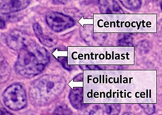
Follicular dendritic cells (FDC) are cells of the immune system found in primary and secondary lymph follicles of the B cell areas of the lymphoid tissue. Unlike dendritic cells (DC), FDCs are not derived from the bone-marrow hematopoietic stem cell, but are of mesenchymal origin. Possible functions of FDC include: organizing lymphoid tissue's cells and microarchitecture, capturing antigen to support B cell, promoting debris removal from germinal centers, and protecting against autoimmunity. Disease processes that FDC may contribute include primary FDC-tumor, chronic inflammatory conditions, HIV-1 infection development, and neuroinvasive scrapie.

<span class="mw-page-title-main">Interleukin 21</span> Mammalian protein found in humans

Interleukin 21 (IL-21) is a protein that in humans is encoded by the IL21 gene.

<span class="mw-page-title-main">CCL18</span> Mammalian protein found in Homo sapiens

Chemokine ligand 18 (CCL18) is a small cytokine belonging to the CC chemokine family. The functions of CCL18 have been well studied in laboratory settings, however the physiological effects of the molecule in living organisms have been difficult to characterize because there is no similar protein in rodents that can be studied. The receptor for CCL18 has been identified in humans only recently, which will help scientists understand the molecule's role in the body.

<span class="mw-page-title-main">BCL6</span> Transcription factor for converting Naive T cells to TFH

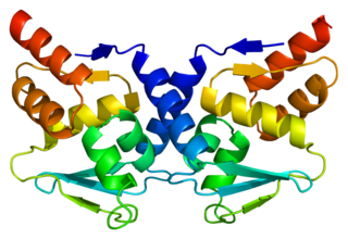
Bcl-6 is a protein that in humans is encoded by the BCL6 gene. BCL6 is a master transcription factor for regulation of T follicular helper cells proliferation. BCL6 has three evolutionary conserved structural domains. The interaction of these domains with corepressors allows for germinal center development and leads to B cell proliferation.

<span class="mw-page-title-main">ICOSLG</span> Protein-coding gene in the species Homo sapiens

ICOS ligand is a protein that in humans is encoded by the ICOSLG gene located at chromosome 21. ICOSLG has also been designated as CD275.

<span class="mw-page-title-main">CD160</span> Protein-coding gene in the species Homo sapiens

CD160 antigen is a protein that in humans is encoded by the CD160 gene.

<span class="mw-page-title-main">HLA-DQB2</span> Protein-coding gene in the species Homo sapiens

HLA class II histocompatibility antigen, DX beta chain is a protein that in humans is encoded by the HLA-DQB2 gene.

<span class="mw-page-title-main">Marginal-zone B cell</span>

Marginal-zone B cells are noncirculating mature B cells that in humans segregate anatomically into the marginal zone (MZ) of the spleen and certain other types of lymphoid tissue. The MZ B cells within this region typically express low-affinity polyreactive B-cell receptors (BCR), high levels of IgM, Toll-like receptors (TLRs), CD21, CD1, CD9, CD27 with low to negligible levels of secreted-IgD, CD23, CD5, and CD11b that help to distinguish them phenotypically from follicular (FO) B cells and B1 B cells.

Within the immune system, Follicular B cells are a type of B cell that reside in primary and secondary lymphoid follicles of secondary and tertiary lymphoid organs, including spleen and lymph nodes. Antibody responses against proteins are believed to involve follicular B cell pathways in secondary lymphoid organs.

<span class="mw-page-title-main">Centroblast</span>

A centroblast generally refers to an activated B cell that is enlarged and is rapidly proliferating in the germinal center of a lymphoid follicle. They are specifically located in the dark zone of the germinal center. Centroblasts form from naive B cells being exposed to follicular dendritic cell cytokines, such as IL-6, IL-15, 8D6, and BAFF. Stimulation from helper T cells is also required for centroblast development. Interaction between CD40 ligand on an activated T helper cell and the B cell CD40 receptor induces centroblasts to express activation-induced cytidine deaminase, leading to somatic hypermutation, allowing the B cell receptor to potentially gain stronger affinity for an antigen. In the absence of FDC and helper T cell stimulation, centroblasts are unable to differentiate and will undergo CD95-mediated apoptosis.

Regulatory B cells (Bregs or Breg cells) represent a small population of B cells that participates in immunomodulation and in the suppression of immune responses. The population of Bregs can be further separated into different human or murine subsets such as B10 cells, marginal zone B cells, Br1 cells, GrB+B cells, CD9+ B cells, and even some plasmablasts or plasma cells. Bregs regulate the immune system by different mechanisms. One of the main mechanisms is the production of anti-inflammatory cytokines such as interleukin 10 (IL-10), IL-35, or transforming growth factor beta (TGF-β). Another known mechanism is the production of cytotoxic Granzyme B. Bregs also express various inhibitory surface markers such as programmed death-ligand 1 (PD-L1), CD39, CD73, and aryl hydrocarbon receptor. The regulatory effects of Bregs were described in various models of inflammation, autoimmune diseases, transplantation reactions, and in anti-tumor immunity.

T-cell receptor revision is a process in the peripheral immune system which is used by mature T cells to alter their original antigenic specificity based on rearranged T cell receptors (TCR). This process can lead either to continuous appearance of potentially self-reactive T cells in the body, not controlled by the central tolerance mechanism in the thymus or better eliminate such self-reactive T cells on the other hand and thus contributing to peripheral tolerance - the extent of each has not been completely understood yet. This process occurs during follicular helper T cell formation in lymph node germinal centers.

<span class="mw-page-title-main">Elina Zúñiga</span> Argentinian immunologist

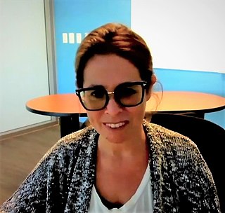
Elina Zúñiga is an Argentinian Immunologist and Professor of Molecular Biology in the Division of Biological Sciences at the University of California, San Diego. Zúñiga has made critical discoveries regarding host-virus interactions in both acute and chronic infections. Using lymphocytic choriomeningitis models (LCMV) and murine cytomegalovirus models, her laboratory at UCSD studies host immune adaptations in chronic viral disease and methods of viral suppression of the immune system in order to develop novel methods and identify novel targets of anti-viral defence. In 2018, Zúñiga was chosen by the American Association of Immunologists to give the international Vanguard Lecture. Zúñiga is also co-founder of the Global Immunotalks series which she and Carla Rothlin started in 2020 as a means to make cutting-edge immunology research freely available and easily accessible to a global audience.

References

  1. Fazilleau; Mark, L; McHeyzer-Williams, LJ; McHeyzer-Williams, MG; et al. (March 2009). "Follicular helper T cells: lineage and location". Immunity. 30 (3): 324–35. doi:10.1016/j.immuni.2009.03.003. PMC   2731675 . PMID   19303387.
  2. Seo; Youn, J; Kim, PH; et al. (May 2009). "IL-21 ensures TGF-beta 1-induced IgA isotype expression in mouse Peyer's patches". J Leukoc Biol. 85 (5): 744–50. doi:10.1189/jlb.0708450. PMID   19168593. S2CID   22358429.
  3. Zaretsky; Taylor, JJ; King, IL; Marshall, FA; Mohrs, M; Pearce, EJ; et al. (May 2009). "T follicular helper cells differentiate from Th2 cells in response to helminth antigens". J Exp Med. 206 (5): 991–9. doi:10.1084/jem.20090303. PMC   2715032 . PMID   19380637.
  4. Kurosaki; Kometani, K; Ise, W (March 2015). "Memory B cells". Nat Rev Immunol. 15 (3): 149–59. doi:10.1038/nri3802. PMID   25677494. S2CID   20825732.
  5. Chtanova; Tangye, SG; Newton, R; Frank, N; Hodge, MR; Rolph, MS; MacKay, CR; et al. (July 2004). "T follicular helper cells express a distinctive transcriptional profile, reflecting their role as non-Th1/Th2 effector cells that provide help for B cells". J Immunol. 173 (1): 68–78. doi: 10.4049/jimmunol.173.1.68 . PMID   15210760.
  6. Nurieva RI, Chung Y, Hwang D, Yang XO, Kang HS, Ma L, Wang YH, Watowich SS, et al. (2008). "Generation of T follicular helper cells is mediated by interleukin-21 but independent of T helper 1, 2, or 17 cell lineages". Immunity. 29 (1): 138–49. doi:10.1016/j.immuni.2008.05.009. PMC   2556461 . PMID   18599325.
  7. Akiba; Takeda, K; Kojima, Y; Usui, Y; Harada, N; Yamazaki, T; Ma, J; Tezuka, K; et al. (Aug 2005). "The role of ICOS in the CXCR5+ follicular B helper T cell maintenance in vivo". J Immunol. 175 (4): 2340–8. doi: 10.4049/jimmunol.175.4.2340 . PMID   16081804.
  8. Bauquet; Jin, H; Paterson, AM; Mitsdoerffer, M; Ho, IC; Sharpe, AH; Kuchroo, VK (Feb 2009). "The costimulatory molecule ICOS regulates the expression of c-Maf and IL-21 in the development of follicular T helper cells and TH-17 cells". Nat Immunol. 10 (2): 167–75. doi:10.1038/ni.1690. PMC   2742982 . PMID   19098919.
  9. Vogelzang; McGuire, HM; Yu, D; Sprent, J; MacKay, CR; King, C (July 2008). "A fundamental role for interleukin-21 in the generation of T follicular helper cells". Immunity. 29 (1): 127–37. doi: 10.1016/j.immuni.2008.06.001 . PMID   18602282.
  10. Ichii; Sakamoto, A; Hatano, M; Okada, S; Toyama, H; Taki, S; Arima, M; Kuroda, Y; Tokuhisa, T; et al. (June 2002). "Role for Bcl-6 in the generation and maintenance of memory CD8+ T cells". Nat Immunol. 3 (6): 558–63. doi:10.1038/ni802. hdl: 20.500.14094/D1002645 . PMID   12021781. S2CID   2552376.
  11. Kim, JR; Lim, HW; Kang, SG; Hillsamer, P; Kim, CH (Feb 4, 2005). "Human CD57+ germinal center-T cells are the major helpers for GC-B cells and induce class switch recombination". BMC Immunology. 6: 3. doi: 10.1186/1471-2172-6-3 . PMC   548684 . PMID   15694005.
  12. McHeyzer-Williams, M; Okitsu, S; Wang, N; McHeyzer-Williams, L (Dec 9, 2011). "Molecular programming of B cell memory". Nature Reviews. Immunology. 12 (1): 24–34. doi:10.1038/nri3128. PMC   3947622 . PMID   22158414.
  13. Yusuf I, Kageyama R, Monticelli L, Johnston RJ, Ditoro D, Hansen K, Barnett B, Crotty S (July 2010). "Germinal center T follicular helper cell IL-4 production is dependent on signaling lymphocytic activation molecule receptor (CD150)". J. Immunol. 185 (1): 190–202. doi:10.4049/jimmunol.0903505. PMC   2913439 . PMID   20525889.
  14. Ramiscal RR, Vinuesa CG (March 2013). "T-cell subsets in the germinal center". Immunol. Rev. 252 (1): 146–55. doi:10.1111/imr.12031. PMID   23405902. S2CID   21787025.
  15. Wollenberg I, Agua-Doce A, Hernández A, Almeida C, Oliveira VG, Faro J, Graca L (November 2011). "Regulation of the germinal center reaction by Foxp3+ follicular regulatory T cells". J. Immunol. 187 (9): 4553–60. doi: 10.4049/jimmunol.1101328 . PMID   21984700.
  16. Schultz, Bruce T.; Teigler, Jeffrey E.; Pissani, Franco; Oster, Alexander F.; Kranias, Gregory; Alter, Galit; Marovich, Mary; Eller, Michael A.; Dittmer, Ulf (2016-01-19). "Circulating HIV-Specific Interleukin-21(+)CD4(+) T Cells Represent Peripheral Tfh Cells with Antigen-Dependent Helper Functions". Immunity. 44 (1): 167–178. doi: 10.1016/j.immuni.2015.12.011 . ISSN   1097-4180. PMID   26795249.
  17. Arifuzzaman M, Rashu R, Leung DT, Hosen MI, Bhuiyan TR, Bhuiyan MS, Rahman MA, Khanam F, Saha A, Charles RC, LaRocque RC, Weil AA, Clements JD, Holmes RK, Calderwood SB, Harris JB, Ryan ET, Qadri F (August 2012). "Antigen-specific memory T cell responses after vaccination with an oral killed cholera vaccine in Bangali children and comparison to responses in patients with naturally acquired cholera". Clinical and Vaccine Immunology. 19 (8): 1304–11. doi:10.1128/CVI.00196-12. PMC   3416086 . PMID   22739692.
  18. Lefebvre JS, Maue AC, Eaton SM, Lanthier PA, Tighe M, Haynes L (2012). "The aged microenvironment contributes to the age-related functional defects of CD4 T cells in mice". Aging Cell. 11 (5): 732–40. doi:10.1111/j.1474-9726.2012.00836.x. PMC   3444657 . PMID   22607653.
  19. Perkey E, Miller RA, Garcia GG (2012). "Ex vivo enzymatic treatment of aged CD4 T cells restores cognate T cell helper function and enhances antibody production in mice". J Immunol. 189 (12): 5582–9. doi:10.4049/jimmunol.1200487. PMC   3518580 . PMID   23136198.
  20. Craft JE. (2012). "Follicular helper T cells in immunity and systemic autoimmunity". Nat Rev Rheumatol. 8 (6): 337–47. doi:10.1038/nrrheum.2012.58. PMC   3604997 . PMID   22549246.