
The danger triangle of the face consists of the area from the corners of the mouth to the bridge of the nose, including the nose and maxilla. Due to the special nature of the blood supply to the human nose and surrounding area, it is possible for retrograde infection from the nasal area to spread to the brain, causing cavernous sinus thrombosis, meningitis, or brain abscess.
Articles related to anatomy include:

The oculomotor nerve, also known as the third cranial nerve, cranial nerve III, or simply CN III, is a cranial nerve that enters the orbit through the superior orbital fissure and innervates extraocular muscles that enable most movements of the eye and that raise the eyelid. The nerve also contains fibers that innervate the intrinsic eye muscles that enable pupillary constriction and accommodation. The oculomotor nerve is derived from the basal plate of the embryonic midbrain. Cranial nerves IV and VI also participate in control of eye movement.
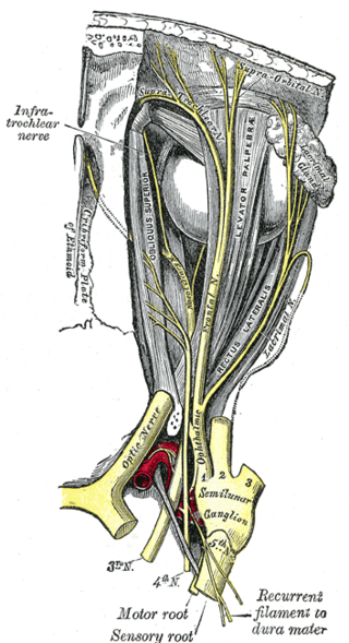
The supraorbital nerve is one of two branches of the frontal nerve, itself a branch of the ophthalmic nerve. The other branch of the frontal nerve is the supratrochlear nerve.

The scalp is the area of the head where head hair grows. It is made up of skin, layers of connective and fibrous tissues, and the membrane of the skull. Anatomically, the scalp is part of the epicranium, a collection of structures covering the cranium. The scalp is bordered by the face at the front, and by the neck at the sides and back. The scientific study of hair and scalp is called trichology.

In anatomy, the orbit is the cavity or socket/hole of the skull in which the eye and its appendages are situated. "Orbit" can refer to the bony socket, or it can also be used to imply the contents. In the adult human, the volume of the orbit is 30 millilitres, of which the eye occupies 6.5 ml. The orbital contents comprise the eye, the orbital and retrobulbar fascia, extraocular muscles, cranial nerves II, III, IV, V, and VI, blood vessels, fat, the lacrimal gland with its sac and duct, the eyelids, medial and lateral palpebral ligaments, cheek ligaments, the suspensory ligament, septum, ciliary ganglion and short ciliary nerves.

The levator palpebrae superioris is the muscle in the orbit that elevates the upper eyelid.

The superior orbital fissure is a foramen or cleft of the skull between the lesser and greater wings of the sphenoid bone. It gives passage to multiple structures, including the oculomotor nerve, trochlear nerve, ophthalmic nerve, abducens nerve, ophthalmic veins, and sympathetic fibres from the cavernous plexus.

The ophthalmic artery (OA) is an artery of the head. It is the first branch of the internal carotid artery distal to the cavernous sinus. Branches of the ophthalmic artery supply all the structures in the orbit around the eye, as well as some structures in the nose, face, and meninges. Occlusion of the ophthalmic artery or its branches can produce sight-threatening conditions.

The ophthalmic nerve (CN V1) is a sensory nerve of the head. It is one of three divisions of the trigeminal nerve (CN V), a cranial nerve. It has three major branches which provide sensory innervation to the eye, and the skin of the upper face and anterior scalp, as well as other structures of the head.

The nasociliary nerve is a branch of the ophthalmic nerve (CN V1) (which is in turn a branch of the trigeminal nerve (CN V)). It is intermediate in size between the other two branches of the ophthalmic nerve, the frontal nerve and lacrimal nerve.
The lacrimal nerve is the smallest of the three main branches of the ophthalmic nerve (CN V1) (itself a branch of the trigeminal nerve (CN V)).

The supratrochlear nerve is a branch of the frontal nerve, itself a branch of the ophthalmic nerve (CN V1) from the trigeminal nerve (CN V). It provides sensory innervation to the skin of the forehead and the upper eyelid.

The superior tarsal muscle is a smooth muscle adjoining the levator palpebrae superioris muscle that helps to raise the upper eyelid.
The supraorbital artery is a branch of the ophthalmic artery. It passes anteriorly within the orbit to exit the orbit through the supraorbital foramen or notch alongside the supraorbital nerve, splitting into two terminal branches which go on to form anastomoses with arteries of the head.

The supratrochlear artery is one of the terminal branches of the ophthalmic artery. It arises within the orbit. It exits the orbit alongside the supratrochlear nerve. It contributes arterial supply to the skin, muscles and pericranium of the forehead.

The following outline is provided as an overview of and topical guide to human anatomy:

The canine space, is a fascial space of the head and neck. It is a thin potential space on the face, and is paired on either side. It is located between the levator anguli oris muscle inferiorly and the levator labii superioris muscle superiorly. The term is derived from the fact that the space is in the region of the canine fossa, and that infections originating from the maxillary canine tooth may spread to involve the space. Infra-orbital is derived from infra- meaning below and orbit which refers to the eye socket.
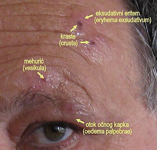
Herpes zoster ophthalmicus (HZO), also known as ophthalmic zoster, is shingles involving the eye or the surrounding area. Common signs include a rash of the forehead with swelling of the eyelid. There may also be eye pain and redness, inflammation of the conjunctiva, cornea or uvea, and sensitivity to light. Fever and tingling of the skin and allodynia near the eye may precede the rash. Complications may include visual impairment, increased pressure within the eye, chronic pain, and stroke.

The ciliary ganglion is a parasympathetic ganglion located just behind the eye in the posterior orbit. Three types of axons enter the ciliary ganglion but only the preganglionic parasympathetic axons synapse there. The entering axons are arranged into three roots of the ciliary ganglion, which join enter the posterior surface of the ganglion.


