History of magnetic resonance imaging

Last updated

The history of magnetic resonance imaging (MRI) includes the work of many researchers who contributed to the discovery of nuclear magnetic resonance (NMR) and described the underlying physics of magnetic resonance imaging, starting early in the twentieth century. MR imaging was invented by Paul C. Lauterbur who developed a mechanism to encode spatial information into an NMR signal using magnetic field gradients in September 1971; he published the theory behind it in March 1973. [1] [2]

Contents

The factors leading to image contrast (differences in tissue relaxation time values) had been described nearly 20 years earlier by physician and scientist Erik Odeblad and Gunnar Lindström. [3] [4] Among many other researchers in the late 1970s and 1980s, Peter Mansfield further refined the techniques used in MR image acquisition and processing, and in 2003 he and Lauterbur were awarded the Nobel Prize in Physiology or Medicine for their contributions to the development of MRI. The first clinical MRI scanners were installed in the early 1980s and significant development of the technology followed in the decades since, leading to its widespread use in medicine today.

Nuclear magnetic resonance

In 1950, spin echoes and free induction decay were first detected by Erwin Hahn [5] [6] and in 1952, Herman Carr produced a one-dimensional NMR spectrum as reported in his Harvard PhD thesis. [7] [8] [9]

The next step (from spectra to imaging) was proposed by Vladislav Ivanov in Soviet Union, who filed in 1960 a patent application for a Magnetic Resonance Imaging device. [10] [11] [12] Ivanov's main contribution was the idea of using magnetic field gradient, combined with a selective frequency excitation/readout, to encode the spatial coordinates. In modern terms, it was only proton-density (not relaxation times) imaging, which was also slow, since only one gradient direction was used at a time and the imaging had to be done slice-by-slice. Nevertheless, it was a true magnetic resonance imaging procedure. Originally rejected as "improbable", Ivanov's application was finally approved in 1984 (with the original priority date). [13]

Relaxation times and early development of MRI

By 1959, Jay Singer had studied blood flow by NMR relaxation time measurements of blood in living humans. [14] [15] Such measurements were not introduced into common medical practice until the mid-1980s, although a patent for a whole-body NMR machine to measure blood flow in the human body was filed by Alexander Ganssen in early 1967. [15] [16] [17] [18] [19]

In the 1960s, the results of work on relaxation, diffusion, and chemical exchange of water in cells and tissues of various types appeared in the scientific literature. [16] In 1967, Ligon reported the measurement of NMR relaxation of water in the arms of living human subjects. [16] In 1968, Jackson and Langham published the first NMR signals from a living animal, an anesthetized rat. [16] [20]

In the 1970s, it was realized that the relaxation times are key determinants of contrast in MRI and can be used to detect and differentiate a range of pathologies. A number of research groups had showed that early cancer cells tended to exhibit longer relaxation times than their corresponding normal cells and as such stimulated initial interest in the idea of detecting cancer with NMR. These early groups include Damadian, [21] Hazlewood and Chang [22] and several others. This also initiated a program to catalog the relaxation times of a wide range of biological tissues, which became one of the main motivations for the development of MRI. [23]

Raymond Damadian's "Apparatus and method for detecting cancer in tissue". Damadian invention.jpg
Raymond Damadian's "Apparatus and method for detecting cancer in tissue".

In a March 1971 paper in the journal Science , [21] Raymond Damadian, an Armenian-American doctor and professor at the Downstate Medical Center State University of New York (SUNY), reported that tumors and normal tissue can be distinguished in vivo by NMR. Damadian's initial methods were flawed for practical use, [24] relying on a point-by-point scan of the entire body and using relaxation rates, which turned out not to be an effective indicator of cancerous tissue. [25] While researching the analytical properties of magnetic resonance, Damadian created a hypothetical magnetic resonance cancer-detecting machine in 1972. He patented such a machine, U.S. Patent 3,789,832 on February 5, 1974. [26] Lawrence Bennett and Dr. Irwin Weisman also found in 1972 that neoplasms display different relaxation times than corresponding normal tissue. [27] [28] Zenuemon Abe and his colleagues applied the patent for a targeted NMR scanner, U.S. Patent 3,932,805 in 1973. [29] They published this technique in 1974. [15] [16] [30] Damadian claims to have invented the MRI. [31]

The U.S. National Science Foundation notes "The patent included the idea of using NMR to 'scan' the human body to locate cancerous tissue." [32] However, it did not describe a method for generating pictures from such a scan or precisely how such a scan might be done. [33] [34]

Imaging

Paul Lauterbur at Stony Brook University expanded on Carr's technique and developed a way to generate the first MRI images, in 2D and 3D, using gradients. In 1973, Lauterbur published the first nuclear magnetic resonance image [1] [35] and the first cross-sectional image of a living mouse in January 1974. [36] In the late 1970s, Peter Mansfield, a physicist and professor at the University of Nottingham, England, developed the echo-planar imaging (EPI) technique that would lead to scans taking seconds rather than hours and produce clearer images than Lauterbur had. [37] Damadian, along with Larry Minkoff and Michael Goldsmith, obtained an image of a tumor in the thorax of a mouse in 1976. [38] They also performed the first MRI body scan of a human being on July 3, 1977, [39] [40] studies they published in 1977. [38] [41] In 1979, Richard S. Likes filed a patent on k-space U.S. Patent 4,307,343 .

MRI Scanner Mark One. The first MRI scanner to be built and used, in Aberdeen Royal Infirmary in Scotland. MRI Scanner Mark One.jpg
MRI Scanner Mark One. The first MRI scanner to be built and used, in Aberdeen Royal Infirmary in Scotland.

Full-body scanning

During the 1970s a team led by John Mallard built the first full-body MRI scanner at the University of Aberdeen. [42] On 28 August 1980 they used this machine to obtain the first clinically useful image of a patient's internal tissues using MRI, which identified a primary tumour in the patient's chest, an abnormal liver, and secondary cancer in his bones. [43] This machine was later used at St Bartholomew's Hospital, in London, from 1983 to 1993. Mallard and his team are credited for technological advances that led to the widespread introduction of MRI. [44]

In 1975, the University of California, San Francisco Radiology Department founded the Radiologic Imaging Laboratory (RIL). [45] With the support of Pfizer, Diasonics, and later Toshiba America MRI, the lab developed new imaging technology and installed systems in the United States and worldwide. [46] In 1981 RIL researchers, including Leon Kaufman and Lawrence Crooks, published Nuclear Magnetic Resonance Imaging in Medicine. In the 1980s the book was considered the definitive introductory textbook to the subject. [47]

In 1980 Paul Bottomley joined the GE Research Center in Schenectady, New York. His team ordered the highest field-strength magnet then available, a 1.5  T system, and built the first high-field device, overcoming problems of coil design, RF penetration and signal-to-noise ratio to build the first whole-body MRI/MRS scanner. [48] The results translated into the highly successful 1.5 T MRI product-line, delivering over 20,000 systems. In 1982, Bottomley performed the first localized MRS in the human heart and brain. After starting a collaboration on heart applications with Robert Weiss at Johns Hopkins, Bottomley returned to the university in 1994 as Russell Morgan Professor and director of the MR Research Division. [49]

Additional techniques

In 1986, Charles L. Dumoulin and Howard R. Hart at General Electric developed MR angiography [50] and Denis Le Bihan, obtained the first images and later patented diffusion MRI. [51] In 1988, Arno Villringer and colleagues demonstrated that susceptibility contrast agents may be employed in perfusion MRI. [52] In 1990, Seiji Ogawa at AT&T Bell labs recognized that oxygen-depleted blood with dHb was attracted to a magnetic field, and discovered the technique that underlies Functional Magnetic Resonance Imaging (fMRI). [53]

In the early 1990s, Peter Basser and Le Bihan working at NIH, [54] and Aaron Filler, Franklyn Howe and colleagues published the first DTI and tractographic brain images. [55] [56] [57] Joseph Hajnal, Young and Graeme Bydder described the use of FLAIR pulse sequence to demonstrate high signal regions in normal white matter in 1992. [58] In the same year, arterial spin labelling was developed by John Detre and Alan P. Koretsky. [59] In 1997, Jürgen R. Reichenbach, E. Mark Haacke and coworkers at Washington University School of Medicine developed Susceptibility weighted imaging. [60]

Advances in semiconductor technology were crucial to the development of practical MRI, which requires a large amount of computational power. [61]

Although MRI is most commonly performed in the clinic at 1.5 T, higher fields such as 3 T for clinical imaging and more recently 7 T for research purposes are gaining popularity because of their increased sensitivity and resolution. In research laboratories, human studies have been performed at 9.4 T (2006) [62] and up to 10.5 T (2019). [63] Non-human animal studies have been performed at up to 21.1 T. [64]

Bedside imaging

In 2020, the United States Food and Drug Administration (USFDA) proffered 510(k)[ clarification needed ] [65] approval of Hyperfine Research's bedside MRI system. The Hyperfine system claims 1/20th the cost, 1/35th the power consumption, and 1/10th the weight of conventional MRI systems. [66] It uses a standard electrical outlet for power. [67]

2003 Nobel Prize

Reflecting the fundamental importance and applicability of MRI in medicine, Paul Lauterbur of Stony Brook University and Sir Peter Mansfield of the University of Nottingham were awarded the 2003 Nobel Prize in Physiology or Medicine for their "discoveries concerning magnetic resonance imaging". The Nobel citation acknowledged Lauterbur's insight of using magnetic field gradients to determine spatial localization, a discovery that allowed the acquisition of 3D and 2D images. Mansfield was credited with introducing the mathematical formalism and developing techniques for efficient gradient utilization and fast imaging. The research that won the Prize was done almost 30 years earlier while Paul Lauterbur was a professor in the Department of Chemistry at Stony Brook University in New York. [1]

Related Research Articles

<span class="mw-page-title-main">Magnetic resonance imaging</span> Medical imaging technique

Magnetic resonance imaging (MRI) is a medical imaging technique used in radiology to form pictures of the anatomy and the physiological processes of the body. MRI scanners use strong magnetic fields, magnetic field gradients, and radio waves to generate images of the organs in the body. MRI does not involve X-rays or the use of ionizing radiation, which distinguishes it from computed tomography (CT) and positron emission tomography (PET) scans. MRI is a medical application of nuclear magnetic resonance (NMR) which can also be used for imaging in other NMR applications, such as NMR spectroscopy.

<span class="mw-page-title-main">Paul Lauterbur</span> American chemist

Paul Christian Lauterbur was an American chemist who shared the Nobel Prize in Physiology or Medicine in 2003 with Peter Mansfield for his work which made the development of magnetic resonance imaging (MRI) possible.

<span class="mw-page-title-main">Peter Mansfield</span> British physicist known for magnetic resonance imaging

Sir Peter Mansfield was a British physicist who was awarded the 2003 Nobel Prize in Physiology or Medicine, shared with Paul Lauterbur, for discoveries concerning Magnetic Resonance Imaging (MRI). Mansfield was a professor at the University of Nottingham.

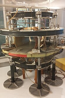
Raymond Vahan Damadian was an American physician, medical practitioner, and inventor of the first NMR scanning machine.

<span class="mw-page-title-main">Diffusion MRI</span> Method of utilizing water in magnetic resonance imaging

Diffusion-weighted magnetic resonance imaging is the use of specific MRI sequences as well as software that generates images from the resulting data that uses the diffusion of water molecules to generate contrast in MR images. It allows the mapping of the diffusion process of molecules, mainly water, in biological tissues, in vivo and non-invasively. Molecular diffusion in tissues is not random, but reflects interactions with many obstacles, such as macromolecules, fibers, and membranes. Water molecule diffusion patterns can therefore reveal microscopic details about tissue architecture, either normal or in a diseased state. A special kind of DWI, diffusion tensor imaging (DTI), has been used extensively to map white matter tractography in the brain.

<span class="mw-page-title-main">Spin echo</span> Response of spin to electromagnetic radiation

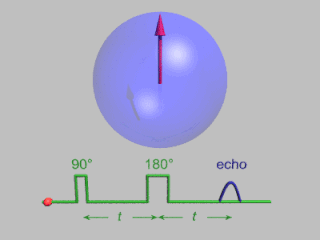
In magnetic resonance, a spin echo or Hahn echo is the refocusing of spin magnetisation by a pulse of resonant electromagnetic radiation. Modern nuclear magnetic resonance (NMR) and magnetic resonance imaging (MRI) make use of this effect.

Kenneth Kin Man Kwong is a Hong Kong-born American nuclear physicist. He is a pioneer in human brain imaging. He received his bachelor's degree in Political Science in 1972 from the University of California, Berkeley. He went on to receive his Ph.D. in physics from the University of California, Riverside studying photon-photon collision interactions.

<span class="mw-page-title-main">Magnetic resonance neurography</span>

Magnetic resonance neurography (MRN) is the direct imaging of nerves in the body by optimizing selectivity for unique MRI water properties of nerves. It is a modification of magnetic resonance imaging. This technique yields a detailed image of a nerve from the resonance signal that arises from in the nerve itself rather than from surrounding tissues or from fat in the nerve lining. Because of the intraneural source of the image signal, the image provides a medically useful set of information about the internal state of the nerve such as the presence of irritation, nerve swelling (edema), compression, pinch or injury. Standard magnetic resonance images can show the outline of some nerves in portions of their courses but do not show the intrinsic signal from nerve water. Magnetic resonance neurography is used to evaluate major nerve compressions such as those affecting the sciatic nerve (e.g. piriformis syndrome), the brachial plexus nerves (e.g. thoracic outlet syndrome), the pudendal nerve, or virtually any named nerve in the body. A related technique for imaging neural tracts in the brain and spinal cord is called magnetic resonance tractography or diffusion tensor imaging.

<span class="mw-page-title-main">Physics of magnetic resonance imaging</span> Overview article

The physics of magnetic resonance imaging (MRI) concerns fundamental physical considerations of MRI techniques and technological aspects of MRI devices. MRI is a medical imaging technique mostly used in radiology and nuclear medicine in order to investigate the anatomy and physiology of the body, and to detect pathologies including tumors, inflammation, neurological conditions such as stroke, disorders of muscles and joints, and abnormalities in the heart and blood vessels among others. Contrast agents may be injected intravenously or into a joint to enhance the image and facilitate diagnosis. Unlike CT and X-ray, MRI uses no ionizing radiation and is, therefore, a safe procedure suitable for diagnosis in children and repeated runs. Patients with specific non-ferromagnetic metal implants, cochlear implants, and cardiac pacemakers nowadays may also have an MRI in spite of effects of the strong magnetic fields. This does not apply on older devices, and details for medical professionals are provided by the device's manufacturer.

<span class="mw-page-title-main">Real-time MRI</span> Type of MRI

Real-time magnetic resonance imaging (RT-MRI) refers to the continuous monitoring ("filming") of moving objects in real time. Because MRI is based on time-consuming scanning of k-space, real-time MRI was possible only with low image quality or low temporal resolution. Using an iterative reconstruction algorithm these limitations have recently been removed: a new method for real-time MRI achieves a temporal resolution of 20 to 30 milliseconds for images with an in-plane resolution of 1.5 to 2.0 mm. Real-time MRI promises to add important information about diseases of the joints and the heart. In many cases MRI examinations may become easier and more comfortable for patients.

<span class="mw-page-title-main">Magnetic resonance imaging of the brain</span>

Magnetic resonance imaging of the brain uses magnetic resonance imaging (MRI) to produce high quality two-dimensional or three-dimensional images of the brain and brainstem as well as the cerebellum without the use of ionizing radiation (X-rays) or radioactive tracers.

<span class="mw-page-title-main">Intravoxel incoherent motion</span> Concept and a method initially introduced and developed by Le Bihan et al

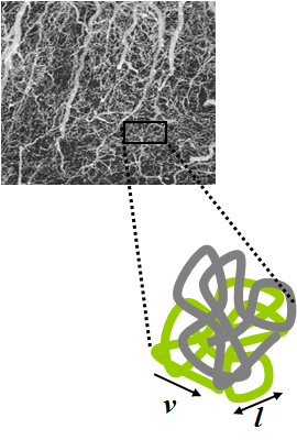
Intravoxel incoherent motion (IVIM) imaging is a concept and a method initially introduced and developed by Le Bihan et al. to quantitatively assess all the microscopic translational motions that could contribute to the signal acquired with diffusion MRI. In this model, biological tissue contains two distinct environments: molecular diffusion of water in the tissue, and microcirculation of blood in the capillary network (perfusion). The concept introduced by D. Le Bihan is that water flowing in capillaries mimics a random walk (Fig.1), as long as the assumption that all directions are represented in the capillaries is satisfied.

<span class="mw-page-title-main">Jürgen Hennig</span> German chemist and medical physicist (born 1951)

Jürgen Klaus Hennig is a German chemist and medical physicist. Internationally he is considered to be one of the pioneers of Magnetic Resonance Imaging for clinical diagnostics. He is the Scientific Director of the Department of Diagnostic Radiology and Chairman of the Magnetic Resonance Development and Application Center (MRDAC) at the University Medical Center Freiburg. In the year 2003 he was awarded the Max Planck Research Award in the category of Biosciences and Medicine.

<span class="mw-page-title-main">Perfusion MRI</span>

Perfusion MRI or perfusion-weighted imaging (PWI) is perfusion scanning by the use of a particular MRI sequence. The acquired data are then post-processed to obtain perfusion maps with different parameters, such as BV, BF, MTT and TTP.

Sodium MRI is a specialised magnetic resonance imaging technique that uses strong magnetic fields, magnetic field gradients, and radio waves to generate images of the distribution of sodium in the body, as opposed to more common forms of MRI that utilise protons present in water (1H-MRI). Like the proton, sodium is naturally abundant in the body, so can be imaged directly without the need for contrast agents or hyperpolarization. Furthermore, sodium ions play a role in important biological processes via their contribution to concentration and electrochemical gradients across cellular membranes, making it of interest as an imaging target in health and disease.

Synthetic MRI is a simulation method in Magnetic Resonance Imaging (MRI), for generating contrast weighted images based on measurement of tissue properties. The synthetic (simulated) images are generated after an MR study, from parametric maps of tissue properties. It is thereby possible to generate several contrast weightings from the same acquisition. This is different from conventional MRI, where the signal acquired from the tissue is used to generate an image directly, often generating only one contrast weighting per acquisition. The synthetic images are similar in appearance to those normally acquired with an MRI scanner.

<span class="mw-page-title-main">MRI sequence</span>

An MRI sequence in magnetic resonance imaging (MRI) is a particular setting of pulse sequences and pulsed field gradients, resulting in a particular image appearance.

Denis Le Bihan is a medical doctor, physicist, member of the Institut de France, member of the French Academy of Technologies and director since 2007 of NeuroSpin, an institution of the Atomic Energy and Alternative Energy Commission (CEA) in Saclay, dedicated to the study of the brain by magnetic resonance imaging (MRI) with a very high magnetic field. Denis Le Bihan has received international recognition for his outstanding work, introducing new imaging methods, particularly for the study of the human brain, as evidenced by the many international awards he has received, such as the Gold Medal of the International Society of Magnetic Resonance in Medicine (2001), the coveted Lounsbery Prize, the Louis D. Prize from the Institut de France, the prestigious Honda Prize (2012), the Louis-Jeantet Prize (2014), the Rhein Foundation Award (2021). His work has focused on the introduction, development and application of highly innovative methods, notably diffusion MRI.

Hyperpolarized 129Xe gas magnetic resonance imaging (MRI) is a medical imaging technique used to visualize the anatomy and physiology of body regions that are difficult to image with standard proton MRI. In particular, the lung, which lacks substantial density of protons, is particularly useful to be visualized with 129Xe gas MRI. This technique has promise as an early-detection technology for chronic lung diseases and imaging technique for processes and structures reliant on dissolved gases. 129Xe is a stable, naturally occurring isotope of xenon with 26.44% isotope abundance. It is one of two Xe isotopes, along with 131Xe, that has non-zero spin, which allows for magnetic resonance. 129Xe is used for MRI because its large electron cloud permits hyperpolarization and a wide range of chemical shifts. The hyperpolarization creates a large signal intensity, and the wide range of chemical shifts allows for identifying when the 129Xe associates with molecules like hemoglobin. 129Xe is preferred over 131Xe for MRI because 129Xe has spin 1/2, a longer T1, and 3.4 times larger gyromagnetic ratio (11.78 MHz/T).

Hyperpolarized gas MRI, also known as hyperpolarized helium-3 MRI or HPHe-3 MRI, is a medical imaging technique that uses hyperpolarized gases to improve the sensitivity and spatial resolution of magnetic resonance imaging (MRI). This technique has many potential applications in medicine, including the imaging of the lungs and other areas of the body with low tissue density.

References

  1. 1 2 3 Lauterbur, P.C. (1973). "Image Formation by Induced Local Interactions: Examples of Employing Nuclear Magnetic Resonance". Nature . 242 (5394): 190–191. Bibcode:1973Natur.242..190L. doi:10.1038/242190a0. S2CID   4176060.
  2. Rinck, Peter A. (2024). Magnetic Resonance in Medicine. A critical introduction. e-Textbook (14th ed.). TRTF – The Round Table Foundation: TwinTree Media. "Magnetic Resonance in Medicine". www.magnetic-resonance.org.
  3. Odeblad, E. & Lindström, G. (1955). "Some preliminary observations on the proton magnetic resonance in biological samples". Acta Radiologica . 43 (6): 469–476. doi: 10.3109/00016925509172514 . PMID   14398444.
  4. Odeblad, Erik; Bhar, Baidya Nath & Lindström, Gunnar (July 1956). "Proton magnetic resonance of human red blood cells in heavy water exchange experiments". Archives of Biochemistry and Biophysics . 63 (1): 221–225. doi:10.1016/0003-9861(56)90025-X. PMID   13341059.
  5. Hahn, E. L. (1950). "Spin echoes". Physical Review . 80 (4): 580–594. Bibcode:1950PhRv...80..580H. doi:10.1103/PhysRev.80.580. S2CID   46554313.
  6. Hahn, E. L. (1950). "Nuclear Induction Due to Free Larmor Precession". Physical Review. 77 (2): 297–298. Bibcode:1950PhRv...77..297H. doi:10.1103/physrev.77.297.2. S2CID   92995835.
  7. Carr, Herman (1952). Free Precession Techniques in Nuclear Magnetic Resonance (PhD). Cambridge, MA: Harvard University. OCLC   76980558.[ page needed ]
  8. Carr, Herman Y. (July 2004). "Field Gradients in Early MRI". Physics Today . 57 (7): 83. Bibcode:2004PhT....57g..83C. doi: 10.1063/1.1784322 .
  9. Carr, Herman (1996). "Free Precession Techniques in Nuclear Magnetic Resonance". In Grant, David M.; Harris, Robin K. (eds.). Encyclopedia of Nuclear Magnetic Resonance. Vol. 1. Hoboken, NJ: Wiley and Sons. p. 253. ISBN   978-0-47195-839-0.[ permanent dead link ]
  10. MacWilliams, Bryon (November 2003). "Russian claims first in magnetic imaging". Nature. 426 (6965): 375. Bibcode:2003Natur.426..375M. doi: 10.1038/426375a . PMID   14647349.
  11. Bateneva, Tatiana (27 October 2003). "Привет Нобелю От Иванова: Как советский лейтенант-ракетчик перегнал Америку" [Regards To Nobel From Ivanov: How a Soviet rocket lieutenant surpassed America]. iNauka (in Russian). Archived from the original on 2005-08-17.
  12. "Патенты автора Иванов Владислав Александрович" [Patents by Vladislav Aleksandrovich Ivanov]. Findpatent.ru (in Russian).
  13. Bateneva, Tatiana (29 October 2003). "Best Regards To Alfred Nobel". iNauka. Archived from the original on 2009-12-13. Retrieved 2009-10-16.
  14. Singer, R.J. (1959). "Blood-flow rates by NMR measurements". Science. 130 (3389): 1652–1653. Bibcode:1959Sci...130.1652S. doi:10.1126/science.130.3389.1652. PMID   17781388. S2CID   42127984.
  15. 1 2 3 "A Short History of Magnetic Resonance Imaging from a European Point of View". European Magnetic Resonance Forum. Archived from the original on 2007-04-13. Retrieved 2016-08-08.
  16. 1 2 3 4 5 Rinck, P. (2017). "Chapter 20 : The history of MR imaging". Magnetic Resonance in Medicine (11th ed.).
  17. de 1566148
  18. Braun, Bianca (June 24, 2013). "First MAGNETOM scanner in the USA in 1983". Siemens. Archived from the original on 2015-03-05.
  19. Haase, Axel (March 2012). "Nachruf auf Alexander Ganssen" [Obituary for Alexander Ganssen]. Physik Journal (in German). 11 (3): 59. Archived from the original on 2016-06-20.
  20. Jackson, J.A. & Langham, W.H. (April 1968). "Whole-body NMR spectrometer". Review of Scientific Instruments . 39 (4): 510–513. Bibcode:1968RScI...39..510J. doi:10.1063/1.1683420. PMID   5641806.
  21. 1 2 Damadian, R. (March 1971). "Tumor detection by nuclear magnetic resonance". Science. 171 (3976): 1151–1153. Bibcode:1971Sci...171.1151D. doi:10.1126/science.171.3976.1151. PMID   5544870. S2CID   31895129.
  22. Hazelwood, C. F.; Chang, D.C.; Medina, D.; Cleveland, G.; Nichols, B. L. (1972). "Distinction between the preneoplastic and neoplastic state of murine mammary glands". Proceedings of the National Academy of Sciences of the United States of America. 69 (6): 1478–1480. arXiv: 1403.0914 . Bibcode:1972PNAS...69.1478H. doi: 10.1073/pnas.69.6.1478 . ISSN   0027-8424. PMC   426730 . PMID   4504364.
  23. Plewes, Donald; Kucharczyk, Walter (2012). "Physics of MRI: A Primer". Journal of Magnetic Resonance Imaging. 35 (5): 1038–1054. doi: 10.1002/jmri.23642 . PMID   22499279. S2CID   206101735.
  24. "The man who did not win". The Sydney Morning Herald . 2003-10-17. Retrieved 2007-08-04.
  25. Stracher, Cameron (2002-06-14). "Scan and Deliver". The Wall Street Journal. Archived from the original on 2007-09-30. Retrieved 2007-08-04.
  26. "Apparatus And Method For Detecting Cancer In Tissue". United States Patent and Trademark Office.
  27. Schooley, Jim (2010). "NBS Examines a Mouse and Opens a New Medical Specialty". NIST . Archived from the original on 2017-10-24.
  28. Weisman, I. D.; Bennett, L. H.; Maxwell, L. R.; Woods, M. W.; Burk, D. (1972-12-22). "Recognition of cancer in vivo by nuclear magnetic resonance". Science. 178 (4067): 1288–1290. Bibcode:1972Sci...178.1288W. doi:10.1126/science.178.4067.1288. ISSN   0036-8075. PMID   4640065. S2CID   8549657.
  29. Abe, Zen-uemon [in Japanese]; Tanaka, Kunio & Hotta, Masao (1974). "Non-invasive measurements of biological information with application of NMR". Transactions of the Society of Instrument and Control Engineers. 10 (3): 290–297. doi: 10.9746/sicetr1965.10.290 .
  30. Tanaka, K.; Yamada, T.; Shimizu, T.; Sano, F.; Abe, Z. (1974). "Fundamental investigations (in vitro) for a non-invasive method of tumor detection by nuclear magnetic resonance". Biotelemetry. 1 (6): 337–350. PMID   4478948.
  31. "The Inventor of the MRI on Real Science Radio". KGOV.com. Archived from the original on 2016-09-27. Retrieved 2016-09-25.
  32. "NSF history". SRI International . Archived from the original on 2012-01-03. Retrieved 2011-11-28.
  33. Maugh, Thomas H. II & Piller, Charles (2003-10-11). "Scientist Claims Exclusion From Nobel Prize for MRI". Los Angeles Times. Retrieved 2013-02-13.
  34. Saryan, Levon A. (2003-11-08). "Does Dr. Raymond Damadian Deserve the Nobel Prize for Medicine?". The Armenian Reporter. Archived from the original on 2012-11-06. Retrieved 2007-08-05 via HighBeam Research.
  35. Filler, A. (October 2009). "Magnetic resonance neurography and diffusion tensor imaging: origins, history, and clinical impact of the first 50,000 cases with an assessment of efficacy and utility in a prospective 5000-patient study group". Neurosurgery . 65 (4 Suppl): A29–43. doi:10.1227/01.NEU.0000351279.78110.00. PMC   2924821 . PMID   19927075.
  36. Lauterbur, P.C. (1974). "Magnetic resonance zeugmatography". Pure and Applied Chemistry . 40 (1–2): 149–157. doi: 10.1351/pac197440010149 . S2CID   53347935.
  37. Mansfield, P.; Grannell, P. (1975). "Diffraction and microscopy in solids and liquids by NMR". Physical Review B . 12 (9): 3618–3634. Bibcode:1975PhRvB..12.3618M. doi:10.1103/physrevb.12.3618.
  38. 1 2 Damadian, R.; Minkoff, L.; Goldsmith, M.; Stanford, M.; Koutcher, J. (1976). "Field focusing nuclear magnetic resonance (FONAR): visualization of a tumor in a live animal". Science. 194 (4272): 1430–1432. Bibcode:1976Sci...194.1430D. doi:10.1126/science.1006309. PMID   1006309.
  39. "First MRI and ultrasound scanning". Benjamin S. Beck. Archived from the original on 2011-11-20.
  40. Wakefield, Julie (June 2000). "The "Indomitable" MRI". Smithsonian . Archived from the original on 2012-09-09.
  41. Hinshaw, W.S.; Bottomley, P.A.; Holland, G.N. (1977). "Radiographic thin-section image of the human wrist by nuclear magnetic resonance". Nature. 270 (5639): 722–723. Bibcode:1977Natur.270..722H. doi:10.1038/270722a0. PMID   593393. S2CID   4183336.
  42. "Celebrated scientist donates medal collection". University of Aberdeen. 21 May 2009.
  43. "John Mallard". British Society for the History of Radiology. Archived from the original on 2017-03-07.
  44. "Mallard MRI body scanner, Aberdeen, Scotland, 1983". The Science Museum . Archived from the original on 2017-04-04.
  45. Crooks, Lawrence E. "Radiologic Imaging Laboratory records, 1977-2000". UCSF Library Archives & Special Collections.
  46. Crooks, Lawrence E. "Radiologic Imaging Laboratory Records". The Online Archive of California .
  47. Froelich, Jerry W. (17 February 1984). "Nuclear Magnetic Resonance (NMR) Imaging". Journal of the American Medical Association . 251 (7): 967–968. doi:10.1001/jama.1984.03340310071034.
  48. Sijbers, J.; Scheunders, P.; Bonnet, N.; Van Dyck, D.; Raman, E. (1996). "Quantification and improvement of the signal-to-noise ratio in a magnetic resonance image acquisition procedure". Magnetic Resonance Imaging . 14 (10): 1157–1163. CiteSeerX   10.1.1.20.3169 . doi:10.1016/S0730-725X(96)00219-6. PMID   9065906.
  49. "Biographical Sketch: Paul A. Bottomley, Ph.D." (PDF). Johns Hopkins University . 2003. Archived from the original (PDF) on July 4, 2010. Retrieved May 20, 2012.
  50. "Blood-flow checker". Popular Science . 230 (4): 12. April 1987.
  51. Le Bihan, D.; Breton, E. (1987). "Method to Measure the Molecular Diffusion and/or Perfusion Parameters of Live Tissue". US Patent # 4,809,701.
  52. Villringer, A.; Rosen, B. R.; Belliveau, J. W.; Ackerman, J. L.; Lauffer, R. B.; Buxton, R. B.; Chao, Y. S.; Wedeen, V. J.; Brady, T. J. (February 1988). "Dynamic imaging with lanthanide chelates in normal brain: contrast due to magnetic susceptibility effects". Magnetic Resonance in Medicine . 6 (2): 164–174. doi:10.1002/mrm.1910060205. ISSN   0740-3194. PMID   3367774. S2CID   41228095.
  53. Faro, Scott H.; Mohamed, Feroze B. (2010-01-15). Bold fMRI. a guide to functional imaging for neuroscientists. Springer. ISBN   978-1-4419-1328-9 . Retrieved 10 June 2015.
  54. Basser, Peter J. (2010). "Invention and Development of Diffusion Tensor MRI (DT-MRI or DTI) at the NIH". Diffusion MRI. pp. 730–740. CiteSeerX   10.1.1.645.9604 . doi:10.1093/med/9780195369779.003.0047. ISBN   978-0-19536-977-9.
  55. Howe, F. A.; Filler, A. G.; Bell, B. A.; Griffiths, J. R. (December 1992). "Magnetic resonance neurography". Magnetic Resonance in Medicine . 28 (2): 328–338. doi:10.1002/mrm.1910280215. ISSN   0740-3194. PMID   1461131. S2CID   36417513.
  56. Filler, A. G.; Howe, F. A.; Hayes, C. E.; Kliot, M.; Winn, H. R.; Bell, B. A.; Griffiths, J. R.; Tsuruda, J. S. (1993-03-13). "Magnetic resonance neurography". The Lancet . 341 (8846): 659–661. doi:10.1016/0140-6736(93)90422-D. ISSN   0140-6736. PMID   8095572. S2CID   24795253.
  57. Filler, Aaron (2009-10-01). "Magnetic Resonance Neurography and Diffusion Tensor Imaging". Neurosurgery. 65 (suppl 4): A29–A43. doi:10.1227/01.neu.0000351279.78110.00. ISSN   0148-396X. PMC   2924821 . PMID   19927075.
  58. Hajnal, J. V.; De Coene, B.; Lewis, P. D.; Baudouin, C. J.; Cowan, F. M.; Pennock, J. M.; Young, I. R.; Bydder, G. M. (July 1992). "High signal regions in normal white matter shown by heavily T2-weighted CSF nulled IR sequences". Journal of Computer Assisted Tomography . 16 (4): 506–513. doi:10.1097/00004728-199207000-00002. ISSN   0363-8715. PMID   1629405. S2CID   42727826.
  59. Koretsky, A.P. (August 2012). "Early development of arterial spin labeling to measure regional brain blood flow by MRI". NeuroImage . 62 (2): 602–607. doi:10.1016/j.neuroimage.2012.01.005. PMC   4199083 . PMID   22245338.
  60. Reichenbach, J.R.; Venkatesan, R.; Schillinger, D.J.; Kido, D.K. & Haacke, E.M. (1997). "Small vessels in the human brain: MR venography with deoxyhemoglobin as an intrinsic contrast agent". Radiology . 204 (1): 272–277. doi:10.1148/radiology.204.1.9205259. PMID   9205259.
  61. Rosenblum, Bruce; Kuttner, Fred (2011). Quantum Enigma: Physics Encounters Consciousness. Oxford University Press. p. 127. ISBN   978-0-19979-295-5.
  62. Vaughan, T.; DelaBarre, L.; Snyder, C.; Tian, J.; Akgun, C.; Shrivastava, D.; Liu, W.; Olson, C.; Adriany, G.; et al. (December 2006). "9.4T human MRI: preliminary results". Magn Reson Med . 56 (6): 1274–1282. doi:10.1002/mrm.21073. PMC   4406343 . PMID   17075852.
  63. Sadeghi‐Tarakameh, Alireza; De la Barre, Lance; Lagore, Russell L.; Torrado‐Carvajal, Angel; Wu, Xiaoping; Grant, Andrea; Adriany, Gregor; Metzger, Gregory J.; Van de Moortele, Pierre‐Francois; Ugurbil, Kamil; Atalar, Ergin (2019-11-21). "In vivo human head MRI at 10.5T: A radiofrequency safety study and preliminary imaging results". Magnetic Resonance in Medicine. 84 (1): 484–496. doi:10.1002/mrm.28093. hdl: 11693/53263 . ISSN   0740-3194. PMC   7695227 . PMID   31751499. S2CID   208226414.
  64. Qian, C.; Masad, I.S.; Rosenberg, J.T.; Elumalai, M.; Brey, W.W.; Grant, S.C.; Gor'kov, P.L. (August 2012). "A volume birdcage coil with an adjustable sliding tuner ring for neuroimaging in high field vertical magnets: ex and in vivo applications at 21.1T". J. Magn. Reson. 221: 110–116. Bibcode:2012JMagR.221..110Q. doi:10.1016/j.jmr.2012.05.016. PMC   4266482 . PMID   22750638.
  65. "510(k) Clearances". U.S. Food and Drug Administration. 2018-04-19. Retrieved 2021-03-05.
  66. Wang, Brian. "First Bedside MRI Provides Images in 2 Minutes Instead of Hours". NextBigFuture.com. Retrieved 2020-02-23.
  67. "US FDA grants 510(k) clearance to Hyperfine Research MRI system". Verdict Medical Devices. 2020-02-13. Retrieved 2020-02-23.