Homing endonuclease

Last updated
Crystal structure of I-CreI bound to its DNA recognition sequence. The enzyme binds as a homodimer; one subunit is depicted in yellow, the other in pink. The enzyme is shown in surface representation; DNA molecule is shown as a collection of spheres, each colored according to its chemical element. I-CreI dimer DNA 4.png
Crystal structure of I-CreI bound to its DNA recognition sequence. The enzyme binds as a homodimer; one subunit is depicted in yellow, the other in pink. The enzyme is shown in surface representation; DNA molecule is shown as a collection of spheres, each colored according to its chemical element.

The homing endonucleases are a collection of endonucleases encoded either as freestanding genes within introns, as fusions with host proteins, or as self-splicing inteins. They catalyze the hydrolysis of genomic DNA within the cells that synthesize them, but do so at very few, or even singular, locations. Repair of the hydrolyzed DNA by the host cell frequently results in the gene encoding the homing endonuclease having been copied into the cleavage site, hence the term 'homing' to describe the movement of these genes. Homing endonucleases can thereby transmit their genes horizontally within a host population, increasing their allele frequency at greater than Mendelian rates.

Contents

Origin and mechanism

Although the origin and function of homing endonucleases is still being researched, the most established hypothesis considers them as selfish genetic elements, [1] similar to transposons, because they facilitate the perpetuation of the genetic elements that encode them independent of providing a functional attribute to the host organism.

Homing endonuclease recognition sequences are long enough to occur randomly only with a very low probability (approximately once every 7×10⁹  bp ), [2] and are normally found in one or very few instances per genome. Generally, owing to the homing mechanism, the gene encoding the endonuclease (the HEG, "homing endonuclease gene") is located within the recognition sequence which the enzyme cuts, thus interrupting the homing endonuclease recognition sequence and limiting DNA cutting only to sites that do not (yet) carry the HEG.

Prior to transmission, one allele carries the gene (HEG+) while the other does not (HEG), and is therefore susceptible to being cut by the enzyme. Once the enzyme is synthesized, it breaks the chromosome in the HEG allele, initiating a response from the cellular DNA repair system. The damage is repaired using recombination, taking the pattern of the opposite, undamaged DNA allele, HEG+, that contains the gene for the endonuclease. Thus, the gene is copied to the allele that initially did not have it and it is propagated through successive generations. [3] This process is called "homing". [3]

Nomenclature

Homing endonucleases are always indicated with a prefix that identifies their genomic origin, followed by a hyphen: "I-" for homing endonucleases encoded within an intron, "PI-" (for "protein insert") for those encoded within an intein. Some authors have proposed using the prefix "F-" ("freestanding") for viral enzymes and other natural enzymes not encoded by introns nor inteins, [4] and "H-" ("hybrid") for enzymes synthesized in a laboratory. [5] Next, a three-letter name is derived from the binominal name of the organism, taking one uppercase letter from the genus name and two lowercase letters from the specific name. (Some mixing is usually done for hybrid enzymes.) Finally, a Roman numeral distinguishes different enzymes found in the same organism:

Comparison to restriction enzymes

Homing endonucleases differ from Type II restriction enzymes in the several respects: [4]

Structural families

A
I-CreI dimer 1.png
B
I-CreI dimer DNA.png


C
I-CreI dimer DNA 2.png
Dimer of the I-CreI homing endonuclease. [9] Alpha helices are shown in green and beta sheets in blue. A: The two small pink spheres in the center of the structure are two metal cations necessary for catalysis. The structure shows the saddle that beta strands create to accommodate the DNA. These strands contain the LAGLIDADG motifs that interact with the DNA minor groove. B & C: DNA atoms are shown as spheres, colored according to chemical element.
LAGLIDADG endonuclease
Identifiers
SymbolLAGLIDADG_1
Pfam PF00961
Pfam clan CL0324
InterPro IPR001982
CATH 1af5
SCOP2 1af5 / SCOPe / SUPFAM
Available protein structures:
Pfam   structures / ECOD  
PDB RCSB PDB; PDBe; PDBj
PDBsum structure summary
See clan entry for related Pfam families.
GIY-YIG endonuclease, catalytic
Identifiers
SymbolGIY-YIG
Pfam PF01541
InterPro IPR000305
PROSITE PS50164
CATH 1mk0
SCOP2 1mk0 / SCOPe / SUPFAM
Available protein structures:
Pfam   structures / ECOD  
PDB RCSB PDB; PDBe; PDBj
PDBsum structure summary

Currently there are six known structural families. Their conserved structural motifs are: [4]

Domain architecture

Hom_end-associated Hint
PDB 1ef0 EBI.jpg
crystal structure of pi-scei miniprecursor
Identifiers
SymbolHom_end_hint
Pfam PF05203
Pfam clan CL0363
InterPro IPR007868
SCOP2 1gpp / SCOPe / SUPFAM
Available protein structures:
Pfam   structures / ECOD  
PDB RCSB PDB; PDBe; PDBj
PDBsum structure summary
Intein motif of the larger LAGLIDADG Hom_end domain.

The yeast homing endonuclease PI-Sce is a LAGLIDADG-type endonuclease encoded as an intein that splices itself out of another protein ( P17255 ). The high-resolution structure reveals two domains: an endonucleolytic centre resembling the C-terminal domain of Hedgehog proteins, and a Hint domain (Hedgehog/Intein) containing the protein-splicing active site. [31]

See also

Related Research Articles

A restriction enzyme, restriction endonuclease, REase, ENase orrestrictase is an enzyme that cleaves DNA into fragments at or near specific recognition sites within molecules known as restriction sites. Restriction enzymes are one class of the broader endonuclease group of enzymes. Restriction enzymes are commonly classified into five types, which differ in their structure and whether they cut their DNA substrate at their recognition site, or if the recognition and cleavage sites are separate from one another. To cut DNA, all restriction enzymes make two incisions, once through each sugar-phosphate backbone of the DNA double helix.

<span class="mw-page-title-main">Protein splicing</span> The post-translational removal of peptide sequences from within a protein sequence

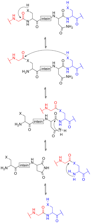
Protein splicing is an intramolecular reaction of a particular protein in which an internal protein segment is removed from a precursor protein with a ligation of C-terminal and N-terminal external proteins on both sides. The splicing junction of the precursor protein is mainly a cysteine or a serine, which are amino acids containing a nucleophilic side chain. The protein splicing reactions which are known now do not require exogenous cofactors or energy sources such as adenosine triphosphate (ATP) or guanosine triphosphate (GTP). Normally, splicing is associated only with pre-mRNA splicing. This precursor protein contains three segments—an N-extein followed by the intein followed by a C-extein. After splicing has taken place, the resulting protein contains the N-extein linked to the C-extein; this splicing product is also termed an extein.

In molecular biology, endonucleases are enzymes that cleave the phosphodiester bond within a polynucleotide chain. Some, such as deoxyribonuclease I, cut DNA relatively nonspecifically, while many, typically called restriction endonucleases or restriction enzymes, cleave only at very specific nucleotide sequences. Endonucleases differ from exonucleases, which cleave the ends of recognition sequences instead of the middle (endo) portion. Some enzymes known as "exo-endonucleases", however, are not limited to either nuclease function, displaying qualities that are both endo- and exo-like. Evidence suggests that endonuclease activity experiences a lag compared to exonuclease activity.

<i>Fok</i>I Restriction enzyme

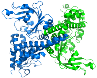
The restriction endonuclease Fok1, naturally found in Flavobacterium okeanokoites, is a bacterial type IIS restriction endonuclease consisting of an N-terminal DNA-binding domain and a non sequence-specific DNA cleavage domain at the C-terminal. Once the protein is bound to duplex DNA via its DNA-binding domain at the 5'-GGATG-3' recognition site, the DNA cleavage domain is activated and cleaves the DNA at two locations, regardless of the nucleotide sequence at the cut site. The DNA is cut 9 nucleotides downstream of the motif on the forward strand, and 13 nucleotides downstream of the motif on the reverse strand, producing two sticky ends with 4-bp overhangs.

In molecular biology, a twintron is an intron-within-intron excised by sequential splicing reactions. A twintron is presumably formed by the insertion of a mobile intron into an existing intron.

<span class="mw-page-title-main">I-CreI</span>

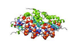
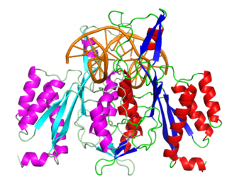
I-CreI is a homing endonuclease whose gene was first discovered in the chloroplast genome of Chlamydomonas reinhardtii, a species of unicellular green algae. It is named for the facts that: it resides in an Intron; it was isolated from Clamydomonas reinhardtii; it was the first (I) such gene isolated from C. reinhardtii. Its gene resides in a group I intron in the 23S ribosomal RNA gene of the C. reinhardtii chloroplast, and I-CreI is only expressed when its mRNA is spliced from the primary transcript of the 23S gene. I-CreI enzyme, which functions as a homodimer, recognizes a 22-nucleotide sequence of duplex DNA and cleaves one phosphodiester bond on each strand at specific positions. I-CreI is a member of the LAGLIDADG family of homing endonucleases, all of which have a conserved LAGLIDADG amino acid motif that contributes to their associative domains and active sites. When the I-CreI-containing intron encounters a 23S allele lacking the intron, I-CreI enzyme "homes" in on the "intron-minus" allele of 23S and effects its parent intron's insertion into the intron-minus allele. Introns with this behavior are called mobile introns. Because I-CreI provides for its own propagation while conferring no benefit on its host, it is an example of selfish DNA.

<i>Eco</i>RV Restriction enzyme

EcoRV is a type II restriction endonuclease isolated from certain strains of Escherichia coli. It has the alternative name Eco32I.

<span class="mw-page-title-main">Group I catalytic intron</span> Large self-splicing ribozymes

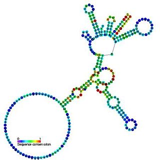
Group I introns are large self-splicing ribozymes. They catalyze their own excision from mRNA, tRNA and rRNA precursors in a wide range of organisms. The core secondary structure consists of nine paired regions (P1-P9). These fold to essentially two domains – the P4-P6 domain and the P3-P9 domain. The secondary structure mark-up for this family represents only this conserved core. Group I introns often have long open reading frames inserted in loop regions.

<span class="mw-page-title-main">Nuclease S1</span> Class of enzymes

Nuclease S1 is an endonuclease enzyme that splits single-stranded DNA (ssDNA) and RNA into oligo- or mononucleotides. This enzyme catalyses the following chemical reaction

<span class="mw-page-title-main">APEX1</span> Protein-coding gene in the species Homo sapiens

DNA-(apurinic or apyrimidinic site) lyase is an enzyme that in humans is encoded by the APEX1 gene.

<span class="mw-page-title-main">R.EcoRII</span> Restriction enzyme

Restriction endonuclease (REase) EcoRII is an enzyme of restriction modification system (RM) naturally found in Escherichia coli, a Gram-negative bacteria. Its molecular mass is 45.2 kDa, being composed of 402 amino acids.

PstI is a type II restriction endonuclease isolated from the Gram negative species, Providencia stuartii.

Meganucleases are endodeoxyribonucleases characterized by a large recognition site ; as a result this site generally occurs only once in any given genome. For example, the 18-base pair sequence recognized by the I-SceI meganuclease would on average require a genome twenty times the size of the human genome to be found once by chance. Meganucleases are therefore considered to be the most specific naturally occurring restriction enzymes.

<span class="mw-page-title-main">Genome editing</span> Type of genetic engineering

Genome editing, or genome engineering, or gene editing, is a type of genetic engineering in which DNA is inserted, deleted, modified or replaced in the genome of a living organism. Unlike early genetic engineering techniques that randomly inserts genetic material into a host genome, genome editing targets the insertions to site-specific locations. The basic mechanism involved in genetic manipulations through programmable nucleases is the recognition of target genomic loci and binding of effector DNA-binding domain (DBD), double-strand breaks (DSBs) in target DNA by the restriction endonucleases, and the repair of DSBs through homology-directed recombination (HDR) or non-homologous end joining (NHEJ).

<span class="mw-page-title-main">Cas9</span> Microbial protein found in Streptococcus pyogenes M1 GAS

Cas9 is a 160 kilodalton protein which plays a vital role in the immunological defense of certain bacteria against DNA viruses and plasmids, and is heavily utilized in genetic engineering applications. Its main function is to cut DNA and thereby alter a cell's genome. The CRISPR-Cas9 genome editing technique was a significant contributor to the Nobel Prize in Chemistry in 2020 being awarded to Emmanuelle Charpentier and Jennifer Doudna.

<i>Eco</i>RI Restriction enzyme

EcoRI is a restriction endonuclease enzyme isolated from species E. coli. It is a restriction enzyme that cleaves DNA double helices into fragments at specific sites, and is also a part of the restriction modification system. The Eco part of the enzyme's name originates from the species from which it was isolated - "E" denotes generic name which is "Escherichia" and "co" denotes species name, "coli" - while the R represents the particular strain, in this case RY13, and the I denotes that it was the first enzyme isolated from this strain.

References

  1. Edgell DR (February 2009). "Selfish DNA: homing endonucleases find a home". Curr Biol. 19 (3): R115–R117. doi: 10.1016/j.cub.2008.12.019 . PMID   19211047. S2CID   2380439.
  2. Jasin M (Jun 1996). "Genetic manipulation of genomonth with rare-cutting endonucleases". Trends Genet. 12 (6): 224–8. doi: 10.1016/0168-9525(96)10019-6 . PMID   8928227.
  3. 1 2 Burt A, Koufopanou V (December 2004). "Homing endonuclease genes: the rise and fall and rise again of a selfish element". Curr Opin Genet Dev. 14 (6): 609–15. doi:10.1016/j.gde.2004.09.010. PMID   15531154.
  4. 1 2 3 Belfort M, Roberts RJ (September 1995). "Homing endonucleases: keeping the house in order". Nucleic Acids Res. 25 (17): 3379–88. doi:10.1093/nar/25.17.3379. PMC   146926 . PMID   9254693.
  5. 1 2 Chevalier BS, Kortemme T, Chadsey MS, Baker D, Monnat RJ, Stoddard BL (October 2002). "Design, activity, and structure of a highly specific artificial endonuclease". Mol. Cell. 10 (4): 895–905. doi: 10.1016/S1097-2765(02)00690-1 . PMID   12419232.
  6. Hirata R, Ohsumk Y, Nakano A, Kawasaki H, Suzuki K, Anraku Y (April 1990). "Molecular structure of a gene, VMA1, encoding the catalytic subunit of H(+)-translocating adenosine triphosphatase from vacuolar membranes of Saccharomyces cerevisiae". J Biol Chem. 265 (12): 6726–33. doi: 10.1016/S0021-9258(19)39210-5 . PMID   2139027.
  7. Kane PM, Yamashiro CT, Wolczyk DF, Neff N, Goebl M, Stevens TH (November 1990). "Protein splicing converts the yeast TFP1 gene product to the 69-kD subunit of the vacuolar H(+)-adenosine triphosphatase". Science. 250 (4981): 651–7. Bibcode:1990Sci...250..651K. doi:10.1126/science.2146742. PMID   2146742.
  8. Perler FB, Comb DG, Jack WE, Moran LS, Qiang B, Kucera RB, Benner J, Slatko BE, Nwankwo DO, Hempstead SK, Carlow CK, Jannasch H (June 1992). "Intervening sequences in an Archaea DNA polymerase gene". PNAS. 89 (12): 5577–81. Bibcode:1992PNAS...89.5577P. doi: 10.1073/pnas.89.12.5577 . PMC   49335 . PMID   1608969.
  9. 1 2 3 Jurica MS, Monnat RJ, Stoddard BL (October 1998). "DNA recognition and cleavage by the LAGLIDADG homing endonuclease I-CreI" (PDF). Mol. Cell. 2 (4): 469–76. doi:10.1016/S1097-2765(00)80146-X. PMID   9809068.
  10. Gimble FS, Wang J (October 1996). "Substrate recognition and induced DNA distortion by the PI-SceI endonuclease, an enzyme generated by protein splicing". J Mol Biol. 263 (2): 163–80. doi:10.1006/jmbi.1996.0567. PMID   8913299.
  11. Argast GM, Stephens KM, Emond MJ, Monnat RJ (July 1998). "I-PpoI and I-CreI homing site sequence degeneracy determined by random mutagenesis and sequential in vitro enrichment". J Mol Biol. 280 (3): 345–53. doi:10.1006/jmbi.1998.1886. PMID   9665841.
  12. Shibata T, Nakagawa K, Morishima N (1995). "Multi-site-specific endonucleases and the initiation of homologous genetic recombination in yeast". Adv Biophys. 31: 77–91. doi:10.1016/0065-227X(95)99384-2. PMID   7625280.
  13. Zimmerly S, Guo H, Eskes R, Yang J, Perlman PS, Lambowitz AM (November 1995). "A group II intron RNA is a catalytic component of a DNA endonuclease involved in intron mobility". Cell. 83 (4): 529–38. doi: 10.1016/0092-8674(95)90092-6 . PMID   7585955. S2CID   10456475.
  14. Linn, Stuart M; Lloyd, R Stephen; Roberts, Richard J (December 1993). Nucleases. Cold Spring Harbor Press. pp. 35–88. ISBN   978-0-87969-426-5.
  15. 1 2 Linn, Stuart M; Lloyd, R Stephen; Roberts, Richard J (December 1993). Nucleases. Cold Spring Harbor Press. pp. 89–109. ISBN   978-0-87969-426-5.
  16. Roberts RJ, Macelis D (January 1997). "REBASE-restriction enzymes and methylases". Nucleic Acids Res. 25 (1): 248–62. doi:10.1093/nar/25.1.248. PMC   146408 . PMID   9016548.
  17. Lambowitz AM, Belfort M (1993). "Introns as mobile genetic elements". Annu Rev Biochem. 62: 587–622. doi:10.1146/annurev.bi.62.070193.003103. PMID   8352597.
  18. Linn, Stuart M; Lloyd, R Stephen; Roberts, Richard J (December 1993). Nucleases. Cold Spring Harbor Press. pp. 111–143. ISBN   978-0-87969-426-5.
  19. Wilson GG (December 1988). "Cloned restriction-modification systems—a review". Gene. 74 (1): 281–9. doi:10.1016/0378-1119(88)90304-6. PMID   3074014.
  20. Heath, P.; et al. (June 1997). "The structure of I-Crel, a group I intron-encoded homing endonuclease". Nature Structural Biology. 4 (6): 468–476. doi:10.1038/nsb0697-468. PMID   9187655. S2CID   12261983.
  21. Duan, X. (May 1997). "Crystal structure of PI-SceI, a homing endonuclease with protein splicing activity". Cell. 89 (4): 555–564. doi: 10.1016/S0092-8674(00)80237-8 . PMID   9160747. S2CID   14156646.
  22. Van Roey, P.; Fox, KM; et al. (July 2001). "Intertwined structure of the DNA-binding domain of intron endonuclease I-TevI with its substrate". EMBO J. 20 (14): 3631–3637. doi:10.1093/emboj/20.14.3631. PMC   125541 . PMID   11447104.
  23. Van Roey, P.; Kowalski, Joseph C.; et al. (July 2002). "Catalytic domain structure and hypothesis for function of GIY-YIG intron endonuclease I-TevI". Nature Structural Biology. 9 (11): 806–811. doi:10.1038/nsb853. PMID   12379841. S2CID   24856337.
  24. Flick, K.; et al. (July 1998). "DNA binding and cleavage by the nuclear intron-encoded homing endonuclease I-PpoI". Nature. 394 (6688): 96–101. Bibcode:1998Natur.394...96F. doi:10.1038/27952. PMID   9665136. S2CID   4427957.
  25. Hafez, M; Hausner, G (August 2012). "Homing endonucleases: DNA scissors on a mission". Genome. 55 (8): 553–69. doi:10.1139/g2012-049. PMID   22891613. S2CID   29183470.
  26. Shen, B.W.; et al. (September 2004). "DNA binding and cleavage by the HNH homing endonuclease I-HmuI". J. Mol. Biol. 342 (1): 43–56. doi:10.1016/j.jmb.2004.07.032. PMID   15313606. S2CID   15990707.
  27. Zhao, L.; et al. (May 2007). "The restriction fold turns to the dark side: a bacterial homing endonuclease with a PD-(D/E)-XK motif". EMBO Journal. 26 (9): 2432–2442. doi:10.1038/sj.emboj.7601672. PMC   1864971 . PMID   17410205.
  28. 1 2 Steczkiewicz, K; Muszewska, A; Knizewski, L; Rychlewski, L; Ginalski, K (August 2012). "Sequence, structure and functional diversity of PD-(D/E)XK phosphodiesterase superfamily". Nucleic Acids Research. 40 (15): 7016–45. doi:10.1093/nar/gks382. PMC   3424549 . PMID   22638584.
  29. Dassa, B.; et al. (March 2009). "Fractured genes: a novel genomic arrangement involving new split inteins and a new homing endonuclease family". Nucleic Acids Research. 37 (8): 2560–2573. doi:10.1093/nar/gkp095. PMC   2677866 . PMID   19264795.
  30. Taylor, GK; Heiter, DF; Pietrokovski, S; Stoddard, BL (December 2011). "Activity, specificity and structure of I-Bth0305I: a representative of a new homing endonuclease family". Nucleic Acids Research. 39 (22): 9705–19. doi:10.1093/nar/gkr669. PMC   3239194 . PMID   21890897.
  31. Moure CM, Gimble FS, Quiocho FA (October 2002). "Crystal structure of the intein homing endonuclease PI-SceI bound to its recognition sequence". Nat. Struct. Biol. 9 (10): 764–70. doi:10.1038/nsb840. PMID   12219083. S2CID   40192379.
This article incorporates text from the public domain Pfam and InterPro: IPR007868
This article incorporates text from the public domain Pfam and InterPro: IPR007869