
The visual cortex of the brain is the area of the cerebral cortex that processes visual information. It is located in the occipital lobe. Sensory input originating from the eyes travels through the lateral geniculate nucleus in the thalamus and then reaches the visual cortex. The area of the visual cortex that receives the sensory input from the lateral geniculate nucleus is the primary visual cortex, also known as visual area 1 (V1), Brodmann area 17, or the striate cortex. The extrastriate areas consist of visual areas 2, 3, 4, and 5.

The thalamus is a large mass of gray matter located in the dorsal part of the diencephalon. Nerve fibers project out of the thalamus to the cerebral cortex in all directions, allowing hub-like exchanges of information. It has several functions, such as the relaying of sensory signals, including motor signals to the cerebral cortex and the regulation of consciousness, sleep, and alertness.

The basal ganglia (BG), or basal nuclei, are a group of subcortical nuclei found in the brains of vertebrates. In humans and some primates, differences exist, primarily in the division of the globus pallidus into external and internal regions, and in the division of the striatum. Positioned at the base of the forebrain and the top of the midbrain, they have strong connections with the cerebral cortex, thalamus, brainstem and other brain areas. The basal ganglia are associated with a variety of functions, including regulating voluntary motor movements, procedural learning, habit formation, conditional learning, eye movements, cognition, and emotion.

The limbic system, also known as the paleomammalian cortex, is a set of brain structures located on both sides of the thalamus, immediately beneath the medial temporal lobe of the cerebrum primarily in the forebrain.

In neuroanatomy, the lateral geniculate nucleus is a structure in the thalamus and a key component of the mammalian visual pathway. It is a small, ovoid, ventral projection of the thalamus where the thalamus connects with the optic nerve. There are two LGNs, one on the left and another on the right side of the thalamus. In humans, both LGNs have six layers of neurons alternating with optic fibers.

The vertebrate cerebrum (brain) is formed by two cerebral hemispheres that are separated by a groove, the longitudinal fissure. The brain can thus be described as being divided into left and right cerebral hemispheres. Each of these hemispheres has an outer layer of grey matter, the cerebral cortex, that is supported by an inner layer of white matter. In eutherian (placental) mammals, the hemispheres are linked by the corpus callosum, a very large bundle of nerve fibers. Smaller commissures, including the anterior commissure, the posterior commissure and the fornix, also join the hemispheres and these are also present in other vertebrates. These commissures transfer information between the two hemispheres to coordinate localized functions.
Timothy John Crow is a British psychiatrist and researcher from Oxford. Much of his research is related to the causes of schizophrenia. He also has an interest in neurology and the evolutionary theory. He is the Honorary Director of the Prince of Wales International Centre for Research into Schizophrenia and Depression. He qualified at the Royal London Hospital in 1964 and obtained a PhD at the University of Aberdeen, Scotland, in 1970. He is a fellow of the Royal Colleges of Physicians and Psychiatrists and the Academy of Medical Sciences. Crow was for twenty years Head of the Division of Psychiatry of the Medical Research Council (MRC) Clinical Research Centre at Northwick Park Hospital and then a member of the External Scientific staff of the MRC in Oxford.
The amygdalofugal pathway is one of the three major efferent pathways of the amygdala, meaning that it is one of the three principal pathways by which fibers leave the amygdala. It leads from the basolateral nucleus and central nucleus of the amygdala. The amygdala is a limbic structure in the medial temporal lobe of the brain. The other main efferent pathways from the amygdala are the stria terminalis and anterior commissure.
In cognitive neuroscience, visual modularity is an organizational concept concerning how vision works. The way in which the primate visual system operates is currently under intense scientific scrutiny. One dominant thesis is that different properties of the visual world require different computational solutions which are implemented in anatomically/functionally distinct regions that operate independently – that is, in a modular fashion.

Vittorio Gallese is professor of Psychobiology at the University of Parma, Italy, and was professor in Experimental Aesthetics at the University of London, UK (2016-2018). He is an expert in neurophysiology, cognitive neuroscience, social neuroscience, and philosophy of mind. Gallese is one of the discoverers of mirror neurons. His research attempts to elucidate the functional organization of brain mechanisms underlying social cognition, including action understanding, empathy, language, mindreading and aesthetic experience.
The Protomap is a primordial molecular map of the functional areas of the mammalian cerebral cortex during early embryonic development, at a stage when neural stem cells are still the dominant cell type. The protomap is a feature of the ventricular zone, which contains the principal cortical progenitor cells, known as radial glial cells. Through a process called 'cortical patterning', the protomap is patterned by a system of signaling centers in the embryo, which provide positional information and cell fate instructions. These early genetic instructions set in motion a development and maturation process that gives rise to the mature functional areas of the cortex, for example the visual, somatosensory, and motor areas. The term protomap was coined by Pasko Rakic. The protomap hypothesis was opposed by the protocortex hypothesis, which proposes that cortical proto-areas initially have the same potential, and that regionalization in large part is controlled by external influences, such as axonal inputs from the thalamus to the cortex. However, a series of papers in the year 2000 and in 2001 provided strong evidence against the protocortex hypothesis, and the protomap hypothesis has been well accepted since then. The protomap hypothesis, together with the related radial unit hypothesis, forms our core understanding of the embryonic development of the cerebral cortex. Once the basic structure is present and cortical neurons have migrated to their final destinations, many other processes contribute to the maturation of functional cortical circuits.
Sleep onset is the transition from wakefulness into sleep. Sleep onset usually transmits into non-rapid eye movement sleep but under certain circumstances it is possible to transit from wakefulness directly into rapid eye movement sleep.
Nonprimary motor cortex is a functionally defined portion of the frontal lobe. It includes two subdivisions, the premotor cortex and the supplementary motor cortex. Largely coincident with the cytoarchitecturally defined area 6 of Brodmann (human), it is located primarily in the rostral portion of the precentral gyrus and caudal portions of the superior frontal gyrus and the middle frontal gyrus, It aids in cerebral control of movement. Anatomically speaking, several nonmprimary areas exist, and make direct connections with the spinal cord.
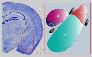
The central nucleus of the amygdala is a nucleus within the amygdala. It "serves as the major output nucleus of the amygdala and participates in receiving and processing pain information."

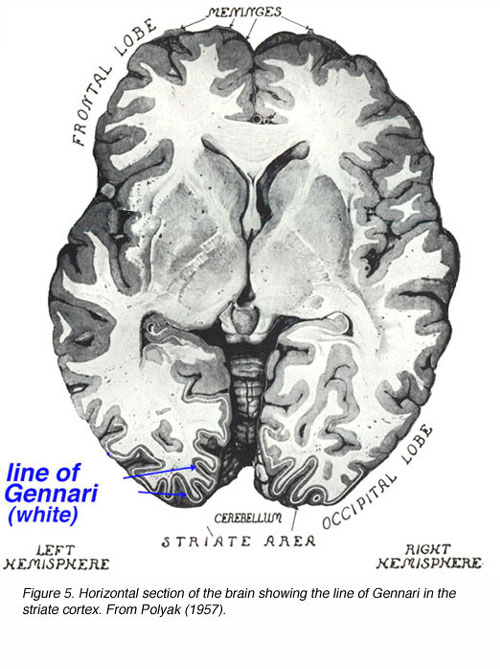
Francesco Gennari was an Italian anatomist. He is known for line of Gennari, a macroscopically white band seen in the cerebral cortex of the occipital lobe, which he observed on 2 February 1776 in the course of examining ice-frozen sections of unstained human brain during his study in medical school. He mentioned it in his book De peculiari structura cerebri, nonnulisque ejus morbis (1782), and referred to it as lineola albidior.
None of the anatomists I happened to read have taught that in addition to the cortical and medullary substance there is in the brain another substance which I am accustomed to call the third substance of this organ. ... it can be found with difficulty or it is somewhat obscure in the anterior part of the brain, but it can be detected more and more clearly in the posterior part of the brain.
Postural control refers to the maintenance of body posture in space. The central nervous system interprets sensory input to produce motor output that maintains upright posture. Sensory information used for postural control largely comes from visual, proprioceptive, and vestibular systems. While the ability to regulate posture in vertebrates was previously thought to be a mostly automatic task, controlled by circuits in the spinal cord and brainstem, it is now clear that cortical areas are also involved, updating motor commands based on the state of the body and environment.
Social cognitive neuroscience is the scientific study of the biological processes underpinning social cognition. Specifically, it uses the tools of neuroscience to study "the mental mechanisms that create, frame, regulate, and respond to our experience of the social world". Social cognitive neuroscience uses the epistemological foundations of cognitive neuroscience, and is closely related to social neuroscience. Social cognitive neuroscience employs human neuroimaging, typically using functional magnetic resonance imaging (fMRI). Human brain stimulation techniques such as transcranial magnetic stimulation and transcranial direct-current stimulation are also used. In nonhuman animals, direct electrophysiological recordings and electrical stimulation of single cells and neuronal populations are utilized for investigating lower-level social cognitive processes.
Edward George Gray (1924–1999) was a British anatomist and neuroscientist who pioneered the investigation of neural tissues with transmission electron microscopy. During his professional career, Gray made a number of profound contributions to our knowledge of synaptic structure. To this day, synapses are classified according to their ultrastructure as Gray type 1 (symmetric) or type 2 (asymmetric), corresponding to inhibitory and excitatory synapses.
Tatsuji Inouye was a Japanese ophthalmologist who studied soldiers wounded during the Russo-Japanese War and is known for his contribution to delineating cortical representation of the visual field, especially in the striate cortex. By comparing the entrance and exit wounds of bullets in the soldiers' heads to their visual deficits (scotomas), he was able to map which portions of the cortex correspond to which portions of the visual field. Importantly, he showed that more cortical area is devoted to the representation of the centre of the visual field compared than to the periphery. He was born in Tokyo and studied medicine at the University of Tokyo. He published his finding first in Leipzig in 1909.
Giovanni Berlucchi is an Italian physiologist, academic, and author. He is a professor Emeritus at the Department of Neurosciences, Biomedicine, and Movement Sciences at the University of Verona.

