Mesolimbic pathway

Last updated

The mesolimbic pathway, sometimes referred to as the reward pathway, is a dopaminergic pathway in the brain. [1] The pathway connects the ventral tegmental area in the midbrain to the ventral striatum of the basal ganglia in the forebrain. The ventral striatum includes the nucleus accumbens and the olfactory tubercle. [2]

Contents

The release of dopamine from the mesolimbic pathway into the nucleus accumbens regulates incentive salience (e.g. motivation and desire for rewarding stimuli) and facilitates reinforcement and reward-related motor function learning; [3] [4] [5] it may also play a role in the subjective perception of pleasure. [3] [5] The dysregulation of the mesolimbic pathway and its output neurons in the nucleus accumbens plays a significant role in the development and maintenance of an addiction. [1] [6] [7] [8]

Anatomy

The mesolimbic pathway and its positioning in relation to the other dopaminergic pathways Mesolimbic pathway.svg
The mesolimbic pathway and its positioning in relation to the other dopaminergic pathways

The mesolimbic pathway is a collection of dopaminergic (i.e., dopamine-releasing) neurons that project from the ventral tegmental area (VTA) to the ventral striatum, which includes the nucleus accumbens (NAcc) and olfactory tubercle. [9] It is one of the component pathways of the medial forebrain bundle, which is a set of neural pathways that mediate brain stimulation reward. [10]

The VTA is located in the midbrain and consists of dopaminergic, GABAergic, and glutamatergic neurons. [11] The dopaminergic neurons in this region receive stimuli from both cholinergic neurons in the pedunculopontine nucleus and the laterodorsal tegmental nucleus as well as glutamatergic neurons in other regions such as the prefrontal cortex. The nucleus accumbens and olfactory tubercle are located in the ventral striatum and are primarily composed of medium spiny neurons. [9] [12] [13] The nucleus accumbens is subdivided into limbic and motor subregions known as the NAcc shell and NAcc core. [11] The medium spiny neurons in the nucleus accumbens receive input from both the dopaminergic neurons of the VTA and the glutamatergic neurons of the hippocampus, amygdala, and medial prefrontal cortex. When they are activated by these inputs, the medium spiny neurons' projections release GABA onto the ventral pallidum. [11]

Function

The mesolimbic pathway regulates incentive salience, motivation, reinforcement learning, and fear, among other cognitive processes. [14] [15] [16]

The mesolimbic pathway is involved in motivational cognition. Depletion of dopamine in this pathway, or lesions at its site of origin, decrease the extent to which an animal is willing to go to obtain a reward (e.g. the number of lever presses for intravenous nicotine delivery in rats or time spent searching for food). Dopaminergic drugs are also able to increase the extent an animal is willing to go to obtain a reward. Moreover, the firing rate of neurons in the mesolimbic pathway increases during anticipation of reward, which may explain craving. [17] Mesolimbic dopamine release was once thought to be the primary mediator of pleasure, but is now believed to have only a minor or secondary role in pleasure perception. [18] [19]

Clinical significance

Mechanisms of addiction

The mesolimbic pathway and a specific set of the pathway's output neurons (e.g. D1-type medium spiny neurons within the nucleus accumbens) play a central role in the neurobiology of addiction. [20] [21] [22] Drug addiction is an illness caused by habitual substance use that induces chemical changes in the brain's circuitry. [23] An addictive drug is defined as a substance that affects the mesolimbic system directly or indirectly by increasing extracellular levels of dopamine. [24]

Common addictive substances such as cocaine, alcohol, and nicotine have been shown to increase extracellular levels of dopamine within the mesolimbic pathway, preferentially within the nucleus accumbens. The mechanisms by which these drugs do so vary depending on the drug prototype. For example, cocaine precludes the re-uptake of synaptic dopamine through blocking the presynaptic dopamine transporter. Another stimulant, amphetamine, reverses the dopamine transporter and induces the release of dopamine from synaptic vesicles. Non-stimulant drugs typically bind with ligand-gated channels or G protein-coupled receptors. Such drugs include alcohol, nicotine, and tetrahydrocannabinol (THC). [25]

Addictive Drugs and their Molecular Interactions [24]
TypeTargetExamples
Alcohol GABAA Receptor, NMDA Receptor Beer, wine, and other beverages
Cannabinoids Cannabinoid Receptor Marijuana
Nicotine Nicotinic Acetylcholine Receptor Tobacco
Opiates μ Opioid Receptor Morphine, heroin
Phencyclidine NMDA Receptor PCP
Stimulants Dopamine Transporter Cocaine, amphetamine, methamphetamine

These dopaminergic activations of the mesolimbic pathway are accompanied by the perception of reward. This stimulus-reward association shows a resistance to extinction and creates an increased motivation to repeat that same behavior that caused it. [26] Additionally, drug intake changes synaptic plasticity in the ventral tegmental area and the nucleus accumbens. Repeated exposure to the drug can lead to lasting changes in the brain that gives rise to addictive behavior. [27] [28]

Relation to other neurological and psychological disorders

The mesolimbic pathway is implicated in schizophrenia, depression, [29] [30] [31] and Parkinson's disease. [32] [33] It is also theorized to be implicated in overuse of digital media, although it could simply be a consequence of a sedentary lifestyle. [34] Each involves distinct structural changes within the mesolimbic pathway. [29]

Other dopamine pathways

See also

Related Research Articles

<span class="mw-page-title-main">Striatum</span> Nucleus in the basal ganglia of the brain

The striatum, or corpus striatum, is a nucleus in the subcortical basal ganglia of the forebrain. The striatum is a critical component of the motor and reward systems; receives glutamatergic and dopaminergic inputs from different sources; and serves as the primary input to the rest of the basal ganglia.

<span class="mw-page-title-main">Dopamine</span> Organic chemical that functions both as a hormone and a neurotransmitter

Dopamine is a neuromodulatory molecule that plays several important roles in cells. It is an organic chemical of the catecholamine and phenethylamine families. Dopamine constitutes about 80% of the catecholamine content in the brain. It is an amine synthesized by removing a carboxyl group from a molecule of its precursor chemical, L-DOPA, which is synthesized in the brain and kidneys. Dopamine is also synthesized in plants and most animals. In the brain, dopamine functions as a neurotransmitter—a chemical released by neurons to send signals to other nerve cells. Neurotransmitters are synthesized in specific regions of the brain, but affect many regions systemically. The brain includes several distinct dopamine pathways, one of which plays a major role in the motivational component of reward-motivated behavior. The anticipation of most types of rewards increases the level of dopamine in the brain, and many addictive drugs increase dopamine release or block its reuptake into neurons following release. Other brain dopamine pathways are involved in motor control and in controlling the release of various hormones. These pathways and cell groups form a dopamine system which is neuromodulatory.

<span class="mw-page-title-main">Nucleus accumbens</span> Region of the basal forebrain

The nucleus accumbens is a region in the basal forebrain rostral to the preoptic area of the hypothalamus. The nucleus accumbens and the olfactory tubercle collectively form the ventral striatum. The ventral striatum and dorsal striatum collectively form the striatum, which is the main component of the basal ganglia. The dopaminergic neurons of the mesolimbic pathway project onto the GABAergic medium spiny neurons of the nucleus accumbens and olfactory tubercle. Each cerebral hemisphere has its own nucleus accumbens, which can be divided into two structures: the nucleus accumbens core and the nucleus accumbens shell. These substructures have different morphology and functions.

<span class="mw-page-title-main">Dopaminergic pathways</span> Projection neurons in the brain that synthesize and release dopamine

Dopaminergic pathways in the human brain are involved in both physiological and behavioral processes including movement, cognition, executive functions, reward, motivation, and neuroendocrine control. Each pathway is a set of projection neurons, consisting of individual dopaminergic neurons.

<span class="mw-page-title-main">Nigrostriatal pathway</span>

The nigrostriatal pathway is a bilateral dopaminergic pathway in the brain that connects the substantia nigra pars compacta (SNc) in the midbrain with the dorsal striatum in the forebrain. It is one of the four major dopamine pathways in the brain, and is critical in the production of movement as part of a system called the basal ganglia motor loop. Dopaminergic neurons of this pathway release dopamine from axon terminals that synapse onto GABAergic medium spiny neurons (MSNs), also known as spiny projection neurons (SPNs), located in the striatum.

<span class="mw-page-title-main">Ventral tegmental area</span> Group of neurons on the floor of the midbrain

The ventral tegmental area (VTA), also known as the ventral tegmental area of Tsai, or simply ventral tegmentum, is a group of neurons located close to the midline on the floor of the midbrain. The VTA is the origin of the dopaminergic cell bodies of the mesocorticolimbic dopamine system and other dopamine pathways; it is widely implicated in the drug and natural reward circuitry of the brain. The VTA plays an important role in a number of processes, including reward cognition and orgasm, among others, as well as several psychiatric disorders. Neurons in the VTA project to numerous areas of the brain, ranging from the prefrontal cortex to the caudal brainstem and several regions in between.

Motivational salience is a cognitive process and a form of attention that motivates or propels an individual's behavior towards or away from a particular object, perceived event or outcome. Motivational salience regulates the intensity of behaviors that facilitate the attainment of a particular goal, the amount of time and energy that an individual is willing to expend to attain a particular goal, and the amount of risk that an individual is willing to accept while working to attain a particular goal.

Neuropharmacology is the study of how drugs affect function in the nervous system, and the neural mechanisms through which they influence behavior. There are two main branches of neuropharmacology: behavioral and molecular. Behavioral neuropharmacology focuses on the study of how drugs affect human behavior (neuropsychopharmacology), including the study of how drug dependence and addiction affect the human brain. Molecular neuropharmacology involves the study of neurons and their neurochemical interactions, with the overall goal of developing drugs that have beneficial effects on neurological function. Both of these fields are closely connected, since both are concerned with the interactions of neurotransmitters, neuropeptides, neurohormones, neuromodulators, enzymes, second messengers, co-transporters, ion channels, and receptor proteins in the central and peripheral nervous systems. Studying these interactions, researchers are developing drugs to treat many different neurological disorders, including pain, neurodegenerative diseases such as Parkinson's disease and Alzheimer's disease, psychological disorders, addiction, and many others.

According to proponents of the concept, sexual addiction, also known as sex addiction, is a state characterized by compulsive participation or engagement in sexual activity, particularly sexual intercourse, despite negative consequences. The concept is contentious; neither of the two major mainstream medical categorization systems recognise sex addiction as a real medical condition, instead categorizing such behavior under labels such as compulsive sexual behavior.

<span class="mw-page-title-main">Medial forebrain bundle</span>

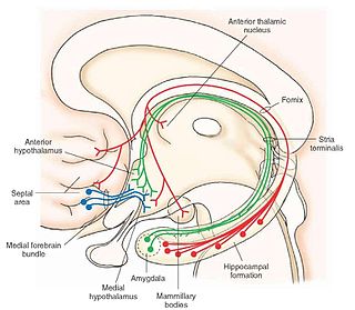
The medial forebrain bundle (MFB) is a neural pathway containing fibers from the basal olfactory regions, the periamygdaloid region and the septal nuclei, as well as fibers from brainstem regions, including the ventral tegmental area and nigrostriatal pathway.

<span class="mw-page-title-main">Medium spiny neuron</span> Type of GABAergic neuron in the striatum

Medium spiny neurons (MSNs), also known as spiny projection neurons (SPNs), are a special type of GABAergic inhibitory cell representing 95% of neurons within the human striatum, a basal ganglia structure. Medium spiny neurons have two primary phenotypes : D1-type MSNs of the direct pathway and D2-type MSNs of the indirect pathway. Most striatal MSNs contain only D1-type or D2-type dopamine receptors, but a subpopulation of MSNs exhibit both phenotypes.

<span class="mw-page-title-main">Reward system</span> Group of neural structures responsible for motivation and desire

The reward system is a group of neural structures responsible for incentive salience, associative learning, and positively-valenced emotions, particularly ones involving pleasure as a core component. Reward is the attractive and motivational property of a stimulus that induces appetitive behavior, also known as approach behavior, and consummatory behavior. A rewarding stimulus has been described as "any stimulus, object, event, activity, or situation that has the potential to make us approach and consume it is by definition a reward". In operant conditioning, rewarding stimuli function as positive reinforcers; however, the converse statement also holds true: positive reinforcers are rewarding.

<span class="mw-page-title-main">Methamphetamine</span> Central nervous system stimulant

Methamphetamine is a potent central nervous system (CNS) stimulant that is mainly used as a recreational drug and less commonly as a second-line treatment for attention deficit hyperactivity disorder and obesity. Methamphetamine was discovered in 1893 and exists as two enantiomers: levo-methamphetamine and dextro-methamphetamine. Methamphetamine properly refers to a specific chemical substance, the racemic free base, which is an equal mixture of levomethamphetamine and dextromethamphetamine in their pure amine forms, but the hydrochloride salt, commonly called crystal meth, is widely used. Methamphetamine is rarely prescribed over concerns involving human neurotoxicity and potential for recreational use as an aphrodisiac and euphoriant, among other concerns, as well as the availability of safer substitute drugs with comparable treatment efficacy such as Adderall and Vyvanse. Dextromethamphetamine is a stronger CNS stimulant than levomethamphetamine.

<span class="mw-page-title-main">FOSB</span> Protein

Protein fosB, also known as FosB and G0/G1 switch regulatory protein 3 (G0S3), is a protein that in humans is encoded by the FBJ murine osteosarcoma viral oncogene homolog B (FOSB) gene.

Behavioral addiction is a form of addiction that involves a compulsion to engage in a rewarding non-substance-related behavior – sometimes called a natural reward – despite any negative consequences to the person's physical, mental, social or financial well-being. Addiction canonically refers to substance abuse; however, the term's connotation has been expanded to include behaviors that may lead to a reward since the 1990s. A gene transcription factor known as ΔFosB has been identified as a necessary common factor involved in both behavioral and drug addictions, which are associated with the same set of neural adaptations in the reward system.

The ventral pallidum (VP) is a structure within the basal ganglia of the brain. It is an output nucleus whose fibres project to thalamic nuclei, such as the ventral anterior nucleus, the ventral lateral nucleus, and the medial dorsal nucleus. The VP is a core component of the reward system which forms part of the limbic loop of the basal ganglia, a pathway involved in the regulation of motivational salience, behavior, and emotions. It is involved in addiction.

<span class="mw-page-title-main">Addiction</span> Disorder resulting in compulsive behaviours

Addiction is generally a neuropsychological disorder defining pervasive and intense urge to engage in maladaptive behaviors providing immediate sensory rewards, despite their harmful consequences. Dependence is generally an addiction that can involve withdrawal issues. Addictive disorder is a category of mental disorders defining important intensities of addictions or dependences, which induce functional disabilities. There are no agreed definitions on these terms – see section on 'definitions'.

Cocaine addiction is the compulsive use of cocaine despite adverse consequences. It arises through epigenetic modification and transcriptional regulation of genes in the nucleus accumbens.

Addiction is a state characterized by compulsive engagement in rewarding stimuli, despite adverse consequences. The process of developing an addiction occurs through instrumental learning, which is otherwise known as operant conditioning.

The rostromedial tegmental nucleus (RMTg), also known as the tail of the ventral tegmental area (tVTA), is a GABAergic nucleus which functions as a "master brake" for the midbrain dopamine system. This region was discovered by the researchers, M. Barrot, J.Kaufling and T. Jhou. It is poorly differentiated from the rest of the ventral tegmental area (VTA) and possesses robust functional and structural links to the dopamine pathways. Notably, both acute and chronic exposure to psychostimulants have been shown to induce FosB and ΔFosB expression in the RMTg; no other drug type has been shown to induce these proteins in the RMTg.

References

  1. 1 2 Dreyer JL (2010). "New insights into the roles of microRNAs in drug addiction and neuroplasticity". Genome Med. 2 (12): 92. doi: 10.1186/gm213 . PMC   3025434 . PMID   21205279.
  2. Ikemoto S (2010). "Brain reward circuitry beyond the mesolimbic dopamine system: a neurobiological theory". Neurosci Biobehav Rev. 35 (2): 129–50. doi:10.1016/j.neubiorev.2010.02.001. PMC   2894302 . PMID   20149820. Recent studies on intracranial self-administration of neurochemicals (drugs) found that rats learn to self-administer various drugs into the mesolimbic dopamine structures–the posterior ventral tegmental area, medial shell nucleus accumbens and medial olfactory tubercle. ... In the 1970s it was recognized that the olfactory tubercle contains a striatal component, which is filled with GABAergic medium spiny neurons receiving glutamatergic inputs form cortical regions and dopaminergic inputs from the VTA and projecting to the ventral pallidum just like the nucleus accumbens
    Figure 3: The ventral striatum and self-administration of amphetamine
  3. 1 2 Malenka RC, Nestler EJ, Hyman SE (2009). Sydor A, Brown RY (eds.). Molecular Neuropharmacology: A Foundation for Clinical Neuroscience (2nd ed.). New York: McGraw-Hill Medical. pp. 147–148, 367, 376. ISBN   978-0-07-148127-4. VTA DA neurons play a critical role in motivation, reward-related behavior (Chapter 15), attention, and multiple forms of memory. This organization of the DA system, wide projection from a limited number of cell bodies, permits coordinated responses to potent new rewards. Thus, acting in diverse terminal fields, dopamine confers motivational salience ("wanting") on the reward itself or associated cues (nucleus accumbens shell region), updates the value placed on different goals in light of this new experience (orbital prefrontal cortex), helps consolidate multiple forms of memory (amygdala and hippocampus), and encodes new motor programs that will facilitate obtaining this reward in the future (nucleus accumbens core region and dorsal striatum). In this example, dopamine modulates the processing of sensorimotor information in diverse neural circuits to maximize the ability of the organism to obtain future rewards. ...
    The brain reward circuitry that is targeted by addictive drugs normally mediates the pleasure and strengthening of behaviors associated with natural reinforcers, such as food, water, and sexual contact. Dopamine neurons in the VTA are activated by food and water, and dopamine release in the NAc is stimulated by the presence of natural reinforcers, such as food, water, or a sexual partner. ...
    The NAc and VTA are central components of the circuitry underlying reward and memory of reward. As previously mentioned, the activity of dopaminergic neurons in the VTA appears to be linked to reward prediction. The NAc is involved in learning associated with reinforcement and the modulation of motoric responses to stimuli that satisfy internal homeostatic needs. The shell of the NAc appears to be particularly important to initial drug actions within reward circuitry; addictive drugs appear to have a greater effect on dopamine release in the shell than in the core of the NAc.
  4. Malenka RC, Nestler EJ, Hyman SE (2009). "Chapter 10: Neural and Neuroendocrine Control of the Internal Milieu". In Sydor A, Brown RY (eds.). Molecular Neuropharmacology: A Foundation for Clinical Neuroscience (2nd ed.). New York: McGraw-Hill Medical. p. 266. ISBN   978-0-07-148127-4. Dopamine acts in the nucleus accumbens to attach motivational significance to stimuli associated with reward.
  5. 1 2 Berridge KC, Kringelbach ML (May 2015). "Pleasure systems in the brain". Neuron. 86 (3): 646–664. doi:10.1016/j.neuron.2015.02.018. PMC   4425246 . PMID   25950633. To summarize: the emerging realization that many diverse pleasures share overlapping brain substrates; better neuroimaging maps for encoding human pleasure in orbitofrontal cortex; identification of hotspots and separable brain mechanisms for generating 'liking' and 'wanting' for the same reward; identification of larger keyboard patterns of generators for desire and dread within NAc, with multiple modes of function; and the realization that dopamine and most 'pleasure electrode' candidates for brain hedonic generators probably did not cause much pleasure after all.
  6. Robison AJ, Nestler EJ (November 2011). "Transcriptional and epigenetic mechanisms of addiction". Nat. Rev. Neurosci. 12 (11): 623–637. doi:10.1038/nrn3111. PMC   3272277 . PMID   21989194. ΔFosB has been linked directly to several addiction-related behaviors ... Importantly, genetic or viral overexpression of ΔJunD, a dominant negative mutant of JunD which antagonizes ΔFosB- and other AP-1-mediated transcriptional activity, in the NAc or OFC blocks these key effects of drug exposure14,22–24. This indicates that ΔFosB is both necessary and sufficient for many of the changes wrought in the brain by chronic drug exposure. ΔFosB is also induced in D1-type NAc MSNs by chronic consumption of several natural rewards, including sucrose, high fat food, sex, wheel running, where it promotes that consumption14,26–30. This implicates ΔFosB in the regulation of natural rewards under normal conditions and perhaps during pathological addictive-like states.
  7. Blum K, Werner T, Carnes S, Carnes P, Bowirrat A, Giordano J, Oscar-Berman M, Gold M (2012). "Sex, drugs, and rock 'n' roll: hypothesizing common mesolimbic activation as a function of reward gene polymorphisms". Journal of Psychoactive Drugs. 44 (1): 38–55. doi:10.1080/02791072.2012.662112. PMC   4040958 . PMID   22641964. It has been found that deltaFosB gene in the NAc is critical for reinforcing effects of sexual reward. Pitchers and colleagues (2010) reported that sexual experience was shown to cause DeltaFosB accumulation in several limbic brain regions including the NAc, medial pre-frontal cortex, VTA, caudate, and putamen, but not the medial preoptic nucleus. Next, the induction of c-Fos, a downstream (repressed) target of DeltaFosB, was measured in sexually experienced and naive animals. The number of mating-induced c-Fos-IR cells was significantly decreased in sexually experienced animals compared to sexually naive controls. Finally, DeltaFosB levels and its activity in the NAc were manipulated using viral-mediated gene transfer to study its potential role in mediating sexual experience and experience-induced facilitation of sexual performance. Animals with DeltaFosB overexpression displayed enhanced facilitation of sexual performance with sexual experience relative to controls. In contrast, the expression of DeltaJunD, a dominant-negative binding partner of DeltaFosB, attenuated sexual experience-induced facilitation of sexual performance, and stunted long-term maintenance of facilitation compared to DeltaFosB overexpressing group. Together, these findings support a critical role for DeltaFosB expression in the NAc in the reinforcing effects of sexual behavior and sexual experience-induced facilitation of sexual performance. ... both drug addiction and sexual addiction represent pathological forms of neuroplasticity along with the emergence of aberrant behaviors involving a cascade of neurochemical changes mainly in the brain's rewarding circuitry.
  8. Olsen CM (December 2011). "Natural rewards, neuroplasticity, and non-drug addictions". Neuropharmacology. 61 (7): 1109–22. doi:10.1016/j.neuropharm.2011.03.010. PMC   3139704 . PMID   21459101.
  9. 1 2 Ikemoto S (2010). "Brain reward circuitry beyond the mesolimbic dopamine system: a neurobiological theory". Neurosci Biobehav Rev. 35 (2): 129–50. doi:10.1016/j.neubiorev.2010.02.001. PMC   2894302 . PMID   20149820. Recent studies on intracranial self-administration of neurochemicals (drugs) found that rats learn to self-administer various drugs into the mesolimbic dopamine structures–the posterior ventral tegmental area, medial shell nucleus accumbens and medial olfactory tubercle. ... In the 1970s it was recognized that the olfactory tubercle contains a striatal component, which is filled with GABAergic medium spiny neurons receiving glutamatergic inputs form cortical regions and dopaminergic inputs from the VTA and projecting to the ventral pallidum just like the nucleus accumbens Figure 3: The ventral striatum and self-administration of amphetamine
  10. You ZB, Chen YQ, Wise RA (2001). "Dopamine and glutamate release in the nucleus accumbens and ventral tegmental area of rat following lateral hypothalamic self-stimulation". Neuroscience. 107 (4): 629–39. doi:10.1016/s0306-4522(01)00379-7. PMID   11720786. S2CID   33615497.
  11. 1 2 3 Pierce RC, Kumaresan V (2006). "The mesolimbic dopamine system: The final common pathway for the reinforcing effect of drugs of abuse?". Neuroscience and Biobehavioral Reviews. 30 (2): 215–38. doi:10.1016/j.neubiorev.2005.04.016. PMID   16099045. S2CID   39886286.
  12. Zhang TA, Maldve RE, Morrisett RA (2006). "Coincident signaling in mesolimbic structures underlying alcohol reinforcement". Biochemical Pharmacology . 72 (8): 919–27. doi:10.1016/j.bcp.2006.04.022. PMID   16764827.
  13. Purves D et al. 2008. Neuroscience. Sinauer 4ed. 754-56
  14. Malenka RC, Nestler EJ, Hyman SE (2009). "Chapter 6: Widely Projecting Systems: Monoamines, Acetylcholine, and Orexin". In Sydor A, Brown RY (eds.). Molecular Neuropharmacology: A Foundation for Clinical Neuroscience (2nd ed.). New York: McGraw-Hill Medical. pp. 147–148, 154–157. ISBN   9780071481274. Neurons from the SNc densely innervate the dorsal striatum where they play a critical role in the learning and execution of motor programs. Neurons from the VTA innervate the ventral striatum (nucleus accumbens), olfactory bulb, amygdala, hippocampus, orbital and medial prefrontal cortex, and cingulate cortex. VTA DA neurons play a critical role in motivation, reward-related behavior, attention, and multiple forms of memory. ... Thus, acting in diverse terminal fields, dopamine confers motivational salience ("wanting") on the reward itself or associated cues (nucleus accumbens shell region), updates the value placed on different goals in light of this new experience (orbital prefrontal cortex), helps consolidate multiple forms of memory (amygdala and hippocampus), and encodes new motor programs that will facilitate obtaining this reward in the future (nucleus accumbens core region and dorsal striatum). ... DA has multiple actions in the prefrontal cortex. It promotes the "cognitive control" of behavior: the selection and successful monitoring of behavior to facilitate attainment of chosen goals. Aspects of cognitive control in which DA plays a role include working memory, the ability to hold information "on line" in order to guide actions, suppression of prepotent behaviors that compete with goal-directed actions, and control of attention and thus the ability to overcome distractions. ... Noradrenergic projections from the LC thus interact with dopaminergic projections from the VTA to regulate cognitive control.
  15. Engert, Veronika; Pruessner, Jens C (9 January 2017). "Dopaminergic and Noradrenergic Contributions to Functionality in ADHD: The Role of Methylphenidate". Current Neuropharmacology. 6 (4): 322–328. doi:10.2174/157015908787386069. ISSN   1570-159X. PMC   2701285 . PMID   19587853.
  16. Pezze, Marie A.; Feldon, Joram (1 December 2004). "Mesolimbic dopaminergic pathways in fear conditioning". Progress in Neurobiology. 74 (5): 301–320. doi:10.1016/j.pneurobio.2004.09.004. ISSN   0301-0082. PMID   15582224. S2CID   36091832.
  17. Salamone, John D.; Correa, Mercè (2012). "The Mysterious Motivational Functions of Mesolimbic Dopamine". Neuron. 76 (3): 470–485. doi:10.1016/j.neuron.2012.10.021. PMC   4450094 . PMID   23141060.
  18. Berridge KC, Kringelbach ML (May 2015). "Pleasure systems in the brain". Neuron. 86 (3): 646–664. doi:10.1016/j.neuron.2015.02.018. PMC   4425246 . PMID   25950633. To summarize: the emerging realization that many diverse pleasures share overlapping brain substrates; better neuroimaging maps for encoding human pleasure in orbitofrontal cortex; identification of hotspots and separable brain mechanisms for generating 'liking' and 'wanting' for the same reward; identification of larger keyboard patterns of generators for desire and dread within NAc, with multiple modes of function; and the realization that dopamine and most 'pleasure electrode' candidates for brain hedonic generators probably did not cause much pleasure after all.
  19. Berridge, Kent C; Kringelbach, Morten L (1 June 2013). "Neuroscience of affect: brain mechanisms of pleasure and displeasure". Current Opinion in Neurobiology. 23 (3): 294–303. doi:10.1016/j.conb.2013.01.017. PMC   3644539 . PMID   23375169.
  20. Robison AJ, Nestler EJ (November 2011). "Transcriptional and epigenetic mechanisms of addiction". Nat. Rev. Neurosci. 12 (11): 623–637. doi:10.1038/nrn3111. PMC   3272277 . PMID   21989194. ΔFosB has been linked directly to several addiction-related behaviors ... Importantly, genetic or viral overexpression of ΔJunD, a dominant negative mutant of JunD which antagonizes ΔFosB- and other AP-1-mediated transcriptional activity, in the NAc or OFC blocks these key effects of drug exposure14,22–24. This indicates that ΔFosB is both necessary and sufficient for many of the changes wrought in the brain by chronic drug exposure. ΔFosB is also induced in D1-type NAc MSNs by chronic consumption of several natural rewards, including sucrose, high fat food, sex, wheel running, where it promotes that consumption14,26–30. This implicates ΔFosB in the regulation of natural rewards under normal conditions and perhaps during pathological addictive-like states.
  21. Blum K, Werner T, Carnes S, Carnes P, Bowirrat A, Giordano J, Oscar-Berman M, Gold M (2012). "Sex, drugs, and rock 'n' roll: hypothesizing common mesolimbic activation as a function of reward gene polymorphisms". Journal of Psychoactive Drugs. 44 (1): 38–55. doi:10.1080/02791072.2012.662112. PMC   4040958 . PMID   22641964. It has been found that deltaFosB gene in the NAc is critical for reinforcing effects of sexual reward. Pitchers and colleagues (2010) reported that sexual experience was shown to cause DeltaFosB accumulation in several limbic brain regions including the NAc, medial pre-frontal cortex, VTA, caudate, and putamen, but not the medial preoptic nucleus. Next, the induction of c-Fos, a downstream (repressed) target of DeltaFosB, was measured in sexually experienced and naive animals. The number of mating-induced c-Fos-IR cells was significantly decreased in sexually experienced animals compared to sexually naive controls. Finally, DeltaFosB levels and its activity in the NAc were manipulated using viral-mediated gene transfer to study its potential role in mediating sexual experience and experience-induced facilitation of sexual performance. Animals with DeltaFosB overexpression displayed enhanced facilitation of sexual performance with sexual experience relative to controls. In contrast, the expression of DeltaJunD, a dominant-negative binding partner of DeltaFosB, attenuated sexual experience-induced facilitation of sexual performance, and stunted long-term maintenance of facilitation compared to DeltaFosB overexpressing group. Together, these findings support a critical role for DeltaFosB expression in the NAc in the reinforcing effects of sexual behavior and sexual experience-induced facilitation of sexual performance. ... both drug addiction and sexual addiction represent pathological forms of neuroplasticity along with the emergence of aberrant behaviors involving a cascade of neurochemical changes mainly in the brain's rewarding circuitry.
  22. Olsen CM (December 2011). "Natural rewards, neuroplasticity, and non-drug addictions". Neuropharmacology. 61 (7): 1109–22. doi:10.1016/j.neuropharm.2011.03.010. PMC   3139704 . PMID   21459101.
  23. Administration (US), Substance Abuse and Mental Health Services; General (US), Office of the Surgeon (November 2016). THE NEUROBIOLOGY OF SUBSTANCE USE, MISUSE, AND ADDICTION. US Department of Health and Human Services.
  24. 1 2 Kandel, Eric R.; Koester, J. D.; Mack, S. H.; Siegelbaum, S. A. (2021). Principles of Neural Science (6e ed.). McGraw Hill.
  25. Adinoff, Bryon (2004). "Neurobiologic Processes in Drug Reward and Addiction". Harvard Review of Psychiatry. 12 (6): 305–320. doi:10.1080/10673220490910844. ISSN   1067-3229. PMC   1920543 . PMID   15764467.
  26. Di Chiara, Gaetano; Bassareo, Valentina (1 February 2007). "Reward system and addiction: what dopamine does and doesn't do". Current Opinion in Pharmacology. Neurosciences. 7 (1): 69–76. doi:10.1016/j.coph.2006.11.003. ISSN   1471-4892. PMID   17174602.
  27. Lüscher, Christian; Malenka, Robert C. (24 February 2011). "Drug-Evoked Synaptic Plasticity in Addiction: From Molecular Changes to Circuit Remodeling". Neuron. 69 (4): 650–663. doi:10.1016/j.neuron.2011.01.017. ISSN   0896-6273. PMC   4046255 . PMID   21338877.
  28. Gipson, Cassandra D.; Kupchik, Yonatan M.; Kalivas, Peter W. (January 2014). "Rapid, transient synaptic plasticity in addiction". Neuropharmacology. 76: 276–286. doi:10.1016/j.neuropharm.2013.04.032. PMC   3762905 . PMID   23639436.
  29. 1 2 Van, den Heuval DMA, Pasterkamp RJ (2008). "Getting connected in the dopamine system". Progress in Neurobiology. 85 (1): 75–93. doi:10.1016/j.pneurobio.2008.01.003. PMID   18304718. S2CID   45462508.{{cite journal}}: CS1 maint: multiple names: authors list (link)
  30. Laviolette SR (2007). "Dopamine modulation of emotional processing in cortical and subcortical neural circuits: evidence for a final common pathway in schizophrenia?". Schizophrenia Bulletin. 33 (4): 971–981. doi:10.1093/schbul/sbm048. PMC   2632330 . PMID   17519393.
  31. Diaz J. 1996. How Drugs Influence Behavior: A Neurobehavorial Approach. Prentice Hall
  32. Nyberg, Eric M.; Tanabe, Jody; Honce, Justin M.; Krmpotich, Theodore; Shelton, Erika; Hedeman, Jessica; Berman, Brian D. (1 May 2015). "Morphologic changes in the mesolimbic pathway in Parkinson's disease motor subtypes". Parkinsonism & Related Disorders. 21 (5): 536–540. doi:10.1016/j.parkreldis.2015.03.008. ISSN   1353-8020. PMC   4424152 . PMID   25817514.
  33. Caminiti, Silvia Paola; Presotto, Luca; Baroncini, Damiano; Garibotto, Valentina; Moresco, Rosa Maria; Gianolli, Luigi; Volonté, Maria Antonietta; Antonini, Angelo; Perani, Daniela (1 January 2017). "Axonal damage and loss of connectivity in nigrostriatal and mesolimbic dopamine pathways in early Parkinson's disease". NeuroImage: Clinical. 14: 734–740. doi:10.1016/j.nicl.2017.03.011. ISSN   2213-1582. PMC   5379906 . PMID   28409113.
  34. "Dopamine, Smartphones & You: A battle for your time". Science in the News. 1 May 2018. Retrieved 10 May 2019.