Related Research Articles

Facial perception is an individual's understanding and interpretation of the face. Here, perception implies the presence of consciousness and hence excludes automated facial recognition systems. Although facial recognition is found in other species, this article focuses on facial perception in humans.

The fusiform gyrus, also known as the lateral occipitotemporal gyrus,is part of the temporal lobe and occipital lobe in Brodmann area 37. The fusiform gyrus is located between the lingual gyrus and parahippocampal gyrus above, and the inferior temporal gyrus below. Though the functionality of the fusiform gyrus is not fully understood, it has been linked with various neural pathways related to recognition. Additionally, it has been linked to various neurological phenomena such as synesthesia, dyslexia, and prosopagnosia.
Visual processing is a term that is used to refer to the brain's ability to use and interpret visual information from the world around us. The process of converting light energy into a meaningful image is a complex process that is facilitated by numerous brain structures and higher level cognitive processes. On an anatomical level, light energy first enters the eye through the cornea, where the light is bent. After passing through the cornea, light passes through the pupil and then lens of the eye, where it is bent to a greater degree and focused upon the retina. The retina is where a group of light-sensing cells, called photoreceptors are located. There are two types of photoreceptors: rods and cones. Rods are sensitive to dim light and cones are better able to transduce bright light. Photoreceptors connect to bipolar cells, which induce action potentials in retinal ganglion cells. These retinal ganglion cells form a bundle at the optic disc, which is a part of the optic nerve. The two optic nerves from each eye meet at the optic chiasm, where nerve fibers from each nasal retina cross which results in the right half of each eye's visual field being represented in the left hemisphere and the left half of each eye's visual fields being represented in the right hemisphere. The optic tract then diverges into two visual pathways, the geniculostriate pathway and the tectopulvinar pathway, which send visual information to the visual cortex of the occipital lobe for higher level processing.

A minimally conscious state (MCS) is a disorder of consciousness distinct from persistent vegetative state and locked-in syndrome. Unlike persistent vegetative state, patients with MCS have partial preservation of conscious awareness. MCS is a relatively new category of disorders of consciousness. The natural history and longer term outcome of MCS have not yet been thoroughly studied. The prevalence of MCS was estimated to be 112,000 to 280,000 adult and pediatric cases.

Affective neuroscience is the study of the neural mechanisms of emotion. This interdisciplinary field combines neuroscience with the psychological study of personality, emotion, and mood. The putative existence of 'basic emotions' and their defining attributes represents a long lasting and yet unsettled issue in the field.
Neuronal tuning refers to the hypothesized property of brain cells by which they selectively represent a particular type of sensory, association, motor, or cognitive information. Some neuronal responses have been hypothesized to be optimally tuned to specific patterns through experience. Neuronal tuning can be strong and sharp, as observed in primary visual cortex, or weak and broad, as observed in neural ensembles. Single neurons are hypothesized to be simultaneously tuned to several modalities, such as visual, auditory, and olfactory. Neurons hypothesized to be tuned to different signals are often hypothesized to integrate information from the different sources. In computational models called neural networks, such integration is the major principle of operation. The best examples of neuronal tuning can be seen in the visual, auditory, olfactory, somatosensory, and memory systems, although due to the small number of stimuli tested the generality of neuronal tuning claims is still an open question.
Visual agnosia is an impairment in recognition of visually presented objects. It is not due to a deficit in vision, language, memory, or intellect. While cortical blindness results from lesions to primary visual cortex, visual agnosia is often due to damage to more anterior cortex such as the posterior occipital and/or temporal lobe(s) in the brain.[2] There are two types of visual agnosia: apperceptive agnosia and associative agnosia.

The inferior temporal gyrus is one of three gyri of the temporal lobe and is located below the middle temporal gyrus, connected behind with the inferior occipital gyrus; it also extends around the infero-lateral border on to the inferior surface of the temporal lobe, where it is limited by the inferior sulcus. This region is one of the higher levels of the ventral stream of visual processing, associated with the representation of objects, places, faces, and colors. It may also be involved in face perception, and in the recognition of numbers.

The lingual gyrus, also known as the medialoccipitotemporal gyrus, is a brain structure that is linked to processing vision, especially related to letters. It is thought to also play a role in analysis of logical conditions and encoding visual memories. It is named after its shape, which is somewhat similar to a tongue. Contrary to the name, the region has little to do with speech.

The colour centre is a region in the brain primarily responsible for visual perception and cortical processing of colour signals received by the eye, which ultimately results in colour vision. The colour centre in humans is thought to be located in the ventral occipital lobe as part of the visual system, in addition to other areas responsible for recognizing and processing specific visual stimuli, such as faces, words, and objects. Many functional magnetic resonance imaging (fMRI) studies in both humans and macaque monkeys have shown colour stimuli to activate multiple areas in the brain, including the fusiform gyrus and the lingual gyrus. These areas, as well as others identified as having a role in colour vision processing, are collectively labelled visual area 4 (V4). The exact mechanisms, location, and function of V4 are still being investigated.

The fusiform face area is a part of the human visual system that is specialized for facial recognition. It is located in the inferior temporal cortex (IT), in the fusiform gyrus.
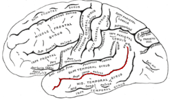
The superior temporal sulcus (STS) is the sulcus separating the superior temporal gyrus from the middle temporal gyrus in the temporal lobe of the brain. A sulcus is a deep groove that curves into the largest part of the brain, the cerebrum, and a gyrus is a ridge that curves outward of the cerebrum.
Visual object recognition refers to the ability to identify the objects in view based on visual input. One important signature of visual object recognition is "object invariance", or the ability to identify objects across changes in the detailed context in which objects are viewed, including changes in illumination, object pose, and background context.

In neuroscience, functional specialization is a theory which suggests that different areas in the brain are specialized for different functions.
Emotional lateralization is the asymmetrical representation of emotional control and processing in the brain. There is evidence for the lateralization of other brain functions as well.
The extrastriate body area (EBA) is a subpart of the extrastriate visual cortex involved in the visual perception of human body and body parts, akin in its respective domain to the fusiform face area, involved in the perception of human faces. The EBA was identified in 2001 by the team of Nancy Kanwisher using fMRI.
The anti-saccade (AS) task is a gross estimation of injury or dysfunction of the frontal lobe, by assessing the brain’s ability to inhibit the reflexive saccade. Saccadic eye movement is primarily controlled by the frontal cortex.
Justine Saade-Sergent was a researcher in the cognitive neuroscience field. She was an associate professor of neurology and neurosurgery at the Montreal Neurological Institute at McGill University from 1979 to 1982.
The Fusiform body area (FBA) is a part of the extrastriate visual cortex, an object representation system involved in the visual processing of human bodies in contrast to body parts. Its function is similar to but distinct from the extrastriate body area (EBA), which perceives bodies in relation body parts, and the fusiform face area (FFA), which is involved in the perception of faces. Marius Peelen and Paul Downing identified this brain region in 2004 through an fMRI study.; in 2005 Rebecca Schwarzlose and a team of cognitive researchers named this brain region the fusiform body area.
The occipital face area (OFA) is a region of the human cerebral cortex which is specialised for face perception. The OFA is located on the lateral surface of the occipital lobe adjacent to the inferior occipital gyrus. The OFA comprises a network of brain regions including the fusiform face area (FFA) and posterior superior temporal sulcus (STS) which support facial processing.
References
- ↑ Sergent J, Ohta S, MacDonald B (1992) Functional neuroanatomy of face and object processing: a positron emission tomography study. Brain 115:15-36
- ↑ Martin, A., Wiggs, C. L., Ungerleider, L. G. & Haxby, J. V. (1996) Neural correlates of category-specific knowledge. Nature 379:649–52.
- ↑ Malach, R. et al. (2002) The topography of high-order human object areas. Trends Cogn. Sci. 6, 176–184