Related Research Articles
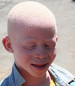
Albinism is a congenital condition characterized in humans by the partial or complete absence of pigment in the skin, hair and eyes. Albinism is associated with a number of vision defects, such as photophobia, nystagmus, and amblyopia. Lack of skin pigmentation makes for more susceptibility to sunburn and skin cancers. In rare cases such as Chédiak–Higashi syndrome, albinism may be associated with deficiencies in the transportation of melanin granules. This also affects essential granules present in immune cells, leading to increased susceptibility to infection.

Melanin is a broad term for a group of natural pigments found in most organisms. The melanin pigments are produced in a specialized group of cells known as melanocytes.

Melanocytes are melanin-producing neural crest-derived cells located in the bottom layer of the skin's epidermis, the middle layer of the eye, the inner ear, vaginal epithelium, meninges, bones, and heart. Melanin is a dark pigment primarily responsible for skin color. Once synthesized, melanin is contained in special organelles called melanosomes which can be transported to nearby keratinocytes to induce pigmentation. Thus darker skin tones have more melanosomes present than lighter skin tones. Functionally, melanin serves as protection against UV radiation. Melanocytes also have a role in the immune system.

In genetics, dominance is the phenomenon of one variant (allele) of a gene on a chromosome masking or overriding the effect of a different variant of the same gene on the other copy of the chromosome. The first variant is termed dominant and the second is called recessive. This state of having two different variants of the same gene on each chromosome is originally caused by a mutation in one of the genes, either new or inherited. The terms autosomal dominant or autosomal recessive are used to describe gene variants on non-sex chromosomes (autosomes) and their associated traits, while those on sex chromosomes (allosomes) are termed X-linked dominant, X-linked recessive or Y-linked; these have an inheritance and presentation pattern that depends on the sex of both the parent and the child. Since there is only one copy of the Y chromosome, Y-linked traits cannot be dominant or recessive. Additionally, there are other forms of dominance, such as incomplete dominance, in which a gene variant has a partial effect compared to when it is present on both chromosomes, and co-dominance, in which different variants on each chromosome both show their associated traits.

Tiger eye or goat eye is a gene causing diluted eye color in horses. There are two variants, Tiger-eye 1 (TE1) and Tiger-eye 2 (TE2), which are both recessive. Horses displaying tiger eye typically have a yellow, orange, or amber iris. Tiger eye has only been found in Puerto Rican Paso Fino horses. Horses of related breeds were tested, and none were found to have either tiger eye allele. No obvious link between eye shade and coat color was seen, making this the first studied gene in horses to affect eye color but not coat color. Tiger eye does not appear to affect vision, and there were no signs of reduced pigment on the retina or retinal pigment epithelium.

Waardenburg syndrome is a group of rare genetic conditions characterised by at least some degree of congenital hearing loss and pigmentation deficiencies, which can include bright blue eyes, a white forelock or patches of light skin. These basic features constitute type 2 of the condition; in type 1, there is also a wider gap between the inner corners of the eyes called telecanthus, or dystopia canthorum. In type 3, which is rare, the arms and hands are also malformed, with permanent finger contractures or fused fingers, while in type 4, the person also has Hirschsprung's disease. There also exist at least two types that can result in central nervous system (CNS) symptoms such as developmental delay and muscle tone abnormalities.
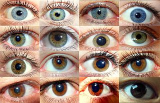
Eye color is a polygenic phenotypic trait determined by two factors: the pigmentation of the eye's iris and the frequency-dependence of the scattering of light by the turbid medium in the stroma of the iris.
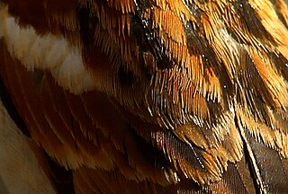
Plumage is a layer of feathers that covers a bird and the pattern, colour, and arrangement of those feathers. The pattern and colours of plumage differ between species and subspecies and may vary with age classes. Within species, there can be different colour morphs. The placement of feathers on a bird is not haphazard, but rather emerge in organized, overlapping rows and groups, and these feather tracts are known by standardized names.

Tyrosinase is an oxidase that is the rate-limiting enzyme for controlling the production of melanin. The enzyme is mainly involved in two distinct reactions of melanin synthesis otherwise known as the Raper Mason pathway. Firstly, the hydroxylation of a monophenol and secondly, the conversion of an o-diphenol to the corresponding o-quinone. o-Quinone undergoes several reactions to eventually form melanin. Tyrosinase is a copper-containing enzyme present in plant and animal tissues that catalyzes the production of melanin and other pigments from tyrosine by oxidation. It is found inside melanosomes which are synthesized in the skin melanocytes. In humans, the tyrosinase enzyme is encoded by the TYR gene.
Albinism-black lock-cell migration disorder is the initialism for the following terms and concepts that describe a condition affecting a person's physical appearance and physiology: (1) A – albinism, (2) B – black lock of hair, (3) C – cell migration disorder of the neurocytes of the gut, and (4) D – sensorineural deafness. The syndrome is caused by mutation in the endothelin B receptor gene (EDNRB).

Heřmanský–Pudlák syndrome is an extremely rare autosomal recessive disorder which results in oculocutaneous albinism, bleeding problems due to a platelet abnormality, and storage of an abnormal fat-protein compound. It is thought to affect around 1 in 500,000 people worldwide, with a significantly higher occurrence in Puerto Ricans, with a prevalence of 1 in 1800. Many of the clinical research studies on the disease have been conducted in Puerto Rico.
Oculocutaneous albinism is a form of albinism involving the eyes, the skin, and the hair. Overall, an estimated 1 in 20,000 people worldwide are born with oculocutaneous albinism. OCA is caused by mutations in several genes that control the synthesis of melanin within the melanocytes. Seven types of oculocutaneous albinism have been described, all caused by a disruption of melanin synthesis and all autosomal recessive disorders. Oculocutaneous albinism is also found in non-human animals.

A white horse is born predominantly white and stays white throughout its life. A white horse has mostly pink skin under its hair coat, and may have brown, blue, or hazel eyes. "True white" horses, especially those that carry one of the dominant white (W) genes, are rare. Most horses that are commonly referred to as "white" are actually "gray" horses whose hair coats are completely white. Gray horses may be born of any color and their hairs gradually turn white as time goes by and take on a white appearance. Nearly all gray horses have dark skin, except under any white markings present at birth. Skin color is the most common method for an observer to distinguish between mature white and gray horses.

P protein, also known as melanocyte-specific transporter protein or pink-eyed dilution protein homolog, is a protein that in humans is encoded by the oculocutaneous albinism II (OCA2) gene. The P protein is believed to be an integral membrane protein involved in small molecule transport, specifically of tyrosine—a precursor of melanin. Certain mutations in OCA2 result in type 2 oculocutaneous albinism. OCA2 encodes the human homologue of the mouse p gene.

Membrane-associated transporter protein (MATP), also known as solute carrier family 45 member 2 (SLC45A2) or melanoma antigen AIM1, is a protein that in humans is encoded by the SLC45A2 gene.

Ocular albinism type 1(OA1) is the most common type of ocular albinism, with a prevalence rate of 1:50,000. It is an inheritable classical Mendelian type X-linked recessive disorder wherein the retinal pigment epithelium lacks pigment while hair and skin appear normal. Since it is usually an X-linked disorder, it occurs mostly in males, while females are carriers unless they are homozygous. About 60 missense and nonsense mutations, insertions, and deletions have been identified in Oa1. Mutations in OA1 have been linked to defective glycosylation and thus improper intracellular transportation.

Amelanism is a pigmentation abnormality characterized by the lack of pigments called melanins, commonly associated with a genetic loss of tyrosinase function. Amelanism can affect fish, amphibians, reptiles, birds, and mammals including humans. The appearance of an amelanistic animal depends on the remaining non-melanin pigments. The opposite of amelanism is melanism, a higher percentage of melanin.

In poultry standards, solid white is coloration of plumage in chickens characterized by a uniform pure white color across all feathers, which is not generally associated with depigmentation in any other part of the body.

Albinism is the congenital absence of melanin in an animal or plant resulting in white hair, feathers, scales and skin and reddish pink or blue eyes. Individuals with the condition are referred to as albinos.
Ocular albinism late onset sensorineural deafness (OASD) is a rare, X-linked recessive disease characterized by intense visual impairments, reduced retinal pigments, translucent pale-blue irises and moderately severe hearing loss from adolescence to middle-age. It is a subtype of Ocular Albinism (OA) that is linked to Ocular albinism type I (OA1). OA1 is the most common form of ocular albinism, affecting at least 1/60,000 males.
References
- 1 2 3 4 5 6 7 8 Online Mendelian Inheritance in Man (OMIM): 203100
- 1 2 3 4 5 6 Lewis RA (1993). "Oculocutaneous Albinism Type 1 – RETIRED CHAPTER, FOR HISTORICAL REFERENCE ONLY". In Adam MP, Ardinger HH, Pagon RA, Wallace SE, Bean LJ, Mirzaa G, Amemiya A, Lewis RA (eds.). GeneReviews. Seattle (WA): University of Washington, Seattle. PMID 20301345.
- 1 2 Hutton, Saunie M.; Spritz, Richard A. (October 2008). "Comprehensive Analysis of Oculocutaneous Albinism among Non-Hispanic Caucasians Shows that OCA1 Is the Most Prevalent OCA Type". Journal of Investigative Dermatology. 128 (10): 2442–2450. doi:10.1038/jid.2008.109. PMC 3515683 . PMID 18463683.
- 1 2 3 4 5 6 7 8 9 10 "Oculocutaneous Albinism". atlasgeneticsoncology.org. Retrieved 2021-04-26.
- 1 2 3 4 5 "Oculocutaneous albinism | Genetic and Rare Diseases Information Center (GARD) – an NCATS Program". rarediseases.info.nih.gov. Retrieved 2021-04-26.
- ↑ "OMIM Entry - * 606933 - TYROSINASE; TYR". www.omim.org. Retrieved 2021-04-26.
- 1 2 "TYR gene: MedlinePlus Genetics". medlineplus.gov. Retrieved 2021-04-26.
- ↑ Ghodsinejad Kalahroudi V, Kamalidehghan B, Arasteh Kani A, Aryani O, Tondar M, Ahmadipour F, et al. (2014-09-12). "Two novel tyrosinase (TYR) gene mutations with pathogenic impact on oculocutaneous albinism type 1 (OCA1)". PLOS ONE. 9 (9): e106656. Bibcode:2014PLoSO...9j6656G. doi: 10.1371/journal.pone.0106656 . PMC 4162572 . PMID 25216246.
- 1 2 Yang, Qi; Yi, Sheng; Li, Mengting; Xie, Bobo; Luo, Jinsi; Wang, Jin; Rong, Xiuliang; Zhang, Qinle; Qin, Zailong; Hang, Limei; Feng, Shihan (2019-06-13). "Genetic analyses of oculocutaneous albinism types 1 and 2 with four novel mutations". BMC Medical Genetics. 20 (1): 106. doi: 10.1186/s12881-019-0842-7 . ISSN 1471-2350. PMC 6567650 . PMID 31196117.
- ↑ Rooryck, Caroline; Morice-Picard, Fanny; Elçioglu, Nursel H.; Lacombe, Didier; Taieb, Alain; Arveiler, Benoît (October 2008). "Molecular diagnosis of oculocutaneous albinism: new mutations in the OCA1-4 genes and practical aspects: Letter to the Editor". Pigment Cell & Melanoma Research. 21 (5): 583–587. doi:10.1111/j.1755-148X.2008.00496.x. PMID 18821858. S2CID 11728943.
- 1 2 3 Rosenmann E, Rosenmann A, Ne'eman Z, Lewin A, Bejarano-Achache I, Blumenfeld A (September 1999). "Prenatal diagnosis of oculocutaneous albinism type I: review and personal experience". Pediatric and Developmental Pathology. 2 (5): 404–14. doi:10.1007/s100249900143. PMID 10441617. S2CID 23369377.
- ↑ "Albinism, Oculocutaneous, Type I | Hereditary Ocular Diseases". disorders.eyes.arizona.edu. Retrieved 2021-04-26.
- 1 2 3 4 Marçon, Carolina Reato; Maia, Marcus (2019). "Albinism: epidemiology, genetics, cutaneous characterization, psychosocial factors". Anais Brasileiros de Dermatologia. 94 (5): 503–520. doi:10.1016/j.abd.2019.09.023. ISSN 0365-0596. PMC 6857599 . PMID 31777350.