Tyrosinase

Last updated
TYR
Identifiers
Aliases TYR , ATN, CMM8, OCA1, OCA1A, OCAIA, SHEP3, tyrosinase, Tyrosinase
External IDs OMIM: 606933 MGI: 98880 HomoloGene: 30969 GeneCards: TYR
Orthologs
SpeciesHumanMouse
Entrez
Ensembl
UniProt
RefSeq (mRNA)

NM_000372

NM_011661
NM_001317397

RefSeq (protein)

NP_000363

NP_001304326
NP_035791

Location (UCSC) Chr 11: 89.18 – 89.3 Mb Chr 7: 87.07 – 87.14 Mb
PubMed search [3] [4]
Wikidata
View/Edit Human View/Edit Mouse

Tyrosinase is an oxidase that is the rate-limiting enzyme for controlling the production of melanin. The enzyme is mainly involved in two distinct reactions of melanin synthesis otherwise known as the Raper Mason pathway. Firstly, the hydroxylation of a monophenol and secondly, the conversion of an o-diphenol to the corresponding o-quinone. o-Quinone undergoes several reactions to eventually form melanin. [5] Tyrosinase is a copper-containing enzyme present in plant and animal tissues that catalyzes the production of melanin and other pigments from tyrosine by oxidation. It is found inside melanosomes which are synthesized in the skin melanocytes. In humans, the tyrosinase enzyme is encoded by the TYR gene. [6]

Contents

Catalyzed reaction

Tyrosinase carries out the oxidation of phenols such as tyrosine and dopamine using dioxygen (O2). In the presence of catechol, benzoquinone is formed (see reaction below). Hydrogens removed from catechol combine with oxygen to form water.

The substrate specificity becomes dramatically restricted in mammalian tyrosinase which uses only L-form of tyrosine or DOPA as substrates, and has restricted requirement for L-DOPA as cofactor. [7]

Active site

Crystallographic structure of a Streptomyces-derived tyrosinase in complex with a so-called "caddie protein". In all models, only the tyrosinase molecule is shown, copper atoms are shown in green and the molecular surface is shown in red. In models D and E, histidine amino acids are shown as a blue line representation. From model E, each copper atom within the active site is indeed complexed with three histidine residues, forming a type 3 copper center. From models C and D, the active site for this protein can be seen to sit within a pillus formed on the molecular surface of the molecule. PPO figure.jpeg
Crystallographic structure of a Streptomyces -derived tyrosinase in complex with a so-called "caddie protein". In all models, only the tyrosinase molecule is shown, copper atoms are shown in green and the molecular surface is shown in red. In models D and E, histidine amino acids are shown as a blue line representation. From model E, each copper atom within the active site is indeed complexed with three histidine residues, forming a type 3 copper center. From models C and D, the active site for this protein can be seen to sit within a pillus formed on the molecular surface of the molecule.
monophenol monooxygenase
Catechol-Quinone.svg
Catechol-Quinone
Identifiers
EC no. 1.14.18.1
CAS no. 9002-10-2
Databases
IntEnz IntEnz view
BRENDA BRENDA entry
ExPASy NiceZyme view
KEGG KEGG entry
MetaCyc metabolic pathway
PRIAM profile
PDB structures RCSB PDB PDBe PDBsum
Gene Ontology AmiGO / QuickGO
Search
PMC articles
PubMed articles
NCBI proteins
Tyrosinase
PDB 1js8 EBI.jpg
Tridimensional structure of a functional unit from octopus hemocyanin
Identifiers
SymbolTyrosinase
Pfam PF00264
Pfam clan CL0205
InterPro IPR002227
PROSITE PDOC00398
SCOP2 1hc2 / SCOPe / SUPFAM
Available protein structures:
Pfam   structures / ECOD  
PDB RCSB PDB; PDBe; PDBj
PDBsum structure summary
Common central domain of tyrosinase
Identifiers
SymbolTyrosinase
Pfam PF00264
InterPro IPR002227
PROSITE PDOC00398
SCOP2 1hc2 / SCOPe / SUPFAM
Available protein structures:
Pfam   structures / ECOD  
PDB RCSB PDB; PDBe; PDBj
PDBsum structure summary

The two copper atoms within the active site of tyrosinase enzymes interact with dioxygen to form a highly reactive chemical intermediate that then oxidizes the substrate. The activity of tyrosinase is similar to catechol oxidase, a related class of copper oxidase. Tyrosinases and catechol oxidases are collectively termed polyphenol oxidases.

Structure

Tyrosinases have been isolated and studied from a wide variety of plant, animal, and fungal species. Tyrosinases from different species are diverse in terms of their structural properties, tissue distribution, and cellular location. [9] No common tyrosinase protein structure occurring across all species has been found. [10] The enzymes found in plant, animal, and fungal tissue frequently differ with respect to their primary structure, size, glycosylation pattern, and activation characteristics. However, all tyrosinases have in common a binuclear, type 3 copper centre within their active sites. Here, two copper atoms are each coordinated with three histidine residues.

This is an alignment structure showing only the conserved region of protein nucleotide sequences of Frogs (Their genebank accession number CAR95491,CAJ82935, BAA02077, BAV78831 and AAC17168), Snakes (Their genebank accession numbers BBC55580, XP032076040 and BBC55647) and Human (Genebank accession number AAA61242) using Clustal Omega. (Note: (*) shows a conserved region,(.) shows more conserved and (:) shows less conserved.) Protein nucleotides.png
This is an alignment structure showing only the conserved region of protein nucleotide sequences of Frogs (Their genebank accession number CAR95491,CAJ82935, BAA02077, BAV78831 and AAC17168), Snakes (Their genebank accession numbers BBC55580, XP032076040 and BBC55647) and Human (Genebank accession number AAA61242) using Clustal Omega. (Note: (*) shows a conserved region,(.) shows more conserved and (:) shows less conserved.)

Plant

In vivo , plant PPOs are expressed as about 64–68 kDa proteins consisting of three domains: a chloroplastic transit peptide (containing a ~4-9 kDa thylakoid signal peptide), a catalytically active domain (~ 37–42 kDa) containing the dinuclear copper center, and a C-terminal domain (~15–19 kDa) shielding the active site. [11]

Mammalian

Mammalian tyrosinase is a single membrane-spanning transmembrane protein. [12] In humans, tyrosinase is sorted into melanosomes [13] and the catalytically active domain of the protein resides within melanosomes. Only a small, enzymatically inessential part of the protein extends into the cytoplasm of the melanocyte.

As opposed to fungal tyrosinase, human tyrosinase is a membrane-bound glycoprotein and has 13% carbohydrate content. [14]

The derived TYR allele (rs2733832) is associated with lighter skin pigmentation in human populations. It is most common in Europe, but is also found at lower, moderate frequencies in Central Asia, the Middle East, North Africa, and among the San and Mbuti Pygmies. [15]

Bacterial

In peatlands, bacterial tyrosinases are proposed to act as key regulators of carbon storage by removing phenolic compounds, which inhibit the degradation of organic carbon. [16]

Gene regulation

The gene for tyrosinase is regulated by the microphthalmia-associated transcription factor (MITF). [17] [18]

The Raper-Mason (melanogenesis) pathway, depicting the steps in melanin synthesis. DHI- 5,6 dihydroxyindole, DHICA - 5,6- dihydroxyphenylalanine, GGT- Gamma-glutamyl transpeptidase, GST- Glutathione-S-transferase; L-Dopa - Levo- Dopa, TRP-2- Tyrosinase- related protein 2 Mason raper pathway.png
The Raper–Mason (melanogenesis) pathway, depicting the steps in melanin synthesis. DHI- 5,6 dihydroxyindole, DHICA - 5,6- dihydroxyphenylalanine, GGT- Gamma-glutamyl transpeptidase, GST- Glutathione-S-transferase; L-Dopa - Levo- Dopa, TRP-2- Tyrosinase- related protein 2
A representative phylogenetic tree cladogram of tyrosinase proteins. Tyrosinase sequences from ten vertebrates species( Genus: Ambystoma, Xenopus, Homo, Elaphe, Thamnophis, Bufo, Rugosa, and Rana) were analyzed. The multiple alignments are generated by the CLUSTAL W program( version 1.7)and the phylogenetic trees were constructed by the Neighbour-joining method without distance correction. So Ambystoma and Xenopus do not cluster with other amphibians. Branches and nodes are drawn according to identical patterns. Tyrosinase phylogeny.png
A representative phylogenetic tree cladogram of tyrosinase proteins. Tyrosinase sequences from ten vertebrates species( Genus: Ambystoma, Xenopus, Homo, Elaphe, Thamnophis, Bufo, Rugosa, and Rana) were analyzed. The multiple alignments are generated by the CLUSTAL W program( version 1.7)and the phylogenetic trees were constructed by the Neighbour-joining method without distance correction. So Ambystoma and Xenopus do not cluster with other amphibians. Branches and nodes are drawn according to identical patterns.

Clinical significance

A mutation in the tyrosinase gene resulting in impaired tyrosinase production leads to type I oculocutaneous albinism, a hereditary disorder that affects one in every 20,000 people. [20]

Tyrosinase activity is very important. If uncontrolled during the synthesis of melanin, it results in increased melanin synthesis. Decreasing tyrosinase activity has been targeted for the improvement or prevention of conditions related to the hyperpigmentation of the skin, such as melasma and age spots. [21]

Several polyphenols, including flavonoids or stilbenoid, substrate analogues, free radical scavengers, and copper chelators, have been known to inhibit tyrosinase. [22] Henceforth, the medical and cosmetic industries are focusing research on tyrosinase inhibitors to treat skin disorders. [5]

Inhibitors

Known Tyrosinase inhibitors are the following: [23]

Genetics

While albinism is common, there have only been a few studies about the genetic mutations in the tyrosinase genes of animals. One of them was on Bubalus bubalis (water buffalo). The tyrosinase mRNA sequence of the wild-type B. bubalis is 1,958 base pairs (bp) with an open reading frame (ORF) of 1,593 bp long, which translates to 530 amino acids. Meanwhile, the tyrosinase gene of the albino B. bubalis (GenBank JN_887463) is truncated at position 477, caused by a point mutation in nucleotide 1431 which converts a Tryptophan (TGG) into a stop codon (TGA), resulting in a shorter and inactive tyrosinase gene. [24] Other albinos have point mutations that appear to inactivate Tyrosinase without truncation (see table and figure for examples).

SpeciesCommon nameAmino Acid mutationGenBankUniprot ID
Bubalus bubalis Water BuffaloW477 -> Stop codonJN_887462J7FBF2
Pelophylax nigromaculatus Pond FrogDeletion of a K228Q04604
Glandirana rugosa Wrinkled FrogG376 -> D376A0A1I9FZH0
Fejervarya kawamurai Rice FrogG57 -> R57A0A1E1G7U0
Mutations in the tyrosinase gene that have been shown to cause albinism in animals. Colored boxes indicate regions in the protein encoded by one of five exons (see figure of gene structure). Positions refer to amino acid positions in protein of each species. Modified after Miura et al. Drawing of ORF with mutations updated.png
Mutations in the tyrosinase gene that have been shown to cause albinism in animals. Colored boxes indicate regions in the protein encoded by one of five exons (see figure of gene structure). Positions refer to amino acid positions in protein of each species. Modified after Miura et al.

Knowing that there are a few studies about the genomic data of the tyrosinase gene, there are only a handful of studies on the mutations in albino amphibians. Miura et al. (2018) investigates the amino acid mutations in the tyrosinase gene in three albino frogs: Pelophylax nigromaculatus (pond frog), Glandirana rugosa (wrinkled frog) and Fejervarya kawamurai (rice frog). In total, five different populations were studied of which three were P. nigromaculatus and one each of G. rugosa and F. kawamurai. In two of the three P. nigromaculatus populations, there was a frameshift mutation because of the insertion of a thymine within exons 1 and 3, and the third population lacked three nucleotides that encoded a Lysine in exon 1. The population of G. rugosa had a missense mutation where there was an amino acid substitution from a Glycine to Aspartic acid, and the mutation of F. kawamurai was also an amino acid substitution from Glycine to Arginine. The mutation for G. rugosa and F. kawamurai occurs in exons 1 and 3. The mutations of the third population of P. nigromaculatus, and the mutations of G. rugosa and F. kawamurai occurred in areas that are highly conserved among vertebrates which could result in a dysfunctional tyrosinase gene. [25]

Substituted amino acids of tyrosinase in albino frogs and corresponding amino acids in other vertebrate species (Miura et al., 2018). kW refers to the rice frog kawamurai Wild type, kA: kawamurai Albino type, rW: rugosa Wild, rA: rugosa Albino, nW nigromaculatus Wild, nA : nigromaculatus Albino collected from Hiroshima. Numbers outside of the parenthesis refer to the amino acid position of the mutated species, and the number in the parenthesis refers to the associated amino acid position in the human sequence. (Miura et al. 2018) Substituted amino acids of tyrosinase in albino frogs.png
Substituted amino acids of tyrosinase in albino frogs and corresponding amino acids in other vertebrate species (Miura et al., 2018). kW refers to the rice frog kawamurai Wild type, kA: kawamurai Albino type, rW: rugosa Wild, rA: rugosa Albino, nW nigromaculatus Wild, nA : nigromaculatus Albino collected from Hiroshima. Numbers outside of the parenthesis refer to the amino acid position of the mutated species, and the number in the parenthesis refers to the associated amino acid position in the human sequence. (Miura et al. 2018)
This is a schematic representation of the intron-exon organization of tyrosinase (TYP) gene in humans (ClinVar: NM_ 000372). Open and closed boxes represent protein-coding and untranslated regions of exons, respectively, with exons labeled by numbers. Intron sizes are indicated by small numbers (in bp). Tyrosinase Human Exon-Intron Representation.png
This is a schematic representation of the intron-exon organization of tyrosinase (TYP) gene in humans (ClinVar: NM_ 000372). Open and closed boxes represent protein-coding and untranslated regions of exons, respectively, with exons labeled by numbers. Intron sizes are indicated by small numbers (in bp).

Evolution

ConSurf uses a series of nine colors from turquoise through white through burgundy to represent conservation grades from variable through conserved, respectively. At right is an alternative color scheme inspired by the earlier (now obsolete) ProteinExplorer's MSA3D (in which grades 4, 5, and 6 use the same color). Consurf tyrosinase protein.png
ConSurf uses a series of nine colors from turquoise through white through burgundy to represent conservation grades from variable through conserved, respectively. At right is an alternative color scheme inspired by the earlier (now obsolete) ProteinExplorer's MSA3D (in which grades 4, 5, and 6 use the same color).

Tyrosinase is a highly conserved protein in animals and apparently arose already in bacteria. The tyrosinase related protein (Tyrp1) and dopachrome tautomerase (Dtc), which encode for protein implicated in melanin synthesis which are the common regulatory elements of exon/intron structure. The development of the three types of vertebrate pigment cells, although different, thus converge at a certain point to allow the expression of members of the tyrosinase family, in order to produce melanin pigments. [27] Tyrosinase family related genes plays an important role in the evolution, genetics, and developmental biology of pigment cells, as well as to approach human disorders associated with defects in their synthesis, regulation or function in vertebrates three types of melanin producing pigment cells are well known since embryonic origin i.e., from the neural crest, neural tube and pineal body. All of them have the capacity to produce melanin pigments. Their biosynthesis is governed by evolutionary conserved enzymes of the tyrosinase family( tyr, tyr1 and tyr2) also called DOPAchrome tautomerase (dct). Among them Tyr plays significance role in melanin production. However, sequenced genome from the different taxa for evolutionary analysis in the depth become more crucial in present study. [28] Similarly, the type-3 copper protein family perform various biological function including pigment formation, innate immunity and oxygen transport. The combine genetic phylogenetic and structural analysis concluded that the original type-3 copper protein possessed a single peptide and grouped into α subclass. The ancestral protein gene underwent to two duplication i.e., first one prior to divergence of unknown eukaryotic lineage and second one before diversification. The prior duplication gave rise to cytosolic form(β) and latter duplication gave membrane bound form (Γ). The structural comparison concluded that active site of α and γ forms are covered by aliphatic amino acids and β form covered with aromatic residue. Thus, the evolution of these gene family is the lineage of multicellular eukaryotes due to loss of one or more of these three subclasses and lineage-specific expansion of one or both of the remaining subclasses. [29] The genomic conserved nucleotide alignments of the tyrosinase among the vertebrate family like frogs, snakes and human suggests that it has evolved from one ancestral tyrosinase gene. The duplication and mutation of this gene is probably responsible for the emergence of a tyrosinase-related gene. [30]

Applications

In the food industry

In the food industry, tyrosinase inhibition is desired as tyrosinase catalyzes the oxidation of phenolic compounds found in fruits and vegetables into quinones, which gives an undesirable taste and color and also decreases the availability of certain essential amino acids as well as the digestibility of the products. As such, highly effective tyrosinase inhibitors are also needed in agriculture and the food industry. [14] Well known tyrosinase inhibitors include kojic acid, [31] tropolone, [32] coumarins, [33] vanillic acid, vanillin, and vanillic alcohol. [34]

In the cosmetic industry

Lighter skin complexion has been associated with youth and beauty across various Asian cultures. Recent research by cosmetic companies has been focused on the development of novel whitening agents that selectively suppress tyrosinase activity to reduce hyperpigmentation while avoiding cytotoxicity of healthy melanocytes. [35] Traditional pharmacological agents such as corticosteroids, hydroquinone, and amino numeric chloride lighten skin through the inhibition of melanocyte maturation. [36] However, these agents are associated with adverse effects. Cosmetic companies have been focused on developing novel whitening agents that selectively suppress the activity of tyrosinase to reduce hyperpigmentation while avoiding melanocyte cytotoxicity as tyrosinase is the rate-limiting step of the melanogenesis pathway.

In insects

Tyrosinase has a wide range of functions in insects, including wound healing, sclerotization, melanin synthesis and parasite encapsulation. As a result, it is an important enzyme as it is the defensive mechanism of insects. Some insecticides are aimed to inhibit tyrosinase. [14]

In mussel-glue inspired polymers

Tyrosinase activated polymerization of peptides, containing cysteine and tyrosine residues, lead to mussel-glue inspired polymers. The tyrosine residues are enzymatically oxidized to dopaquinones, to which thiols of cysteine could link by an intermolecular Michael-addition. The resulting polymers adsorb strongly to various surfaces with high adhesion energies. [37] [38]

Related Research Articles

<span class="mw-page-title-main">Albinism in humans</span> Condition characterized by partial or complete absence of pigment in the skin, hair and eyes

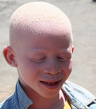
Albinism is a congenital condition characterized in humans by the partial or complete absence of pigment in the skin, hair and eyes. Albinism is associated with a number of vision defects, such as photophobia, nystagmus, and amblyopia. Lack of skin pigmentation makes for more susceptibility to sunburn and skin cancers. In rare cases such as Chédiak–Higashi syndrome, albinism may be associated with deficiencies in the transportation of melanin granules. This also affects essential granules present in immune cells, leading to increased susceptibility to infection.

<span class="mw-page-title-main">Melanin</span> Group of natural pigments found in most organisms

Melanin consist of oligomers or polymers arranged in a disordered manner which among other functions provide the pigments of many organisms. Melanin pigments are produced in a specialized group of cells known as melanocytes. They have been described as "among the last remaining biological frontiers with the unknown".

<span class="mw-page-title-main">Melanocyte</span> Melanin-producing cells of the skin

Melanocytes are melanin-producing neural crest-derived cells located in the bottom layer of the skin's epidermis, the middle layer of the eye, the inner ear, vaginal epithelium, meninges, bones, and heart. Melanin is a dark pigment primarily responsible for skin color. Once synthesized, melanin is contained in special organelles called melanosomes which can be transported to nearby keratinocytes to induce pigmentation. Thus darker skin tones have more melanosomes present than lighter skin tones. Functionally, melanin serves as protection against UV radiation. Melanocytes also have a role in the immune system.

<span class="mw-page-title-main">Melanosome</span> Organelle found in animal cells used for the synthesis, storage and transport of melanin

A melanosome is an organelle found in animal cells and is the site for synthesis, storage and transport of melanin, the most common light-absorbing pigment found in the animal kingdom. Melanosomes are responsible for color and photoprotection in animal cells and tissues.

<span class="mw-page-title-main">Food browning</span> Food process

Browning is the process of food turning brown due to the chemical reactions that take place within. The process of browning is one of the chemical reactions that take place in food chemistry and represents an interesting research topic regarding health, nutrition, and food technology. Though there are many different ways food chemically changes over time, browning in particular falls into two main categories: enzymatic versus non-enzymatic browning processes.

<span class="mw-page-title-main">Hypopigmentation</span> Area of skin becoming lighter than the baseline skin color

Hypopigmentation is characterized specifically as an area of skin becoming lighter than the baseline skin color, but not completely devoid of pigment. This is not to be confused with depigmentation, which is characterized as the absence of all pigment. It is caused by melanocyte or melanin depletion, or a decrease in the amino acid tyrosine, which is used by melanocytes to make melanin. Some common genetic causes include mutations in the tyrosinase gene or OCA2 gene. As melanin pigments tend to be in the skin, eye, and hair, these are the commonly affected areas in those with hypopigmentation.

<span class="mw-page-title-main">Skin whitening</span> Practice of using chemical substances to lighten the skin

Skin whitening, also known as skin lightening and skin bleaching, is the practice of using chemical substances in an attempt to lighten the skin or provide an even skin color by reducing the melanin concentration in the skin. Several chemicals have been shown to be effective in skin whitening, while some have proven to be toxic or have questionable safety profiles. This includes mercury compounds which may cause neurological problems and kidney problems.

Catechol oxidase is a copper oxidase that contains a type 3 di-copper cofactor and catalyzes the oxidation of ortho-diphenols into ortho-quinones coupled with the reduction of molecular oxygen to water. It is present in a variety of species of plants and fungi including Ipomoea batatas and Camellia sinensis. Metalloenzymes with type 3 copper centers are characterized by their ability to reversibly bind dioxygen at ambient conditions. In plants, catechol oxidase plays a key role in enzymatic browning by catalyzing the oxidation of catechol to o-quinone in the presence of oxygen, which can rapidly polymerize to form the melanin that grants damaged fruits their dark brown coloration.

<span class="mw-page-title-main">Agouti-signaling protein</span> Protein-coding gene in the species Homo sapiens

Agouti-signaling protein is a protein that in humans is encoded by the ASIP gene. It is responsible for the distribution of melanin pigment in mammals. Agouti interacts with the melanocortin 1 receptor to determine whether the melanocyte produces phaeomelanin, or eumelanin. This interaction is responsible for making distinct light and dark bands in the hairs of animals such as the agouti, which the gene is named after. In other species such as horses, agouti signalling is responsible for determining which parts of the body will be red or black. Mice with wildtype agouti will be grey-brown, with each hair being partly yellow and partly black. Loss of function mutations in mice and other species cause black fur coloration, while mutations causing expression throughout the whole body in mice cause yellow fur and obesity.

<span class="mw-page-title-main">White horse</span> Horse coat color

A white horse is born predominantly white and stays white throughout its life. A white horse has mostly pink skin under its hair coat, and may have brown, blue, or hazel eyes. "True white" horses, especially those that carry one of the dominant white (W) genes, are rare. Most horses that are commonly referred to as "white" are actually "gray" horses whose hair coats are completely white. Gray horses may be born of any color and their hairs gradually turn white as time goes by and take on a white appearance. Nearly all gray horses have dark skin, except under any white markings present at birth. Skin color is the most common method for an observer to distinguish between mature white and gray horses.

<span class="mw-page-title-main">Microphthalmia-associated transcription factor</span> Mammalian protein found in Homo sapiens

Microphthalmia-associated transcription factor also known as class E basic helix-loop-helix protein 32 or bHLHe32 is a protein that in humans is encoded by the MITF gene.

Polyphenol oxidase, an enzyme involved in fruit browning, is a tetramer that contains four atoms of copper per molecule.

<span class="mw-page-title-main">Labrador Retriever coat colour genetics</span> Genetics behind Labrador Retriever coat colour

The genetic basis of coat colour in the Labrador Retriever has been found to depend on several distinct genes. The interplay among these genes is used as an example of epistasis.

<span class="mw-page-title-main">TYRP1</span> Enzyme

Tyrosinase-related protein 1, also known as TYRP1, is an intermembrane enzyme which in humans is encoded by the TYRP1 gene.

<span class="mw-page-title-main">P protein</span> Protein-coding gene in humans

P protein, also known as melanocyte-specific transporter protein or pink-eyed dilution protein homolog, is a protein that in humans is encoded by the oculocutaneous albinism II (OCA2) gene. The P protein is believed to be an integral membrane protein involved in small molecule transport, specifically of tyrosine—a precursor of melanin. Certain mutations in OCA2 result in type 2 oculocutaneous albinism. OCA2 encodes the human homologue of the mouse p gene.

<span class="mw-page-title-main">Membrane-associated transporter protein</span> Protein

Membrane-associated transporter protein (MATP), also known as solute carrier family 45 member 2 (SLC45A2) or melanoma antigen AIM1, is a protein that in humans is encoded by the SLC45A2 gene.

Oculocutaneous albinism type I or type 1A is an autosomal recessive skin disease. This subtype of oculocutaneous albinism is caused when the gene for tyrosinase does not function properly.

<span class="mw-page-title-main">Ocular albinism type 1</span> Most common type of ocular albinism

Ocular albinism type 1(OA1) is the most common type of ocular albinism, with a prevalence rate of 1:50,000. It is an inheritable classical Mendelian type X-linked recessive disorder wherein the retinal pigment epithelium lacks pigment while hair and skin appear normal. Since it is usually an X-linked disorder, it occurs mostly in males, while females are carriers unless they are homozygous. About 60 missense and nonsense mutations, insertions, and deletions have been identified in Oa1. Mutations in OA1 have been linked to defective glycosylation and thus improper intracellular transportation.

<span class="mw-page-title-main">Amelanism</span> Pigmentation abnormality

Amelanism is a pigmentation abnormality characterized by the lack of pigments called melanins, commonly associated with a genetic loss of tyrosinase function. Amelanism can affect fish, amphibians, reptiles, birds, and mammals including humans. The appearance of an amelanistic animal depends on the remaining non-melanin pigments. The opposite of amelanism is melanism, a higher percentage of melanin.

<span class="mw-page-title-main">Albinism</span> Disorder causing lack of pigmentation

Albinism is the congenital absence of melanin in an animal or plant resulting in white hair, feathers, scales and skin and reddish pink or blue eyes. Individuals with the condition are referred to as albinos.

References

  1. 1 2 3 GRCh38: Ensembl release 89: ENSG00000077498 - Ensembl, May 2017
  2. 1 2 3 GRCm38: Ensembl release 89: ENSMUSG00000004651 - Ensembl, May 2017
  3. "Human PubMed Reference:". National Center for Biotechnology Information, U.S. National Library of Medicine.
  4. "Mouse PubMed Reference:". National Center for Biotechnology Information, U.S. National Library of Medicine.
  5. 1 2 Kumar CM, Sathisha UV, Dharmesh S, Rao AG, Singh SA (Mar 2011). "Interaction of sesamol (3,4-methylenedioxyphenol) with tyrosinase and its effect on melanin synthesis". Biochimie. 93 (3): 562–9. doi:10.1016/j.biochi.2010.11.014. PMID   21144881.
  6. Barton DE, Kwon BS, Francke U (Jul 1988). "Human tyrosinase gene, mapped to chromosome 11 (q14----q21), defines second region of homology with mouse chromosome 7". Genomics. 3 (1): 17–24. doi:10.1016/0888-7543(88)90153-X. PMID   3146546.
  7. Hearing VJ, Ekel TM, Montague PM, Nicholson JM (Feb 1980). "Mammalin tyrosinase. Stoichiometry and measurement of reaction products". Biochimica et Biophysica Acta (BBA) - Enzymology. 611 (2): 251–68. doi:10.1016/0005-2744(80)90061-3. PMID   6766744.
  8. PDB: 1WX3 ; Matoba Y, Kumagai T, Yamamoto A, Yoshitsu H, Sugiyama M (2006). "Crystallographic evidence that the dinuclear copper center of tyrosinase is flexible during catalysis". J. Biol. Chem. 281 (13): 8981–8990. doi: 10.1074/jbc.M509785200 . PMID   16436386.
  9. Mayer AM (Nov 2006). "Polyphenol oxidases in plants and fungi: going places? A review". Phytochemistry. 67 (21): 2318–31. Bibcode:2006PChem..67.2318M. doi:10.1016/j.phytochem.2006.08.006. PMID   16973188.
  10. Jaenicke E, Decker H (Apr 2003). "Tyrosinases from crustaceans form hexamers". The Biochemical Journal. 371 (Pt 2): 515–23. doi:10.1042/BJ20021058. PMC   1223273 . PMID   12466021.
  11. Mayer AM (November 2006). "Polyphenol oxidases in plants and fungi: going places? A review". Phytochemistry. 67 (21): 2318–31. Bibcode:2006PChem..67.2318M. doi:10.1016/j.phytochem.2006.08.006. PMID   16973188.
  12. Kwon BS, Haq AK, Pomerantz SH, Halaban R (Nov 1987). "Isolation and sequence of a cDNA clone for human tyrosinase that maps at the mouse c-albino locus". Proceedings of the National Academy of Sciences of the United States of America. 84 (21): 7473–7. Bibcode:1987PNAS...84.7473K. doi: 10.1073/pnas.84.21.7473 . PMC   299318 . PMID   2823263.
  13. Theos AC, Tenza D, Martina JA, Hurbain I, Peden AA, Sviderskaya EV, Stewart A, Robinson MS, Bennett DC, Cutler DF, Bonifacino JS, Marks MS, Raposo G (Nov 2005). "Functions of adaptor protein (AP)-3 and AP-1 in tyrosinase sorting from endosomes to melanosomes". Molecular Biology of the Cell. 16 (11): 5356–72. doi:10.1091/mbc.E05-07-0626. PMC   1266432 . PMID   16162817.
  14. 1 2 3 Kim YJ, Uyama H (Aug 2005). "Tyrosinase inhibitors from natural and synthetic sources: structure, inhibition mechanism and perspective for the future". Cellular and Molecular Life Sciences. 62 (15): 1707–23. doi:10.1007/s00018-005-5054-y. PMID   15968468. S2CID   8280251.
  15. "Allele Frequency For Polymorphic Site: rs2733832". ALFRED. Archived from the original on 20 August 2016. Retrieved 23 June 2016.
  16. Panis F, Krachler RF, Krachler R, Rompel A (June 2021). "Expression, Purification, and Characterization of a Well-Adapted Tyrosinase from Peatlands Identified by Partial Community Analysis". Environmental Science & Technology. 55 (16): 11445–11454. Bibcode:2021EnST...5511445P. doi:10.1021/acs.est.1c02514. PMC   8375020 . PMID   34156250.
  17. Hou L, Panthier JJ, Arnheiter H (Dec 2000). "Signaling and transcriptional regulation in the neural crest-derived melanocyte lineage: interactions between KIT and MITF". Development. 127 (24): 5379–89. doi:10.1242/dev.127.24.5379. PMID   11076759.
  18. Hoek KS, Schlegel NC, Eichhoff OM, Widmer DS, Praetorius C, Einarsson SO, Valgeirsdottir S, Bergsteinsdottir K, Schepsky A, Dummer R, Steingrimsson E (Dec 2008). "Novel MITF targets identified using a two-step DNA microarray strategy". Pigment Cell & Melanoma Research. 21 (6): 665–76. doi: 10.1111/j.1755-148X.2008.00505.x . PMID   19067971.
  19. M. S. Eller, K. Ostrom, and B. A. Gilchrest, “DNA damage enhances melanogenesis,” Proceedings of the National Academy of Sciences of the United States of America, vol. 93, no. 3, pp. 1087–1092, 1996
  20. Witkop CJ (Oct 1979). "Albinism: hematologic-storage disease, susceptibility to skin cancer, and optic neuronal defects shared in all types of oculocutaneous and ocular albinism". The Alabama Journal of Medical Sciences. 16 (4): 327–30. PMID   546241.
  21. Ando H, Kondoh H, Ichihashi M, Hearing VJ (Apr 2007). "Approaches to identify inhibitors of melanin biosynthesis via the quality control of tyrosinase". The Journal of Investigative Dermatology. 127 (4): 751–61. doi: 10.1038/sj.jid.5700683 . PMID   17218941.
  22. Chang TS (Jun 2009). "An updated review of tyrosinase inhibitors". International Journal of Molecular Sciences. 10 (6): 2440–75. doi: 10.3390/ijms10062440 . PMC   2705500 . PMID   19582213.
  23. Pillaiyar T, Manickam M, Namasivayam V (December 2017). "Skin whitening agents: medicinal chemistry perspective of tyrosinase inhibitors". Journal of Enzyme Inhibition and Medicinal Chemistry. 32 (1): 403–425. doi:10.1080/14756366.2016.1256882. PMC   6010116 . PMID   28097901.
  24. Damé, Maria Cecília Florisbal; Xavier, Gildenor Medeiros; Oliveira-Filho, José Paes; Borges, Alexandre Secorun; Oliveira, Henrique Nunes; Riet-Correa, Franklin; Schild, Ana Lucia (2012-07-20). "A nonsense mutation in the tyrosinase gene causes albinism in water buffalo". BMC Genetics. 13: 62. doi: 10.1186/1471-2156-13-62 . ISSN   1471-2156. PMC   3411452 . PMID   22817390.
  25. 1 2 Miura, Ikuo; Tagami, Masataka; Fujitani, Takeshi; Ogata, Mitsuaki (2018-02-10). "Spontaneous tyrosinase mutations identified in albinos of three wild frog species". Genes & Genetic Systems. 92 (4): 189–196. doi: 10.1266/ggs.16-00061 . ISSN   1880-5779. PMID   28674275.
  26. Kim, Young-Hyun; Park, Sang-Je; Choe, Se-Hee; Lee, Ja-Rang; Cho, Hyeon-Mu; Kim, Sun-Uk; Kim, Ji-Su; Sim, Bo-Woong; Song, Bong-Seok; Lee, Youngjeon; Jin, Yeung Bae; Hong, Jung-Joo; Jeong, Kang-Jin; Kang, Philyong; Baek, Seung-Ho; Lee, Sang-Rae; Huh, Jae-Won; Chang, Kyu-Tae (2017). "Identification and characterization of the tyrosinase gene ( TYR ) and its transcript variants ( TYR_1 and TYR_2 ) in the crab-eating macaque ( Macaca fascicularis )". Gene. 630: 21–27. doi: 10.1016/j.gene.2017.07.047 . PMID   28756020.
  27. Agnes Camacho-Hubner, Christelle Richard, Friedrich Beermann (20 Feb 2022). "Genomic structure and evolutionary conservation of the tyrosinase gene family from Fugu". Gene. 285 (1–2): 59–68. doi:10.1016/s0378-1119(02)00411-0. PMID   12039032.{{cite journal}}: CS1 maint: multiple names: authors list (link)
  28. Rosaria Esposito, Salvatore D'Aniello, Paola Squarzoni, Maria Rosa Pezzotti, Filomena Ristoratore and Antonietta Spagnuolo (2012). "New Insights into the Evolution of Metazoan Tyrosinase Gene Family". PLOS ONE. 7 (4): e35731. Bibcode:2012PLoSO...735731E. doi: 10.1371/journal.pone.0035731 . PMC   3334994 . PMID   22536431.{{cite journal}}: CS1 maint: multiple names: authors list (link)
  29. Aguilera, Felipe; McDougall, Carmel; Degnan, Bernard M. (2013-05-01). "Origin, evolution and classification of type-3 copper proteins: lineage-specific gene expansions and losses across the Metazoa". BMC Evolutionary Biology. 13 (1): 96. Bibcode:2013BMCEE..13...96A. doi: 10.1186/1471-2148-13-96 . ISSN   1471-2148. PMC   3658974 . PMID   23634722.
  30. F. Murisier and F. Beermann (2006). "Genetics of pigment cells:lessons from the tyrosinase gene family". Histology and Histopathology. 21 (5): 567–578. PMID   16493586.
  31. Mendes E, Perry Mde J, Francisco AP (May 2014). "Design and discovery of mushroom tyrosinase inhibitors and their therapeutic applications". Expert Opinion on Drug Discovery. 9 (5): 533–54. doi:10.1517/17460441.2014.907789. PMID   24708040. S2CID   12589166.
  32. Rescigno A, Sollai F, Pisu B, Rinaldi A, Sanjust E (Aug 2002). "Tyrosinase inhibition: general and applied aspects". Journal of Enzyme Inhibition and Medicinal Chemistry. 17 (4): 207–18. doi: 10.1080/14756360210000010923 . PMID   12530473.
  33. Sollai F, Zucca P, Sanjust E, Steri D, Rescigno A (December 2008). "Umbelliferone and esculetin: inhibitors or substrates for polyphenol oxidases?". Biological & Pharmaceutical Bulletin. 31 (12): 2187–93. doi: 10.1248/bpb.31.2187 . hdl: 11584/105440 . PMID   19043197.
  34. Rescigno A, Casañola-Martin GM, Sanjust E, Zucca P, Marrero-Ponce Y (March 2011). "Vanilloid derivatives as tyrosinase inhibitors driven by virtual screening-based QSAR models". Drug Testing and Analysis. 3 (3): 176–81. doi:10.1002/dta.187. PMID   21125547.
  35. Qian, W., Liu, W., Zhu, D., Cao, Y., Tang, A., Gong, G., Su, H."Natural skin‑whitening compounds for the treatment of melanogenesis (Review)". Experimental and Therapeutic Medicine 20.1 (2020): 173-185.
  36. Lajis AFB and Ariff AB: Discovery of new depigmenting compounds and their efficacy to treat hyperpigmentation: Evidence from in vitro study. J Cosmet Dermatol. 18:703–727. 2019.
  37. Horsch J, Wilke P, Pretzler M, Seuss M, Melnyk I, Remmler D, et al. (November 2018). "Polymerizing Like Mussels Do: Toward Synthetic Mussel Foot Proteins and Resistant Glues". Angewandte Chemie. 57 (48): 15728–15732. doi:10.1002/anie.201809587. PMC   6282983 . PMID   30246912.
  38. Arias S, Amini S, Horsch J, Pretzler M, Rompel A, Melnyk I, et al. (October 2020). "Toward Artificial Mussel-Glue Proteins: Differentiating Sequence Modules for Adhesion and Switchable Cohesion". Angewandte Chemie. 59 (42): 18495–18499. doi:10.1002/anie.202008515. PMC   7590116 . PMID   32596967.