
The retina is the innermost, light-sensitive layer of tissue of the eye of most vertebrates and some molluscs. The optics of the eye create a focused two-dimensional image of the visual world on the retina, which then processes that image within the retina and sends nerve impulses along the optic nerve to the visual cortex to create visual perception. The retina serves a function which is in many ways analogous to that of the film or image sensor in a camera.

Retinol, also called vitamin A1, is a fat-soluble vitamin in the vitamin A family that is found in food and used as a dietary supplement. Retinol or other forms of vitamin A are needed for vision, cellular development, maintenance of skin and mucous membranes, immune function and reproductive development. Dietary sources include fish, dairy products, and meat. As a supplement it is used to treat and prevent vitamin A deficiency, especially that which results in xerophthalmia. It is taken by mouth or by injection into a muscle. As an ingredient in skin-care products, it is used to reduce wrinkles and other effects of skin aging.

Eyes are organs of the visual system. They provide living organisms with vision, the ability to receive and process visual detail, as well as enabling several photo response functions that are independent of vision. Eyes detect light and convert it into electro-chemical impulses in neurons (neurones). In higher organisms, the eye is a complex optical system which collects light from the surrounding environment, regulates its intensity through a diaphragm, focuses it through an adjustable assembly of lenses to form an image, converts this image into a set of electrical signals, and transmits these signals to the brain through complex neural pathways that connect the eye via the optic nerve to the visual cortex and other areas of the brain. Eyes with resolving power have come in ten fundamentally different forms, and 96% of animal species possess a complex optical system. Image-resolving eyes are present in molluscs, chordates and arthropods.

Retinitis pigmentosa (RP) is a genetic disorder of the eyes that causes loss of vision. Symptoms include trouble seeing at night and decreasing peripheral vision. As peripheral vision worsens, people may experience "tunnel vision". Complete blindness is uncommon. Onset of symptoms is generally gradual and often begins in childhood.

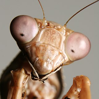
A compound eye is a visual organ found in arthropods such as insects and crustaceans. It may consist of thousands of ommatidia, which are tiny independent photoreception units that consist of a cornea, lens, and photoreceptor cells which distinguish brightness and color. The image perceived by this arthropod eye is a combination of inputs from the numerous ommatidia, which are oriented to point in slightly different directions. Compared with single-aperture eyes, compound eyes have poor image resolution; however, they possess a very large view angle and the ability to detect fast movement and, in some cases, the polarization of light. Because a compound eye is made up of a collection of ommatidia, each with its own lens, light will enter each ommatidium instead of using a single entrance point. The individual light receptors behind each lens are then turned on and off due to a series of changes in the light intensity during movement or when an object is moving, creating a flicker-effect known as the flicker frequency, which is the rate at which the ommatidia are turned on and off– this facilitates faster reaction to movement; honey bees respond in 0.01s compared with 0.05s for humans.

A photoreceptor cell is a specialized type of neuroepithelial cell found in the retina that is capable of visual phototransduction. The great biological importance of photoreceptors is that they convert light into signals that can stimulate biological processes. To be more specific, photoreceptor proteins in the cell absorb photons, triggering a change in the cell's membrane potential.

Rod cells are photoreceptor cells in the retina of the eye that can function in lower light better than the other type of visual photoreceptor, cone cells. Rods are usually found concentrated at the outer edges of the retina and are used in peripheral vision. On average, there are approximately 92 million rod cells in the human retina. Rod cells are more sensitive than cone cells and are almost entirely responsible for night vision. However, rods have little role in color vision, which is the main reason why colors are much less apparent in dim light.

The fovea centralis is a small, central pit composed of closely packed cones in the eye. It is located in the center of the macula lutea of the retina.

A retinal ganglion cell (RGC) is a type of neuron located near the inner surface of the retina of the eye. It receives visual information from photoreceptors via two intermediate neuron types: bipolar cells and retina amacrine cells. Retina amacrine cells, particularly narrow field cells, are important for creating functional subunits within the ganglion cell layer and making it so that ganglion cells can observe a small dot moving a small distance. Retinal ganglion cells collectively transmit image-forming and non-image forming visual information from the retina in the form of action potential to several regions in the thalamus, hypothalamus, and mesencephalon, or midbrain.
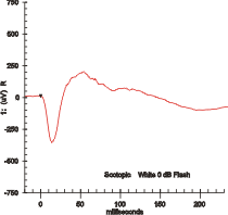
Electroretinography measures the electrical responses of various cell types in the retina, including the photoreceptors, inner retinal cells, and the ganglion cells. Electrodes are placed on the surface of the cornea or on the skin beneath the eye to measure retinal responses. Retinal pigment epithelium (RPE) responses are measured with an EOG test with skin-contact electrodes placed near the canthi. During a recording, the patient's eyes are exposed to standardized stimuli and the resulting signal is displayed showing the time course of the signal's amplitude (voltage). Signals are very small, and typically are measured in microvolts or nanovolts. The ERG is composed of electrical potentials contributed by different cell types within the retina, and the stimulus conditions can elicit stronger response from certain components.
A simple eye refers to a form of eye or an optical arrangement composed of a single lens and without an elaborate retina such as occurs in most vertebrates. In this sense "simple eye" is distinct from a multi-lensed "compound eye", and is not necessarily at all simple in the usual sense of the word.
Visual phototransduction is the sensory transduction process of the visual system by which light is detected to yield nerve impulses in the rod cells and cone cells in the retina of the eye in humans and other vertebrates. It relies on the visual cycle, a sequence of biochemical reactions in which a molecule of retinal bound to opsin undergoes photoisomerization, initiates a cascade that signals detection of the photon, and is indirectly restored to its photosensitive isomer for reuse. Phototransduction in some invertebrates such as fruit flies relies on similar processes.
Intrinsically photosensitive retinal ganglion cells (ipRGCs), also called photosensitive retinal ganglion cells (pRGC), or melanopsin-containing retinal ganglion cells (mRGCs), are a type of neuron in the retina of the mammalian eye. The presence of ipRGCs was first suspected in 1927 when rodless, coneless mice still responded to a light stimulus through pupil constriction, This implied that rods and cones are not the only light-sensitive neurons in the retina. Yet research on these cells did not advance until the 1980s. Recent research has shown that these retinal ganglion cells, unlike other retinal ganglion cells, are intrinsically photosensitive due to the presence of melanopsin, a light-sensitive protein. Therefore, they constitute a third class of photoreceptors, in addition to rod and cone cells.

In neurobiology, lateral inhibition is the capacity of an excited neuron to reduce the activity of its neighbors. Lateral inhibition disables the spreading of action potentials from excited neurons to neighboring neurons in the lateral direction. This creates a contrast in stimulation that allows increased sensory perception. It is also referred to as lateral antagonism and occurs primarily in visual processes, but also in tactile, auditory, and even olfactory processing. Cells that utilize lateral inhibition appear primarily in the cerebral cortex and thalamus and make up lateral inhibitory networks (LINs). Artificial lateral inhibition has been incorporated into artificial sensory systems, such as vision chips, hearing systems, and optical mice. An often under-appreciated point is that although lateral inhibition is visualised in a spatial sense, it is also thought to exist in what is known as "lateral inhibition across abstract dimensions." This refers to lateral inhibition between neurons that are not adjacent in a spatial sense, but in terms of modality of stimulus. This phenomenon is thought to aid in colour discrimination.

The pigmented layer of retina or retinal pigment epithelium (RPE) is the pigmented cell layer just outside the neurosensory retina that nourishes retinal visual cells, and is firmly attached to the underlying choroid and overlying retinal visual cells.
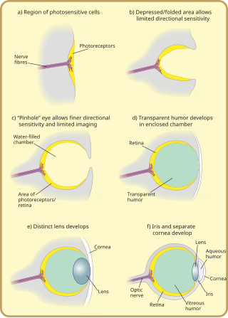
Many scientists have found the evolution of the eye attractive to study because the eye distinctively exemplifies an analogous organ found in many animal forms. Simple light detection is found in bacteria, single-celled organisms, plants and animals. Complex, image-forming eyes have evolved independently several times.
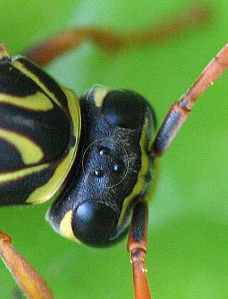
In the compound eye of invertebrates such as insects and crustaceans, the pseudopupil appears as a dark spot which moves across the eye as the animal is rotated. This occurs because the ommatidia that one observes "head-on" absorb the incident light, while those to one side reflect it. The pseudopupil therefore reveals which ommatidia are aligned with the axis along which the observer is viewing.

Apposition eyes are the most common form of eye, and are presumably the ancestral form of compound eye. They are found in all arthropod groups, although they may have evolved more than once within this phylum. Some annelids and bivalves also have apposition eyes. They are also possessed by Limulus, the horseshoe crab, and there are suggestions that other chelicerates developed their simple eyes by reduction from a compound starting point. Some caterpillars appear to have evolved compound eyes from simple eyes in the opposite fashion.
Sevenless (sev) is a gene in the fruit fly Drosophila melanogaster that encodes a receptor tyrosine kinase protein essential to the development of the R7 photoreceptor cells in the Drosophila embryonic eye. The Drosophila ommatidium contains 8 distinct retinula or R cells, each of which has a different spectral sensitivity. The R7 photo receptor, located in each of several ommatidium in the fly's compound eye, is used to detect ultraviolet light. The R8 photoreceptor contains an activator of the RTK for on a precursor R7 cell, called the bride of sevenless (BOSS). The binding of BOSS to sevenless stimulates a complex series of reactions involving the RTK (sevenless), MAP kinases, Ras and many more molecules to differentiate that precursor R7 photo receptor to a fully functional R7 photo receptor that can see UV light. Much of this knowledge was gained by examining flies with a mutant sevenless which still produced a fully functional R7 photoreceptor when a dominant Ras was injected into the mutant R7 precursor.
Cephalopods, as active marine predators, possess sensory organs specialized for use in aquatic conditions. They have a camera-type eye which consists of an iris, a circular lens, vitreous cavity, pigment cells, and photoreceptor cells that translate light from the light-sensitive retina into nerve signals which travel along the optic nerve to the brain. For the past 140 years, the camera-type cephalopod eye has been compared with the vertebrate eye as an example of convergent evolution, where both types of organisms have independently evolved the camera-eye trait and both share similar functionality. Contention exists on whether this is truly convergent evolution or parallel evolution. Unlike the vertebrate camera eye, the cephalopods' form as invaginations of the body surface, and consequently the cornea lies over the top of the eye as opposed to being a structural part of the eye. Unlike the vertebrate eye, a cephalopod eye is focused through movement, much like the lens of a camera or telescope, rather than changing shape as the lens in the human eye does. The eye is approximately spherical, as is the lens, which is fully internal.


