The cerebral cortex, also known as the cerebral mantle, is the outer layer of neural tissue of the cerebrum of the brain in humans and other mammals. It is the largest site of neural integration in the central nervous system. and plays a key role in attention, perception, awareness, thought, memory, language, and consciousness. The cerebral cortex is part of the brain responsible for cognition.

Neuropil is any area in the nervous system composed of mostly unmyelinated axons, dendrites and glial cell processes that forms a synaptically dense region containing a relatively low number of cell bodies. The most prevalent anatomical region of neuropil is the brain which, although not completely composed of neuropil, does have the largest and highest synaptically concentrated areas of neuropil in the body. For example, the neocortex and olfactory bulb both contain neuropil.
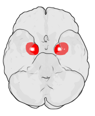
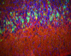
The amygdala is a paired nuclear complex present in the cerebral hemispheres of vertebrates. It is considered part of the limbic system. In primates, it is located medially within the temporal lobes. It consists of many nuclei, each made up of further subnuclei. The subdivision most commonly made is into the basolateral, central, cortical, and medial nuclei together with the intercalated cell clusters. The amygdala has a primary role in the processing of memory, decision-making, and emotional responses. The amygdala was first identified and named by Karl Friedrich Burdach in 1822.

The limbic system, also known as the paleomammalian cortex, is a set of brain structures located on both sides of the thalamus, immediately beneath the medial temporal lobe of the cerebrum primarily in the forebrain.

A Brodmann area is a region of the cerebral cortex, in the human or other primate brain, defined by its cytoarchitecture, or histological structure and organization of cells. The concept was first introduced by the German anatomist Korbinian Brodmann in the early 20th century. Brodmann mapped the human brain based on the varied cellular structure across the cortex and identified 52 distinct regions, which he numbered 1 to 52. These regions, or Brodmann areas, correspond with diverse functions including sensation, motor control, and cognition.
Proisocortex or pro-isocortex is one of two subtypes of cortical areas in the areas belonging to the neocortex. The other subtype is termed the true isocortex. Proisocortical areas are transitional areas placed between areas of true isocortex and areas of periallocortex. The histological structure of proisocortex is also transitional between true isocortex and either peripaleocortex or periarchicortex, depending on with which subtype of periallocortex the given proisocortical area borders.

The neocortex, also called the neopallium, isocortex, or the six-layered cortex, is a set of layers of the mammalian cerebral cortex involved in higher-order brain functions such as sensory perception, cognition, generation of motor commands, spatial reasoning and language. The neocortex is further subdivided into the true isocortex and the proisocortex.

The brain is the central organ of the human nervous system, and with the spinal cord makes up the central nervous system. The brain consists of the cerebrum, the brainstem and the cerebellum. It controls most of the activities of the body, processing, integrating, and coordinating the information it receives from the sense organs, and making decisions as to the instructions sent to the rest of the body. The brain is contained in, and protected by, the skull bones of the head.

The triune brain is a model of the evolution of the vertebrate forebrain and behavior, proposed by the American physician and neuroscientist Paul D. MacLean in the 1960s. The triune brain consists of the reptilian complex, the paleomammalian complex, and the neomammalian complex (neocortex), viewed each as independently conscious, and as structures sequentially added to the forebrain in the course of evolution. According to the model, the basal ganglia are in charge of our primal instincts, the limbic system is in charge of our emotions and the neocortex is responsible for objective or rational thoughts.

A cortical minicolumn (also called cortical microcolumn) is a vertical column through the cortical layers of the brain. Neurons within the microcolumn "receive common inputs, have common outputs, are interconnected, and may well constitute a fundamental computational unit of the cerebral cortex". Minicolumns comprise perhaps 80–120 neurons, except in the primate primary visual cortex (V1), where there are typically more than twice the number. There are about 2×10⁸ minicolumns in humans. From calculations, the diameter of a minicolumn is about 28–40 μm. Minicolumns grow from progenitor cells within the embryo and contain neurons within multiple layers (2–6) of the cortex.

The insular cortex is a portion of the cerebral cortex folded deep within the lateral sulcus within each hemisphere of the mammalian brain.

The archicortex, or archipallium, is the phylogenetically second oldest region of the brain's cerebral cortex. It is often considered contiguous with the olfactory cortex, but its extent varies among species. In older species, such as fish, the archipallium makes up most of the cerebrum. Amphibians develop an archipallium and paleopallium.
The allocortex, or heterogenetic cortex, and neocortex are the two types of cerebral cortex in the brain. In the human brain, the allocortex is the much smaller area of cortex taking up just 10%; the neocortex takes up the remaining 90%. It is characterized by having just three or four cortical layers, in contrast with the six layers of the neocortex. There are three subtypes of allocortex: the paleocortex, the archicortex, and the periallocortex—a transitional zone between the neocortex and the allocortex.

The posterior cingulate cortex (PCC) is the caudal part of the cingulate cortex, located posterior to the anterior cingulate cortex. This is the upper part of the "limbic lobe". The cingulate cortex is made up of an area around the midline of the brain. Surrounding areas include the retrosplenial cortex and the precuneus.
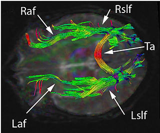
The amygdalofugal pathway is one of the three major efferent pathways of the amygdala, meaning that it is one of the three principal pathways by which fibers leave the amygdala. It leads from the basolateral nucleus and central nucleus of the amygdala. The amygdala is a limbic structure in the medial temporal lobe of the brain. The other main efferent pathways from the amygdala are the stria terminalis and anterior commissure.

In anatomy of animals, the paleocortex, or paleopallium, is a region within the telencephalon in the vertebrate brain. This type of cortical tissue consists of three cortical laminae. In comparison, the neocortex has six layers and the archicortex has three or four layers. Because the number of laminae that compose a type of cortical tissue seems to be directly proportional to both the information-processing capabilities of that tissue and its phylogenetic age, paleocortex is thought to be an intermediate between the archicortex and the neocortex in both aspects.

In neuroanatomy, pallium refers to the layers of grey and white matter that cover the upper surface of the cerebrum in vertebrates. The non-pallial part of the telencephalon builds the subpallium. In basal vertebrates, the pallium is a relatively simple three-layered structure, encompassing 3–4 histogenetically distinct domains, plus the olfactory bulb.
Disconnection syndrome is a general term for a collection of neurological symptoms caused – via lesions to associational or commissural nerve fibres – by damage to the white matter axons of communication pathways in the cerebrum, independent of any lesions to the cortex. The behavioral effects of such disconnections are relatively predictable in adults. Disconnection syndromes usually reflect circumstances where regions A and B still have their functional specializations except in domains that depend on the interconnections between the two regions.
Mesocortex is the transitional areas of the cerebral cortex, formed at borders between true isocortex and true allocortex. Parts of mesocortex that lie closer to the true isocortex and have more resemblance to the isocortex in their cytoarchitectonics and histology, are called proisocortex. Parts of mesocortex that lie closer to the true allocortex and have more resemblance to the allocortex in their cytoarchitectonics and histology, are called periallocortex.
The neurocircuitry that underlies executive function processes and emotional and motivational processes are known to be distinct in the brain. However, there are brain regions that show overlap in function between the two cognitive systems. Brain regions that exist in both systems are interesting mainly for studies on how one system affects the other. Examples of such cross-modal functions are emotional regulation strategies such as emotional suppression and emotional reappraisal, the effect of mood on cognitive tasks, and the effect of emotional stimulation of cognitive tasks.