Placental abruption

Last updated
Placental abruption
Other namesAbruptio placentae
Blausen 0737 PlacentalAbruption.png
Drawing of internal and external bleeding from placental abruption
Specialty Obstetrics
Symptoms Vaginal bleeding, low abdominal pain, dangerously low blood pressure [1]
Complications Mother: disseminated intravascular coagulopathy, kidney failure [2]
Baby: low birthweight, preterm delivery, stillbirth [2]
Usual onset24 to 26 weeks of pregnancy [2]
CausesUnclear [2]
Risk factors Smoking, preeclampsia, prior abruption [2]
Diagnostic method Based on symptoms, ultrasound [1]
Differential diagnosis Placenta previa, bloody show, chorioamnionitis [3]
Treatment Bed rest, delivery [1]
Medication Corticosteroids [1]
Frequency~0.7% of pregnancies [2]

Placental abruption is when the placenta separates early from the uterus, in other words separates before childbirth. [2] It occurs most commonly around 25 weeks of pregnancy. [2] Symptoms may include vaginal bleeding, lower abdominal pain, and dangerously low blood pressure. [1] Complications for the mother can include disseminated intravascular coagulopathy and kidney failure. [2] Complications for the baby can include fetal distress, low birthweight, preterm delivery, and stillbirth. [2] [3]

Contents

The cause of placental abruption is not entirely clear. [2] Risk factors include smoking, pre-eclampsia, prior abruption (most important and predictive risk factor), trauma during pregnancy, cocaine use, and previous cesarean section. [2] [1] Diagnosis is based on symptoms and supported by ultrasound. [1] It is classified as a complication of pregnancy. [1]

For small abruption, bed rest may be recommended, while for more significant abruptions or those that occur near term, delivery may be recommended. [1] [4] If everything is stable, vaginal delivery may be tried, otherwise cesarean section is recommended. [1] In those less than 36 weeks pregnant, corticosteroids may be given to speed development of the baby's lungs. [1] Treatment may require blood transfusion or emergency hysterectomy. [2]

Placental abruption occurs in about 1 in 200 pregnancies. [5] Along with placenta previa and uterine rupture it is one of the most common causes of vaginal bleeding in the later part of pregnancy. [6] Placental abruption is the reason for about 15% of infant deaths around the time of birth. [2] The condition was described at least as early as 1664. [7]

Signs and symptoms

In the early stages of placental abruption, there may be no symptoms. [1] When symptoms develop, they tend to develop suddenly. Common symptoms include:

Vaginal bleeding, if it occurs, may be bright red or dark. [1]

A placental abruption caused by arterial bleeding at the center of the placenta leads to sudden development of severe symptoms and life-threatening conditions including fetal heart rate abnormalities, severe maternal hemorrhage, and disseminated intravascular coagulation (DIC). Those abruptions caused by venous bleeding at the periphery of the placenta develop more slowly and cause small amounts of bleeding, intrauterine growth restriction, and oligohydramnios (low levels of amniotic fluid). [9]

Risk factors

Risk factors for placental abruption include disease, trauma, history, anatomy, and exposure to substances. The risk of placental abruption increases sixfold after severe maternal trauma. Anatomical risk factors include uncommon uterine anatomy (e.g. bicornuate uterus), uterine synechiae, and leiomyoma. Substances that increase risk of placental abruption include cocaine and tobacco when consumed during pregnancy, especially the third trimester. History of placental abruption or previous Caesarian section increases the risk by a factor of 2.3. [11] [13] [14] [15] [9]

Pathophysiology

Gross pathology of a uterus which has been opened to show a placental abruption, with a hematoma separating the placenta from the uterus. Gross pathology of placental abruption.jpg
Gross pathology of a uterus which has been opened to show a placental abruption, with a hematoma separating the placenta from the uterus.

In the vast majority of cases, placental abruption is caused by the maternal vessels tearing away from the decidua basalis, not the fetal vessels. The underlying cause is often unknown. A small number of abruptions are caused by trauma that stretches the uterus. Because the placenta is less elastic than the uterus, it tears away when the uterine tissue stretches suddenly. When anatomical risk factors are present, the placenta does not attach in a place that provides adequate support, and it may not develop appropriately or be separated as it grows. Cocaine use during the third trimester has a 10% chance of causing abruption. Though the exact mechanism is not known, cocaine and tobacco cause systemic vasoconstriction, which can severely restrict the placental blood supply (hypoperfusion and ischemia), or otherwise disrupt the vasculature of the placenta, causing tissue necrosis, bleeding, and therefore abruption. [9]

In most cases, placental disease and abnormalities of the spiral arteries develop throughout the pregnancy and lead to necrosis, inflammation, vascular problems, and ultimately, abruption. Because of this, most abruptions are caused by bleeding from the arterial supply, not the venous supply. Production of thrombin via massive bleeding causes the uterus to contract and leads to DIC. [9]

The accumulating blood pushes between the layers of the decidua, pushing the uterine wall and placenta apart. When the placenta is separated, it is unable to exchange waste, nutrients, and oxygen, a necessary function for the fetus's survival. The fetus dies when it no longer receives enough oxygen and nutrients to survive. [9]

Diagnosis

Ultrasound showing placental abruption Ultrasound Scan ND 370.jpg
Ultrasound showing placental abruption

Placental abruption is suspected when a pregnant mother has sudden localized abdominal pain with or without bleeding. The fundus may be monitored because a rising fundus can indicate bleeding. An ultrasound may be used to rule out placenta praevia but is not diagnostic for abruption. [8] The diagnosis is one of exclusion, meaning other possible sources of vaginal bleeding or abdominal pain have to be ruled out in order to diagnose placental abruption. [5] Of note, use of magnetic resonance imaging has been found to be highly sensitive in depicting placental abruption, and may be considered if no ultrasound evidence of placental abruption is present, especially if the diagnosis of placental abruption would change management. [16]

Classification

Based on severity:

Prevention

Although the risk of placental abruption cannot be eliminated, it can be reduced. Avoiding tobacco, alcohol and cocaine during pregnancy decreases the risk. Staying away from activities which have a high risk of physical trauma is also important. Women who have high blood pressure or who have had a previous placental abruption and want to conceive must be closely supervised by a doctor. [17]

The risk of placental abruption can be reduced by maintaining a good diet including taking folate, regular sleep patterns and correction of pregnancy-induced hypertension.[ citation needed ]

Use of aspirin before 16 weeks of pregnancy to prevent pre-eclampsia also appears effective at preventing placental abruption. [18]

Management

Treatment depends on the amount of blood loss and the status of the fetus. [19] If the fetus is less than 36 weeks, and neither mother or fetus are in any distress, then they may simply be monitored in hospital until a change in condition or fetal maturity whichever comes first.

Immediate delivery of the fetus may be indicated if the fetus is mature or if the fetus or mother is in distress. Blood volume replacement to maintain blood pressure and blood plasma replacement to maintain fibrinogen levels may be needed. Vaginal birth is usually preferred over Caesarean section unless there is fetal distress. Caesarean section carries an increased risk in cases of disseminated intravascular coagulation. The mother should be monitored for 7 days for postpartum hemorrhage. Excessive bleeding from uterus may necessitate hysterectomy. The mother may be given Rhogam if she is Rh negative.

Prognosis

The prognosis of this complication depends on whether treatment is received by the patient, on the quality of treatment, and on the severity of the abruption. Outcomes for the baby also depend on the gestational age. [5]

In the Western world, maternal deaths due to placental abruption are rare. The fetal prognosis is worse than the maternal prognosis; approximately 12% of fetuses affected by placental abruption die. 77% of fetuses that die from placental abruption die before birth; the remainder die due to complications of preterm birth. [9]

Without any form of medical intervention, as often happens in many parts of the world, placental abruption has a high maternal mortality rate.[ citation needed ]

Mother

Baby

Epidemiology

Placental abruption occurs in approximately 0.2–1% of all pregnancies. [8] Though different causes change when abruption is most likely to occur, the majority of placental abruptions occur before 37 weeks gestation, and 12–14% occur before 32 weeks gestation. [8] [9]

Related Research Articles

Obstetrics is the field of study concentrated on pregnancy, childbirth and the postpartum period. As a medical specialty, obstetrics is combined with gynecology under the discipline known as obstetrics and gynecology (OB/GYN), which is a surgical field.

<span class="mw-page-title-main">Amniocentesis</span> Sampling of amniotic fluid done mainly to detect fetal chromosomal abnormalities

Amniocentesis is a medical procedure used primarily in the prenatal diagnosis of genetic conditions. It has other uses such as in the assessment of infection and fetal lung maturity. Prenatal diagnostic testing, which includes amniocentesis, is necessary to conclusively diagnose the majority of genetic disorders, with amniocentesis being the gold-standard procedure after 15 weeks' gestation.

Fetal distress, also known as non-reassuring fetal status, is a condition during pregnancy or labor in which the fetus shows signs of inadequate oxygenation. Due to its imprecision, the term "fetal distress" has fallen out of use in American obstetrics. The term "non-reassuring fetal status" has largely replaced it. It is characterized by changes in fetal movement, growth, heart rate, and presence of meconium stained fluid.

<span class="mw-page-title-main">Placenta praevia</span> Medical condition

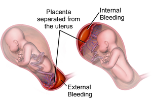
Placenta praevia is when the placenta attaches inside the uterus but in a position near or over the cervical opening. Symptoms include vaginal bleeding in the second half of pregnancy. The bleeding is bright red and tends not to be associated with pain. Complications may include placenta accreta, dangerously low blood pressure, or bleeding after delivery. Complications for the baby may include fetal growth restriction.

Antepartum bleeding, also known as antepartum haemorrhage (APH) or prepartum hemorrhage, is genital bleeding during pregnancy after the 28th week of pregnancy up to delivery.

Bloody show or show is the passage of a small amount of blood or blood-tinged mucus through the vagina near the end of pregnancy. It is caused by thinning and dilation of the cervix, leading to detachment of the cervical mucus plug that seals the cervix during pregnancy and tearing of small cervical blood vessels, and is one of the signs that labor may be imminent. The bloody show may be expelled from the vagina in pieces or altogether and often appears as a jelly-like piece of mucus stained with blood. Although the bloody show may be alarming at first, it is not a concern of patient health after 37 weeks gestation.

Obstetrical bleeding is bleeding in pregnancy that occurs before, during, or after childbirth. Bleeding before childbirth is that which occurs after 24 weeks of pregnancy. Bleeding may be vaginal or less commonly into the abdominal cavity. Bleeding which occurs before 24 weeks is known as early pregnancy bleeding.

<span class="mw-page-title-main">Uterine rupture</span> Medical condition

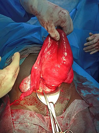
Uterine rupture is when the muscular wall of the uterus tears during pregnancy or childbirth. Symptoms, while classically including increased pain, vaginal bleeding, or a change in contractions, are not always present. Disability or death of the mother or baby may result.

<span class="mw-page-title-main">Prelabor rupture of membranes</span> Medical condition

Prelabor rupture of membranes (PROM), previously known as premature rupture of membranes, is breakage of the amniotic sac before the onset of labor. Women usually experience a painless gush or a steady leakage of fluid from the vagina. Complications in the baby may include premature birth, cord compression, and infection. Complications in the mother may include placental abruption and postpartum endometritis.

<span class="mw-page-title-main">Complications of pregnancy</span> Medical condition

Complications of pregnancy are health problems that are related to, or arise during pregnancy. Complications that occur primarily during childbirth are termed obstetric labor complications, and problems that occur primarily after childbirth are termed puerperal disorders. While some complications improve or are fully resolved after pregnancy, some may lead to lasting effects, morbidity, or in the most severe cases, maternal or fetal mortality.

<span class="mw-page-title-main">Abdominal pregnancy</span> Medical condition

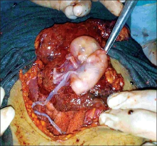
An abdominal pregnancy is a rare type of ectopic pregnancy where the embryo or fetus is growing and developing outside the womb in the abdomen, but not in the Fallopian tube, ovary or broad ligament.

<span class="mw-page-title-main">Placenta accreta spectrum</span> Medical condition

Placenta accreta occurs when all or part of the placenta attaches abnormally to the myometrium. Three grades of abnormal placental attachment are defined according to the depth of attachment and invasion into the muscular layers of the uterus:

  1. Accreta – chorionic villi attached to the myometrium, rather than being restricted within the decidua basalis.
  2. Increta – chorionic villi invaded into the myometrium.
  3. Percreta – chorionic villi invaded through the perimetrium.
<span class="mw-page-title-main">Uterine atony</span> Loss of tone in the uterine musculature

Uterine atony is the failure of the uterus to contract adequately following delivery. Contraction of the uterine muscles during labor compresses the blood vessels and slows flow, which helps prevent hemorrhage and facilitates coagulation. Therefore, a lack of uterine muscle contraction can lead to an acute hemorrhage, as the vasculature is not being sufficiently compressed. Uterine atony is the most common cause of postpartum hemorrhage, which is an emergency and potential cause of fatality. Across the globe, postpartum hemorrhage is among the top five causes of maternal death. Recognition of the warning signs of uterine atony in the setting of extensive postpartum bleeding should initiate interventions aimed at regaining stable uterine contraction.

<span class="mw-page-title-main">Vasa praevia</span> Condition in which fetal blood vessels cross or run near the internal opening of the uterus.

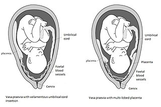
Vasa praevia is a condition in which fetal blood vessels cross or run near the internal opening of the uterus. These vessels are at risk of rupture when the supporting membranes rupture, as they are unsupported by the umbilical cord or placental tissue.

<span class="mw-page-title-main">Velamentous cord insertion</span> Velamentous placenta

Velamentous cord insertion is a complication of pregnancy where the umbilical cord is inserted in the fetal membranes. It is a major cause of antepartum hemorrhage that leads to loss of fetal blood and associated with high perinatal mortality. In normal pregnancies, the umbilical cord inserts into the middle of the placental mass and is completely encased by the amniotic sac. The vessels are hence normally protected by Wharton's jelly, which prevents rupture during pregnancy and labor. In velamentous cord insertion, the vessels of the umbilical cord are improperly inserted in the chorioamniotic membrane, and hence the vessels traverse between the amnion and the chorion towards the placenta. Without Wharton's jelly protecting the vessels, the exposed vessels are susceptible to compression and rupture.

<span class="mw-page-title-main">Placental disease</span> Medical condition

A placental disease is any disease, disorder, or pathology of the placenta.

An obstetric labor complication is a difficulty or abnormality that arises during the process of labor or delivery.

<span class="mw-page-title-main">Chorionic hematoma</span> Medical condition

Chorionic hematoma is the pooling of blood (hematoma) between the chorion, a membrane surrounding the embryo, and the uterine wall. It occurs in about 3.1% of all pregnancies, it is the most common sonographic abnormality and the most common cause of first trimester bleeding.

<span class="mw-page-title-main">Circumvallate placenta</span> Medical condition

Circumvallate placenta is a rare condition affecting about 1-2% of pregnancies, in which the amnion and chorion fetal membranes essentially "double back" on the fetal side around the edges of the placenta. After delivery, a circumvallate placenta has a thick ring of membranes on its fetal surface. Circumvallate placenta is a placental morphological abnormality associated with increased fetal morbidity and mortality due to the restricted availability of nutrients and oxygen to the developing fetus.

Early pregnancy bleeding refers to vaginal bleeding before 14 weeks of gestational age. If the bleeding is significant, hemorrhagic shock may occur. Concern for shock is increased in those who have loss of consciousness, chest pain, shortness of breath, or shoulder pain.

References

  1. 1 2 3 4 5 6 7 8 9 10 11 12 13 "Abruptio Placentae - Gynecology and Obstetrics". Merck Manuals Professional Edition. October 2017. Retrieved 9 December 2017.
  2. 1 2 3 4 5 6 7 8 9 10 11 12 13 14 15 16 17 18 19 20 21 22 23 Tikkanen, M (February 2011). "Placental abruption: epidemiology, risk factors and consequences". Acta Obstetricia et Gynecologica Scandinavica. 90 (2): 140–9. doi: 10.1111/j.1600-0412.2010.01030.x . PMID   21241259. S2CID   10871832.
  3. 1 2 Saxena, Richa (2014). Bedside Obstetrics & Gynecology. JP Medical Ltd. pp. 205–209. ISBN   9789351521037.
  4. Gibbs, Ronald S. (2008). Danforth's Obstetrics and Gynecology. Lippincott Williams & Wilkins. p. 385. ISBN   9780781769372.
  5. 1 2 3 4 5 6 7 8 9 Sheffield, [edited by] F. Gary Cunningham, Kenneth J. Leveno, Steven L. Bloom, Catherine Y. Spong, Jodi S. Dashe, Barbara L. Hoffman, Brian M. Casey, Jeanne S. (2014). Williams obstetrics (24th ed.). ISBN   978-0071798938.{{cite book}}: |first1= has generic name (help)CS1 maint: multiple names: authors list (link)
  6. Hofmeyr, GJ; Qureshi, Z (October 2016). "Preventing deaths due to haemorrhage". Best Practice & Research. Clinical Obstetrics & Gynaecology. 36: 68–82. doi:10.1016/j.bpobgyn.2016.05.004. PMID   27450867.
  7. The Journal of the Indiana State Medical Association. The Association. 1956. p. 1564.
  8. 1 2 3 4 5 6 7 8 9 Li, Yang; Tian, Yuan; Liu, Ning; Chen, Yang; Wu, Fuju (2019-03-01). "Analysis of 62 placental abruption cases: Risk factors and clinical outcomes". Taiwanese Journal of Obstetrics and Gynecology. 58 (2): 223–226. doi: 10.1016/j.tjog.2019.01.010 . ISSN   1028-4559. PMID   30910143.
  9. 1 2 3 4 5 6 7 "Placental abruption: Clinical features and diagnosis". www.uptodate.com. Retrieved 2016-06-04.
  10. 1 2 3 4 "Placenta and Placental Problems | Doctor". Patient.info. 2011-03-18. Retrieved 2012-10-23.
  11. 1 2 3 Ananth, CV; Savitz, DA; Williams, MA (August 1996). "Placental abruption and its association with hypertension and prolonged rupture of membranes: a methodologic review and meta-analysis". Obstetrics and Gynecology. 88 (2): 309–18. doi:10.1016/0029-7844(96)00088-9. PMID   8692522. S2CID   21246925.
  12. Balkawade NU, Shinde MA (October 2012). "Study of Length of Umbilical Cord and Fetal Outcome: A Study of 1,000 Deliveries". Journal of Obstetrics and Gynecology of India. 62 (5): 520–525. doi:10.1007/s13224-012-0194-0. PMC   3526711 . PMID   24082551.
  13. Ananth, C (1999). "Incidence of placental abruption in relation to cigarette smoking and hypertensive disorders during pregnancy: A meta-analysis of observational studies". Obstetrics & Gynecology. 93 (4): 622–8. doi:10.1016/S0029-7844(98)00408-6. PMID   10214847.
  14. Cressman, AM; Natekar, A; Kim, E; Koren, G; Bozzo, P (July 2014). "Cocaine abuse during pregnancy". Journal of Obstetrics and Gynaecology Canada. 36 (7): 628–31. doi:10.1016/S1701-2163(15)30543-0. PMID   25184982.
  15. Klar, M; Michels, KB (September 2014). "Cesarean section and placental disorders in subsequent pregnancies--a meta-analysis". Journal of Perinatal Medicine. 42 (5): 571–83. doi:10.1515/jpm-2013-0199. PMID   24566357. S2CID   21151164.
  16. Masselli, G; Brunelli, R; Di Tola, M; Anceschi, M; Gualdi, G (April 2011). "MR imaging in the evaluation of placental abruption: correlation with sonographic findings". Radiology. 259 (1): 222–30. doi: 10.1148/radiol.10101547 . PMID   21330568.
  17. "Placental abruption: Prevention". MayoClinic.com. 2012-01-10. Retrieved 2012-10-23.
  18. Roberge, S; Bujold, E; Nicolaides, KH (May 2018). "Meta-analysis on the effect of aspirin use for prevention of preeclampsia on placental abruption and antepartum hemorrhage". American Journal of Obstetrics and Gynecology. 218 (5): 483–489. doi: 10.1016/j.ajog.2017.12.238 . PMID   29305829.
  19. Merriam, Audrey; D'Alton, Mary E. (2018-01-01), Copel, Joshua A.; D'Alton, Mary E.; Feltovich, Helen; Gratacós, Eduard (eds.), "96 - Placental Abruption", Obstetric Imaging: Fetal Diagnosis and Care (Second Edition), Elsevier, pp. 426–429.e1, doi:10.1016/b978-0-323-44548-1.00096-6, ISBN   978-0-323-44548-1 , retrieved 2020-12-10
  20. Pitaphrom, A; Sukcharoen, N (October 2006). "Pregnancy outcomes in placental abruption". Journal of the Medical Association of Thailand = Chotmaihet Thangphaet. 89 (10): 1572–8. PMID   17128829.