Gestational hypertension

Last updated
Pregnancy-induced hypertension
Hypertrophic decidual vasculopathy intermed mag.jpg
Micrograph showing hypertrophic decidual vasculopathy, the histomorphologic correlate of gestational hypertension. H&E stain.
Specialty Obstetrics   OOjs UI icon edit-ltr-progressive.svg

Gestational hypertension or pregnancy-induced hypertension (PIH) is the development of new hypertension in a pregnant woman after 20 weeks' gestation without the presence of protein in the urine or other signs of pre-eclampsia. [1] Gestational hypertension is defined as having a blood pressure greater than 140/90 on two occasions at least 6 hours apart. [1]

Contents

Signs and symptoms

No single diagnostic test currently exists to predict the likelihood of developing gestational hypertension. High blood pressure is the major sign in diagnosing gestational hypertension. Some women with gestational hypertension may present asymptomatic, but a number of symptoms are associated with the condition. [2]

Symptoms

Risk factors

Maternal causes

Pregnancy

Family history

Diagnosis

Conditions

There exist several hypertensive states of pregnancy:

Gestational hypertension
Gestational hypertension is usually defined as having a blood pressure higher than 140/90 measured on two separate occasions, more than 6 hours apart, without the presence of protein in the urine and diagnosed after 20 weeks of gestation. [5]
Pre-eclampsia
Pre-eclampsia is gestational hypertension plus proteinuria (>300 mg of protein in a 24-hour urine sample). Severe pre-eclampsia involves a blood pressure greater than 160/110, with additional medical signs and symptoms. HELLP syndrome is a type of pre-eclampsia. It is a combination of three medical conditions: hemolytic anemia, elevated liver enzymes and low platelet count.[ citation needed ]
Eclampsia
This is when tonic-clonic seizures appear in a pregnant woman with high blood pressure and proteinuria.

Pre-eclampsia and eclampsia are sometimes treated as components of a common syndrome. [6]

Treatment

There is no specific treatment, but is monitored closely to rapidly identify pre-eclampsia and its life-threatening complications (HELLP syndrome and eclampsia).[ citation needed ]

Drug treatment options are limited, as many antihypertensives may negatively affect the fetus. ACE inhibitors, angiotensin receptor blockers, and direct renin inhibitors are contraindicated in pregnancy as they are teratogenic. Methyldopa, hydralazine, nifedipine, and labetalol are most commonly used for severe pregnancy hypertension. [7]

The fetus is at increased risk for a variety of life-threatening conditions, including pulmonary hypoplasia (immature lungs). If the dangerous complications appear after the fetus has reached a point of viability, even though still immature, then an early delivery may be warranted to save the lives of both mother and baby. An appropriate plan for labor and delivery includes selection of a hospital with provisions for advanced life support of newborn babies.[ citation needed ]

Evolutionary considerations

Humans

Gestational hypertension is one of the most common disorders seen in human pregnancies. [8] Though relatively benign on its own, in roughly half of the cases of gestational hypertension the disorder progresses into pre-eclampsia, a dangerous condition that can prove fatal to expectant mothers. [9] However, gestational hypertension is a condition that is fairly rare to see in other animals. For years, it has been the belief of the scientific community that gestational hypertension and pre-eclampsia were relatively unique to humans, although there has been some recent evidence that other primates can also develop similar conditions, albeit due to different underlying mechanisms. [8] The underlying cause of gestational hypertension in humans is commonly believed to be an improperly implanted placenta. Humans have evolved to have a very invasive placenta to facilitate better oxygen transfer from the mother to the fetus, to support the growth of its large brain. [10]

Origins of the placenta

Placental circulation Gray39.png
Placental circulation

The origins of gestational hypertension may lie with the development of humans' hemochorial placenta. A hemochorial placenta optimizes the amount of oxygen and nutrients that can be absorbed into the fetal blood supply, while at the same time ensuring rapid diffusion of wastes away from the fetus. This hemochorial placenta differs from lower primates' epitheliochorial placentae in the way that it allows the fetal tissues to interact directly with the mother's blood. The hemochorial placenta thereby promotes more rapid diffusion to and from the fetal blood supply. [11]

In animals with epitheliochorial placentae such as horses and pigs, the greatest resistance to maternal blood flow in the vascular system was found within the placenta. However, in animals with hemochorial placental structures such as rodents and primates, the vascular resistance in the placenta was low, leading scientists to the conclusion that the greatest resistance to maternal blood flow is found elsewhere in the maternal vascular system. [12] The high vascular resistance outside of the placenta leads to higher maternal blood pressure throughout the body.[ citation needed ]

The fetal cells that implant into the uterine wall are known as the trophoblast. The hemochorial placenta bathes the fetal trophoblast in maternal blood by forming lacunae, or lakes, of the mother's blood that surround fetal tissue. The lacunae are filled by the spiral arteries, which means that the mother's blood pressure is the driving force behind the introduction of new blood, which contains both oxygen and food for the fetus, to the system. [13] It is thought that humans need the increased diffusion provided by the hemochorial placenta in order to grow the large brains compared to their body size that distinguish them from other primates. [14]

Incorrect placental implantation

It is thought that "failings" in normal hemochorial placental structure lead to pre-eclampsia and gestational hypertension. [15] The human placenta implants "earlier, deeper, and more extensively" into the uterine wall, which can potentially lead to many problems that are found in human pregnancies, but not as much in other animals. Miscarriage and pre-eclampsia are both very rare in other species, but are two of the most common pregnancy-related diseases in humans. [16] The genetic roots of gestational hypertension and pre-eclampsia are certain, as women with a family history of the condition are three times more likely to develop it when they are pregnant. [17]

One of the potential causes of gestational hypertension and pre-eclampsia is when the trophoblast does not invade far enough into the uterine lining. [18] When the fetus's trophoblast does not fully extend into the uterine wall, the spiral arteries do not become fully converted into low-resistance channels. [16] It has been found that this incomplete conversion of spiral arteries increases the resistance to uterine blood flow during pregnancy, and that this occurrence was associated with gestational hypertension. [19] One potential cause of this incomplete breach of the spiral arteries that leads to gestational hypertension is a mistaken immune response by the maternal tissue, reaction to the alien fetal tissue. [20] Therefore, it is clear that the complication of gestational hypertension has roots in the early implantation of the fetus in the uterine wall, an implantation technique that is unique to humans.[ citation needed ]

The highly invasive placenta that is found in humans is thought to be linked to humans' high circulating levels of the hormones CG and hCG. It has been shown that the higher the levels of these hormones, the deeper the trophoblast's invasion into the uterine wall. Instances of gestational hypertension and pre-eclampsia have been shown to occur when the invasion of the uterine wall is not deep enough, because of lower CG and hCG levels in the mother. [21]

Evolutionary tradeoff

Despite these risks for gestational hypertension, the hemochorial placenta has been favored because of its advantages in the way that it aids in diffusion from mother to fetus later in pregnancy. The bipedal posture that has allowed humans to walk upright has also led to a reduced cardiac output, and it has been suggested that this is what necessitated humans' aggressive early placental structures. [22] Increased maternal blood pressure can attempt to make up for lower cardiac output, ensuring that the fetus's growing brain receives enough oxygen and nutrients. [21] The benefits of being able to walk upright and run on land have outweighed the disadvantages that come from bipedalism, including the placental diseases of pregnancy, such as gestational hypertension. Similarly, the advantages of having a large brain size have outweighed the deleterious effects of having a placenta that does not always convert the spiral arteries effectively, leaving humans vulnerable to contracting gestational hypertension. It is speculated that this was not the case with Neanderthals, and that they died out because their cranial capacity increased too much, and their placentae were not equipped to handle the fetal brain development, leading to widespread pre-eclampsia and maternal and fetal death. [23]

Gestational hypertension in the early stages of pregnancy (trimester 1) has been shown to improve the health of the child both in its first year of life, and its later life. [24] However, when the disease develops later in the pregnancy (subsequent trimesters), or turns into pre-eclampsia, there begin to be detrimental health effects for the fetus, including low birth-weight. [9] It has been proposed that fetal genes designed to increase the mother's blood pressure are so beneficial that they outweigh the potential negative effects that can come from pre-eclampsia. [24] It has also been suggested that gestational hypertension and pre-eclampsia have remained active traits due to the cultural capacity of humans, and the tendency for midwives or helpers to aid in delivering babies. [25]

Relevance of evolutionary history

It is the goal of evolutionary medicine to find treatments for diseases that are informed by the evolutionary history of a disease. It has been suggested that gestational hypertension is linked to insulin resistance during pregnancy. [26] Both the increase in blood sugar that can lead to gestational diabetes and the increase in blood pressure that can lead to gestational hypertension are mechanisms that mean to optimize the amount of nutrients that can be passed from maternal tissue to fetal tissue. It has been suggested that techniques used to combat insulin insensitivity might also prove beneficial to those with gestational hypertension. [26] Measures to avoid insulin resistance include avoiding obesity before pregnancy, minimizing weight gain during pregnancy, eating foods with low glycemic indices, and exercising. [26]

Related Research Articles

<span class="mw-page-title-main">Placenta</span> Organ that connects the fetus to the uterine wall

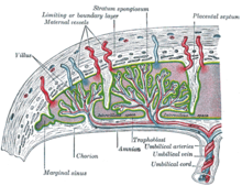
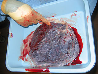
The placenta is a temporary embryonic and later fetal organ that begins developing from the blastocyst shortly after implantation. It plays critical roles in facilitating nutrient, gas and waste exchange between the physically separate maternal and fetal circulations, and is an important endocrine organ, producing hormones that regulate both maternal and fetal physiology during pregnancy. The placenta connects to the fetus via the umbilical cord, and on the opposite aspect to the maternal uterus in a species-dependent manner. In humans, a thin layer of maternal decidual (endometrial) tissue comes away with the placenta when it is expelled from the uterus following birth. Placentas are a defining characteristic of placental mammals, but are also found in marsupials and some non-mammals with varying levels of development.

<span class="mw-page-title-main">Eclampsia</span> Pre-eclampsia characterized by the presence of seizures

Eclampsia is the onset of seizures (convulsions) in a woman with pre-eclampsia. Pre-eclampsia is a hypertensive disorder of pregnancy that presents with three main features: new onset of high blood pressure, large amounts of protein in the urine or other organ dysfunction, and edema. If left untreated, pre-eclampsia can result in long-term consequences for the mother, namely increased risk of cardiovascular diseases and associated complications. In more severe cases, it may be fatal for both the mother and the fetus. The diagnostic criteria for pre-eclampsia is high blood pressure occurring after 20 weeks gestation or during the second half of pregnancy. Most often it occurs during the 3rd trimester of pregnancy and may occur before, during, or after delivery. The seizures are of the tonic–clonic type and typically last about a minute. Following the seizure, there is either a period of confusion or coma. Other complications include aspiration pneumonia, cerebral hemorrhage, kidney failure, pulmonary edema, HELLP syndrome, coagulopathy, placental abruption and cardiac arrest.

<span class="mw-page-title-main">Pre-eclampsia</span> Hypertension occurring during pregnancy

Pre-eclampsia is a multi-system disorder specific to pregnancy, characterized by the onset of high blood pressure and often a significant amount of protein in the urine. When it arises, the condition begins after 20 weeks of pregnancy. In severe cases of the disease there may be red blood cell breakdown, a low blood platelet count, impaired liver function, kidney dysfunction, swelling, shortness of breath due to fluid in the lungs, or visual disturbances. Pre-eclampsia increases the risk of undesirable as well as lethal outcomes for both the mother and the fetus including preterm labor. If left untreated, it may result in seizures at which point it is known as eclampsia.

Fetal distress, also known as non-reassuring fetal status, is a condition during pregnancy or labor in which the fetus shows signs of inadequate oxygenation. Due to its imprecision, the term "fetal distress" has fallen out of use in American obstetrics. The term "non-reassuring fetal status" has largely replaced it. It is characterized by changes in fetal movement, growth, heart rate, and presence of meconium stained fluid.

<span class="mw-page-title-main">Trophoblast</span> Early embryonic structure that gives rise to the placenta

The trophoblast is the outer layer of cells of the blastocyst. Trophoblasts are present four days after fertilization in humans. They provide nutrients to the embryo and develop into a large part of the placenta. They form during the first stage of pregnancy and are the first cells to differentiate from the fertilized egg to become extraembryonic structures that do not directly contribute to the embryo. After blastulation, the trophoblast is contiguous with the ectoderm of the embryo and is referred to as the trophectoderm. After the first differentiation, the cells in the human embryo lose their totipotency because they can no longer form a trophoblast. They become pluripotent stem cells.

<span class="mw-page-title-main">Placental abruption</span> Medical condition

Placental abruption is when the placenta separates early from the uterus, in other words separates before childbirth. It occurs most commonly around 25 weeks of pregnancy. Symptoms may include vaginal bleeding, lower abdominal pain, and dangerously low blood pressure. Complications for the mother can include disseminated intravascular coagulopathy and kidney failure. Complications for the baby can include fetal distress, low birthweight, preterm delivery, and stillbirth.

<span class="mw-page-title-main">Complications of pregnancy</span> Medical condition

Complications of pregnancy are health problems that are related to, or arise during pregnancy. Complications that occur primarily during childbirth are termed obstetric labor complications, and problems that occur primarily after childbirth are termed puerperal disorders. While some complications improve or are fully resolved after pregnancy, some may lead to lasting effects, morbidity, or in the most severe cases, maternal or fetal mortality.

<span class="mw-page-title-main">Intrauterine hypoxia</span> Medical condition when the fetus is deprived of sufficient oxygen

Intrauterine hypoxia occurs when the fetus is deprived of an adequate supply of oxygen. It may be due to a variety of reasons such as prolapse or occlusion of the umbilical cord, placental infarction, maternal diabetes and maternal smoking. Intrauterine growth restriction may cause or be the result of hypoxia. Intrauterine hypoxia can cause cellular damage that occurs within the central nervous system. This results in an increased mortality rate, including an increased risk of sudden infant death syndrome (SIDS). Oxygen deprivation in the fetus and neonate have been implicated as either a primary or as a contributing risk factor in numerous neurological and neuropsychiatric disorders such as epilepsy, attention deficit hyperactivity disorder, eating disorders and cerebral palsy.

<span class="mw-page-title-main">Cytotrophoblast</span>

"Cytotrophoblast" is the name given to both the inner layer of the trophoblast or the cells that live there. It is interior to the syncytiotrophoblast and external to the wall of the blastocyst in a developing embryo.

Placental insufficiency or utero-placental insufficiency is the failure of the placenta to deliver sufficient nutrients to the fetus during pregnancy, and is often a result of insufficient blood flow to the placenta. The term is also sometimes used to designate late decelerations of fetal heart rate as measured by cardiotocography or an NST, even if there is no other evidence of reduced blood flow to the placenta, normal uterine blood flow rate being 600mL/min.

Placenta-specific protein 1 is a small, secreted cell surface protein encoded on the X-chromosome by the PLAC1 gene. Since its discovery in 1999, PLAC1 has been found to play a role in placental development and maintenance, several gestational disorders including preeclampsia, fetal development and a large number of cancers.

<span class="mw-page-title-main">Velamentous cord insertion</span> Velamentous placenta

Velamentous cord insertion is a complication of pregnancy where the umbilical cord is inserted in the fetal membranes. It is a major cause of antepartum hemorrhage that leads to loss of fetal blood and associated with high perinatal mortality. In normal pregnancies, the umbilical cord inserts into the middle of the placental mass and is completely encased by the amniotic sac. The vessels are hence normally protected by Wharton's jelly, which prevents rupture during pregnancy and labor. In velamentous cord insertion, the vessels of the umbilical cord are improperly inserted in the chorioamniotic membrane, and hence the vessels traverse between the amnion and the chorion towards the placenta. Without Wharton's jelly protecting the vessels, the exposed vessels are susceptible to compression and rupture.

<span class="mw-page-title-main">Placental disease</span> Medical condition

A placental disease is any disease, disorder, or pathology of the placenta.

Reproductive immunology refers to a field of medicine that studies interactions between the immune system and components related to the reproductive system, such as maternal immune tolerance towards the fetus, or immunological interactions across the blood-testis barrier. The concept has been used by fertility clinics to explain fertility problems, recurrent miscarriages and pregnancy complications observed when this state of immunological tolerance is not successfully achieved. Immunological therapy is a method for treating many cases of previously "unexplained infertility" or recurrent miscarriage.

Immune tolerance in pregnancy or maternal immune tolerance is the immune tolerance shown towards the fetus and placenta during pregnancy. This tolerance counters the immune response that would normally result in the rejection of something foreign in the body, as can happen in cases of spontaneous abortion. It is studied within the field of reproductive immunology.

<span class="mw-page-title-main">Circumvallate placenta</span> Medical condition

Circumvallate placenta is a rare condition affecting about 1-2% of pregnancies, in which the amnion and chorion fetal membranes essentially "double back" on the fetal side around the edges of the placenta. After delivery, a circumvallate placenta has a thick ring of membranes on its fetal surface. Circumvallate placenta is a placental morphological abnormality associated with increased fetal morbidity and mortality due to the restricted availability of nutrients and oxygen to the developing fetus.

<span class="mw-page-title-main">High-risk pregnancy</span> Medical condition

A high-risk pregnancy is one where the mother or the fetus has an increased risk of adverse outcomes compared to uncomplicated pregnancies. No concrete guidelines currently exist for distinguishing “high-risk” pregnancies from “low-risk” pregnancies; however, there are certain studied conditions that have been shown to put the mother or fetus at a higher risk of poor outcomes. These conditions can be classified into three main categories: health problems in the mother that occur before she becomes pregnant, health problems in the mother that occur during pregnancy, and certain health conditions with the fetus.

Hypertensive disease of pregnancy, also known as maternal hypertensive disorder, is a group of high blood pressure disorders that include preeclampsia, preeclampsia superimposed on chronic hypertension, gestational hypertension, and chronic hypertension.

<span class="mw-page-title-main">Pregnancy Outcome Prediction study</span>

The Pregnancy Outcome Prediction (POP) Study is a prospective cohort study of 4,512 women who have never given birth, recruited at the Rosie Hospital between January 2008 and July 2012.

Extravillous trophoblasts(EVTs), are one form of differentiated trophoblast cells of the placenta. They are invasive mesenchymal cells which function to establish critical tissue connection in the developing placental-uterine interface. EVTs derive from progenitor cytotrophoblasts (CYTs), as does the other main trophoblast subtype, syncytiotrophoblast (SYN). They are sometimes called intermediate trophoblast.

References

  1. 1 2 "40". Williams obstetrics (24th ed.). McGraw-Hill Professional. 2014. ISBN   9780071798938.
  2. "Gestational Hypertension". www.stanfordchildrens.org. Stanford Medicine Children's Health. Retrieved 2023-10-07.
  3. 1 2 3 4 "Gestational Hypertension". Stanford Children's Health. Retrieved 2017-11-30.
  4. Garg AX, Nevis IF, McArthur E, Sontrop JM, Koval JJ, Lam NN, Hildebrand AM, Reese PP, Storsley L, Gill JS, Segev DL, Habbous S, Bugeja A, Knoll GA, Dipchand C, Monroy-Cuadros M, Lentine KL (January 2015). "Gestational hypertension and preeclampsia in living kidney donors". N. Engl. J. Med. 372 (2): 124–33. doi:10.1056/NEJMoa1408932. PMC   4362716 . PMID   25397608.
  5. Lo, JO; Mission, JF; Caughey, AB (April 2013). "Hypertensive disease of pregnancy and maternal mortality". Current Opinion in Obstetrics and Gynecology. 25 (2): 124–32. doi:10.1097/gco.0b013e32835e0ef5. PMID   23403779. S2CID   246228.
  6. " preeclampsia/eclampsia " at Dorland's Medical Dictionary
  7. Brown CM, Garovic VD (March 2014). "Drug Treatment of Hypertension in Pregnancy". Drugs. 74 (3): 283–296. doi:10.1007/s40265-014-0187-7. PMC   4558097 . PMID   24554373.
  8. 1 2 Abrams ET, Rutherford JN (2011). "Framing postpartum hemorrhage as a consequence of human placental biology: an evolutionary and comparative perspective". American Anthropologist. 113 (3): 417–30. doi:10.1111/j.1548-1433.2011.01351.x. PMC   3168987 . PMID   21909154.
  9. 1 2 Barton JR, O'brien JM, Bergauer NK, Jacques DL, Sibai BM (April 2001). "Mild gestational hypertension remote from term: progression and outcome". Am. J. Obstet. Gynecol. 184 (5): 979–83. doi:10.1067/mob.2001.112905. PMID   11303208.
  10. Rosenberg KR, Trevathan WR (December 2007). "An anthropological perspective on the evolutionary context of preeclampsia in humans". J. Reprod. Immunol. 76 (1–2): 91–7. doi:10.1016/j.jri.2007.03.011. PMID   17499857.
  11. Campbell, Bernard Grant. "Reproduction and the Placenta." Human Evolution: An Introduction to Man's Adaptations. New York: Aldine De Gruyter, 1998. 317-20.
  12. Moll W, Künzel W (January 1973). "The blood pressure in arteries entering the placentae of guinea pigs, rats, rabbits, and sheep". Pflügers Arch. 338 (2): 125–31. doi:10.1007/bf00592748. PMID   4734441. S2CID   24904753.
  13. Ahokas RA, McKinney ET (2009). "Development and Physiology of the Placenta and Membranes". The Global Library of Women's Medicine. doi:10.3843/GLOWM.10101. ISSN   1756-2228.
  14. Martin RD (August 2003). "Human reproduction: a comparative background for medical hypotheses". J. Reprod. Immunol. 59 (2): 111–35. doi:10.1016/s0165-0378(03)00042-1. PMID   12896817.
  15. Cross JC (2003). "The Genetics of Pre-eclampsia: A Feto-placental or Maternal Problem?". Clinical Genetics. 64 (2): 96–103. doi:10.1034/j.1399-0004.2003.00127.x. PMID   12859402. S2CID   23691148.
  16. 1 2 Jauniaux E, Poston L, Burton GJ (2006). "Placental-related diseases of pregnancy: Involvement of oxidative stress and implications in human evolution". Hum. Reprod. Update. 12 (6): 747–55. doi:10.1093/humupd/dml016. PMC   1876942 . PMID   16682385.
  17. Duckitt K, Harrington D (March 2005). "Risk factors for pre-eclampsia at antenatal booking: systematic review of controlled studies". BMJ. 330 (7491): 565. doi:10.1136/bmj.38380.674340.E0. PMC   554027 . PMID   15743856.
  18. Norwitz ER (October 2006). "Defective implantation and placentation: laying the blueprint for pregnancy complications". Reprod. Biomed. Online. 13 (4): 591–9. doi: 10.1016/s1472-6483(10)60649-9 . PMID   17007686.
  19. Olofsson P, Laurini RN, Marsál K (May 1993). "A high uterine artery pulsatility index reflects a defective development of placental bed spiral arteries in pregnancies complicated by hypertension and fetal growth retardation". Eur. J. Obstet. Gynecol. Reprod. Biol. 49 (3): 161–8. doi:10.1016/0028-2243(93)90265-e. PMID   8405630.
  20. Robertson WB, Brosens I, Dixon G (1976). "Maternal uterine vascular lesions in the hypertensive complications of pregnancy". Perspect Nephrol Hypertens. 5: 115–27. PMID   1005030.
  21. 1 2 Cole LA (November 2009). "hCG and hyperglycosylated hCG in the establishment and evolution of hemochorial placentation". J. Reprod. Immunol. 82 (2): 112–18. doi:10.1016/j.jri.2009.04.007. PMID   19560212.
  22. Rockwell LC, Vargas E, Moore LG (2003). "Human physiological adaptation to pregnancy: inter- and intraspecific perspectives". Am. J. Hum. Biol. 15 (3): 330–41. doi:10.1002/ajhb.10151. PMID   12704709. S2CID   19806255.
  23. Chaline J (August 2003). "Increased cranial capacity in hominid evolution and preeclampsia". J. Reprod. Immunol. 59 (2): 137–52. doi:10.1016/s0165-0378(03)00043-3. PMID   12896818.
  24. 1 2 Hollegaard B, Byars SG, Lykke J, Boomsma JJ (2013). "Parent-offspring conflict and the persistence of pregnancy-induced hypertension in modern humans". PLOS ONE. 8 (2): e56821. Bibcode:2013PLoSO...856821H. doi: 10.1371/journal.pone.0056821 . PMC   3581540 . PMID   23451092.
  25. Rosenberg Karen R.; Trevathan Wenda R. (2007). "An Anthropological Perspective on the Evolutionary Context of Preeclampsia in Humans". Journal of Reproductive Immunology. 76 (1–2): 91–97. doi:10.1016/j.jri.2007.03.011. PMID   17499857.
  26. 1 2 3 Solomon CG, Seely EW (February 2001). "Brief review: hypertension in pregnancy : a manifestation of the insulin resistance syndrome?". Hypertension. 37 (2): 232–9. doi: 10.1161/01.hyp.37.2.232 . PMID   11230277.