Related Research Articles

The cerebral cortex, also known as the cerebral mantle, is the outer layer of neural tissue of the cerebrum of the brain in humans and other mammals. It is separated into two cortices, by the longitudinal fissure that divides the cerebrum into the left and right cerebral hemispheres. The two hemispheres are joined beneath the cortex by the corpus callosum. The cerebral cortex is the largest site of neural integration in the central nervous system. It plays a key role in attention, perception, awareness, thought, memory, language, and consciousness.
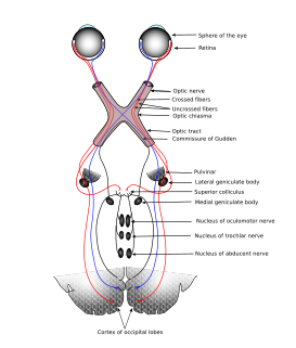
The sensory nervous system is a part of the nervous system responsible for processing sensory information. A sensory system consists of sensory neurons, neural pathways, and parts of the brain involved in sensory perception. Commonly recognized sensory systems are those for vision, hearing, touch, taste, smell, and balance. In short, senses are transducers from the physical world to the realm of the mind where we interpret the information, creating our perception of the world around us.

A Brodmann area is a region of the cerebral cortex, in the human or other primate brain, defined by its cytoarchitecture, or histological structure and organization of cells.

The parietal lobe is one of the four major lobes of the cerebral cortex in the brain of mammals. The parietal lobe is positioned above the temporal lobe and behind the frontal lobe and central sulcus.
The temporal lobe is involved in processing sensory input into derived meanings for the appropriate retention of visual memory, language comprehension, and emotion association.
The cerebrum or telencephalon is a large part of the brain containing the cerebral cortex, as well as several subcortical structures, including the hippocampus, basal ganglia, and olfactory bulb. In the human brain, the cerebrum is the uppermost region of the central nervous system. The prosencephalon or forebrain is the embryonic structure from which the cerebrum develops prenatally. In mammals, the dorsal telencephalon, or pallium, develops into the cerebral cortex, and the ventral telencephalon, or subpallium, becomes the basal ganglia. The cerebrum is also divided into approximately symmetric left and right cerebral hemispheres.

Afferent nerve fibers refer to axonal projections that arrive at a particular brain region, as opposed to efferent projections that exit the region. These terms have a slightly different meaning in the context of the peripheral nervous system (PNS) and central nervous system (CNS).
The limbic lobe is an arc-shaped region of cortex on the medial surface of each cerebral hemisphere of the mammalian brain, consisting of parts of the frontal, parietal and temporal lobes. The term is ambiguous, with some authors including the paraterminal gyrus, the subcallosal area, the cingulate gyrus, the parahippocampal gyrus, the dentate gyrus, the hippocampus and the subiculum; while the Terminologia Anatomica includes the cingulate sulcus, the cingulate gyrus, the isthmus of cingulate gyrus, the fasciolar gyrus, the parahippocampal gyrus, the parahippocampal sulcus, the dentate gyrus, the fimbrodentate sulcus, the fimbria of hippocampus, the collateral sulcus, and the rhinal sulcus, and omits the hippocampus.

The postcentral gyrus is a prominent gyrus in the lateral parietal lobe of the human brain. It is the location of the primary somatosensory cortex, the main sensory receptive area for the sense of touch. Like other sensory areas, there is a map of sensory space in this location, called the sensory homunculus.

The inferior frontal gyrus(IFG), (gyrus frontalis inferior), is the lowest positioned gyrus of the frontal gyri, of the frontal lobe, and is part of the prefrontal cortex.

Brodmann areas 41 and 42 are parts of the primary auditory cortex.

The primary somatosensory cortex is located in the postcentral gyrus, and is part of the somatosensory system. It was initially defined from surface stimulation studies of Wilder Penfield, and parallel surface potential studies of Bard, Woolsey, and Marshall. Although initially defined to be roughly the same as Brodmann areas 3, 1 and 2, more recent work by Kaas has suggested that for homogeny with other sensory fields only area 3 should be referred to as "primary somatosensory cortex", as it receives the bulk of the thalamocortical projections from the sensory input fields.

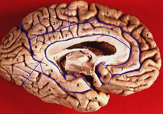
The lobes of the brain were originally a purely anatomical classification, but have been shown also to be related to different brain functions. The cerebrum, the largest portion of the human brain, is divided into lobes, but so is the cerebellum. If not specified, the expression "lobes of the brain" refers to the cerebrum.

A cortical homunculus is a distorted representation of the human body, based on a neurological "map" of the areas and proportions of the human brain dedicated to processing motor functions, or sensory functions, for different parts of the body. The word homunculus is Latin for "little man", and was a term used in alchemy and folklore long before scientific literature began using it. A cortical homunculus, or "cortex man", illustrates the concept of a representation of the body lying within the brain. Nerve fibres—conducting somatosensory information from all over the body—terminate in various areas of the parietal lobe in the cerebral cortex, forming a representational map of the body.

The inferior temporal gyrus is placed below the middle temporal gyrus, and is connected behind with the inferior occipital gyrus; it also extends around the infero-lateral border on to the inferior surface of the temporal lobe, where it is limited by the inferior sulcus. This region is one of the higher levels of the ventral stream of visual processing, associated with the representation of complex object features, such as global shape. It may also be involved in face perception, and in the recognition of numbers.

In neuroanatomy, a sulcus is a depression or groove in the cerebral cortex. It surrounds a gyrus, creating the characteristic folded appearance of the brain in humans and other mammals. The larger sulci are usually called fissures.
A topographic map is the ordered projection of a sensory surface, like the retina or the skin, or an effector system, like the musculature, to one or more structures of the central nervous system. Topographic maps can be found in all sensory systems and in many motor systems.
The sensory cortex can refer informally to the primary somatosensory cortex, or it can be used as a term for the primary and secondary cortices of the different senses : the visual cortex on the occipital lobes, the auditory cortex on the temporal lobes, the primary olfactory cortex on the uncus of the piriform region of the temporal lobes, the gustatory cortex on the insular lobe, and the primary somatosensory cortex on the anterior parietal lobes. Just posterior to the primary somatosensory cortex lies the somatosensory association cortex, which integrates sensory information from the primary somatosensory cortex to construct an understanding of the object being felt. Inferior to the frontal lobes are found the olfactory bulbs, which receive sensory input from the olfactory nerves and route those signals throughout the brain. Not all olfactory information is routed to the olfactory cortex: some neural fibers are routed to the supraorbital region of the frontal lobe, while others are routed directly to limbic structures. The direct limbic connection makes the olfactory sense unique.
Somatotopy is the point-for-point correspondence of an area of the body to a specific point on the central nervous system. Typically, the area of the body corresponds to a point on the primary somatosensory cortex. This cortex is typically represented as a sensory homunculus which orients the specific body parts and their respective locations upon the homunculus. Areas such as the appendages, digits, penis, and face can draw their sensory locations upon the somatosensory cortex. The areas which are finely controlled have larger portions of the somatosensory cortex whereas areas which are coarsely controlled have smaller portions. Areas such as the viscera do not have sensory locations on the post central gyrus.
References
- ↑ Kandel, Eric (2013). Principles of Neural Science. McGraw-Hill. p. 344. ISBN 978-0-07-139011-8.
- ↑ Kandel, Eric (2013). Principles of Neural Science. McGraw-Hill. p. 727. ISBN 978-0-07-139011-8.
- ↑ Simmons, W. Kyle; Rapuano, Kristina M.; Kallman, Seth J.; Ingeholm, John E.; Miller, Bernard; Gotts, Stephen J.; Avery, Jason A.; Hall, Kevin D.; Hall, Kevin D. (Nov 2013). "Category-specific integration of homeostatic signals in caudal, but not rostral, human insula". Nat. Neurosci. 16 (11): 551–1552. doi:10.1038/nn.3535. PMC 3835665 . PMID 24077565.
- ↑ Marieb, Elaine N.; Hoehn, Katja (2008). Anatomy & Physiology, Third Edition . Boston: Benjamin Cummings/Pearson. pp. 391–395. ISBN 0-8053-0094-5.