Related Research Articles

Enzymes are proteins that act as biological catalysts by accelerating chemical reactions. The molecules upon which enzymes may act are called substrates, and the enzyme converts the substrates into different molecules known as products. Almost all metabolic processes in the cell need enzyme catalysis in order to occur at rates fast enough to sustain life. Metabolic pathways depend upon enzymes to catalyze individual steps. The study of enzymes is called enzymology and the field of pseudoenzyme analysis recognizes that during evolution, some enzymes have lost the ability to carry out biological catalysis, which is often reflected in their amino acid sequences and unusual 'pseudocatalytic' properties.
A protein phosphatase is a phosphatase enzyme that removes a phosphate group from the phosphorylated amino acid residue of its substrate protein. Protein phosphorylation is one of the most common forms of reversible protein posttranslational modification (PTM), with up to 30% of all proteins being phosphorylated at any given time. Protein kinases (PKs) are the effectors of phosphorylation and catalyse the transfer of a γ-phosphate from ATP to specific amino acids on proteins. Several hundred PKs exist in mammals and are classified into distinct super-families. Proteins are phosphorylated predominantly on Ser, Thr and Tyr residues, which account for 79.3, 16.9 and 3.8% respectively of the phosphoproteome, at least in mammals. In contrast, protein phosphatases (PPs) are the primary effectors of dephosphorylation and can be grouped into three main classes based on sequence, structure and catalytic function. The largest class of PPs is the phosphoprotein phosphatase (PPP) family comprising PP1, PP2A, PP2B, PP4, PP5, PP6 and PP7, and the protein phosphatase Mg2+- or Mn2+-dependent (PPM) family, composed primarily of PP2C. The protein Tyr phosphatase (PTP) super-family forms the second group, and the aspartate-based protein phosphatases the third. The protein pseudophosphatases form part of the larger phosphatase family, and in most cases are thought to be catalytically inert, instead functioning as phosphate-binding proteins, integrators of signalling or subcellular traps. Examples of membrane-spanning protein phosphatases containing both active (phosphatase) and inactive (pseudophosphatase) domains linked in tandem are known, conceptually similar to the kinase and pseudokinase domain polypeptide structure of the JAK pseudokinases. A complete comparative analysis of human phosphatases and pseudophosphatases has been completed by Manning and colleagues, forming a companion piece to the ground-breaking analysis of the human kinome, which encodes the complete set of ~536 human protein kinases.

In biochemistry, a kinase is an enzyme that catalyzes the transfer of phosphate groups from high-energy, phosphate-donating molecules to specific substrates. This process is known as phosphorylation, where the high-energy ATP molecule donates a phosphate group to the substrate molecule. As a result, kinase produces a phosphorylated substrate and ADP. Conversely, it is referred to as dephosphorylation when the phosphorylated substrate donates a phosphate group and ADP gains a phosphate group. These two processes, phosphorylation and dephosphorylation, occur four times during glycolysis.

In cell biology, protein kinase A (PKA) is a family of serine-threonine kinase whose activity is dependent on cellular levels of cyclic AMP (cAMP). PKA is also known as cAMP-dependent protein kinase. PKA has several functions in the cell, including regulation of glycogen, sugar, and lipid metabolism. It should not be confused with 5'-AMP-activated protein kinase.

In biochemistry, a phosphatase is an enzyme that uses water to cleave a phosphoric acid monoester into a phosphate ion and an alcohol. Because a phosphatase enzyme catalyzes the hydrolysis of its substrate, it is a subcategory of hydrolases. Phosphatase enzymes are essential to many biological functions, because phosphorylation and dephosphorylation serve diverse roles in cellular regulation and signaling. Whereas phosphatases remove phosphate groups from molecules, kinases catalyze the transfer of phosphate groups to molecules from ATP. Together, kinases and phosphatases direct a form of post-translational modification that is essential to the cell's regulatory network.
CAMK, also written as CaMK or CCaMK, is an abbreviation for the Ca2+/calmodulin-dependent protein kinase class of enzymes. CAMKs are activated by increases in the concentration of intracellular calcium ions (Ca2+) and calmodulin. When activated, the enzymes transfer phosphates from ATP to defined serine or threonine residues in other proteins, so they are serine/threonine-specific protein kinases. Activated CAMK is involved in the phosphorylation of transcription factors and therefore, in the regulation of expression of responding genes. CAMK also works to regulate the cell life cycle (i.e. programmed cell death), rearrangement of the cell's cytoskeletal network, and mechanisms involved in the learning and memory of an organism.

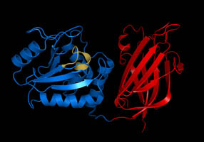
A catalytic triad is a set of three coordinated amino acids that can be found in the active site of some enzymes. Catalytic triads are most commonly found in hydrolase and transferase enzymes. An acid-base-nucleophile triad is a common motif for generating a nucleophilic residue for covalent catalysis. The residues form a charge-relay network to polarise and activate the nucleophile, which attacks the substrate, forming a covalent intermediate which is then hydrolysed to release the product and regenerate free enzyme. The nucleophile is most commonly a serine or cysteine amino acid, but occasionally threonine or even selenocysteine. The 3D structure of the enzyme brings together the triad residues in a precise orientation, even though they may be far apart in the sequence.

Protein tyrosine phosphatases (EC 3.1.3.48, systematic name protein-tyrosine-phosphate phosphohydrolase) are a group of enzymes that remove phosphate groups from phosphorylated tyrosine residues on proteins:
Phosphatase and tensin homolog (PTEN) is a phosphatase in humans and is encoded by the PTEN gene. Mutations of this gene are a step in the development of many cancers, specifically glioblastoma, lung cancer, breast cancer, and prostate cancer. Genes corresponding to PTEN (orthologs) have been identified in most mammals for which complete genome data are available.
In molecular biology, biochemistry and cell signaling the kinome of an organism is the complete set of protein kinases encoded in its genome. Kinases are usually enzymes that catalyze phosphorylation reactions and fall into several groups and families, e.g., those that phosphorylate the amino acids serine and threonine, those that phosphorylate tyrosine and some that can phosphorylate both, such as the MAP2K and GSK families. The term was first used in 2002 by Gerard Manning and colleagues in twin papers analyzing the 518 human protein kinases, and refers to both protein kinases and protein pseudokinases and their evolution of protein kinases throughout the eukaryotes. Other kinomes have been determined for rice, several fungi, nematodes, and insects, sea urchins, Dictyostelium discoideum, and the process of infection by Mycobacterium tuberculosis. Although the primary sequence of protein kinases shows substantial divergence between unrelated eukaryotes, and amino acid differences in catalytic motifs have permitted their separation of kinomes into canonical and pseudokinase subtypes, the variation found in the amino acid motifs adjacent to the site of actual phosphorylation of substrates by eukaryotic kinases is much smaller.

Dual specificity protein phosphatase 1 is an enzyme that in humans is encoded by the DUSP1 gene.

Serine/threonine-protein phosphatase 2A 56 kDa regulatory subunit alpha isoform is an enzyme that in humans is encoded by the PPP2R5A gene.

Serine/threonine kinase 11 (STK11) also known as liver kinase B1 (LKB1) or renal carcinoma antigen NY-REN-19 is a protein kinase that in humans is encoded by the STK11 gene.

Inositol (1,4,5) trisphosphate 3-kinase (EC 2.7.1.127), abbreviated here as ITP3K, is an enzyme that facilitates a phospho-group transfer from adenosine triphosphate to 1D-myo-inositol 1,4,5-trisphosphate. This enzyme belongs to the family of transferases, specifically those transferring phosphorus-containing groups (phosphotransferases) with an alcohol group as acceptor. The systematic name of this enzyme class is ATP:1D-myo-inositol-1,4,5-trisphosphate 3-phosphotransferase. ITP3K catalyzes the transfer of the gamma-phosphate from ATP to the 3-position of inositol 1,4,5-trisphosphate to form inositol 1,3,4,5-tetrakisphosphate. ITP3K is highly specific for the 1,4,5-isomer of IP3, and it exclusively phosphorylates the 3-OH position, producing Ins(1,3,4,5)P4, also known as inositol tetrakisphosphate or IP4.

Tribbles homolog 3 is a protein that in humans is encoded by the TRIB3 gene.

Tribbles homolog 1 is a protein kinase that in humans is encoded by the TRIB1 gene. Orthologs of this protein pseudokinase (pseudoenzyme) can be found almost ubiquitously throughout the animal kingdom. It exerts its biological functions through binding to signalling proteins of the MAPKK level of the MAPK pathway, therefore eliciting a regulatory role in the function of this pathway which mediates proliferation, apoptosis and differentiation in cells. Tribbles-1 is encoded by the trib1 gene, which in humans can be found on chromosome 8 at position 24.13 on the longest arm (q). Recent crystal structures show that Tribbles 1 has an unusual 3D structure, containing a 'broken' C-helix region, a binding site for ubiquitinated substrates such as C/EBPalpha and a key regulatory C-tail region. Like TRIB2 and TRIB3, TRIB1 has recently been considered as a potential allosteric drug target.

Tribbles homolog 2 is an atypical protein kinase that is encoded in human by the TRIB2 gene. TRIB2 is a pseudokinase member of the (pseudoenzyme) class of signaling/scaffold proteins, possessing very low vestigial catalytic output in vitro and critical scaffolding signaling functions in cells. It is known to signal to canonical MAPK and AKT pathways and to regulate the ubiquitination of substrates with important functions in cell proliferation that control the cell ccyle. It has also been associated with various diseases, especially in human and murine blood and solid tumor models. Like TRIB1 and TRIB3, TRIB2 has recently been considered as a potential allosteric drug target, and its three dimensional structure has been solved with the aid of stabilizing nanobodies corroborating the potential for new approaches for drug targeting outside the highly degraded ATP site and is a putative regulator of cancer-associated signalling and survival through AKT pSer473 modulation. Recent work has established a convincing link between targetable overexpression of TRIB2 and prostate cancer drug responses

Protein phosphatase 1 (PP1) belongs to a certain class of phosphatases known as protein serine/threonine phosphatases. This type of phosphatase includes metal-dependent protein phosphatases (PPMs) and aspartate-based phosphatases. PP1 has been found to be important in the control of glycogen metabolism, muscle contraction, cell progression, neuronal activities, splicing of RNA, mitosis, cell division, apoptosis, protein synthesis, and regulation of membrane receptors and channels.
The phosphatome of an organism is the set of phosphatase genes in its genome. Phosphatases are enzymes that catalyze the removal of phosphate from biomolecules. Over half of all cellular proteins are modified by phosphorylation which typically controls their functions. Protein phosphorylation is controlled by the opposing actions of protein phosphatases and protein kinases. Most phosphorylation sites are not linked to a specific phosphatase, so the phosphatome approach allows a global analysis of dephosphorylation, screening to find the phosphatase responsible for a given reaction, and comparative studies between different phosphatases, similar to how protein kinase research has been impacted by the kinome approach.
Pseudokinases are catalytically-deficient pseudoenzyme variants of protein kinases that are represented in all kinomes across the kingdoms of life. Pseudokinases have both physiological and pathophysiological functions.
References
- ↑ Ribeiro AJ, Das S, Dawson N, Zaru R, Orchard S, Thornton JM, Orengo C, Zeqiraj E, Murphy JM, Eyers PA (Aug 2019). "Emerging concepts in pseudoenzyme classification, evolution, and signaling". Science Signaling. 12 (594): eaat9797. doi:10.1126/scisignal.aat9797. PMID 31409758.
- ↑ Kwon A, Scott S, Taujale R, Yeung W, Kochut KJ, Eyers PA, Kannan N (April 2019). "Tracing the origin and evolution of pseudokinases across the tree of life". Science Signaling. 12 (578): eaav3810. doi:10.1126/scisignal.aav3810. PMC 6997932 . PMID 31015289.
- ↑ Jeffery CJ (Feb 2019). "The demise of catalysis, but new functions arise: pseudoenzymes as the phoenixes of the protein world". Biochemical Society Transactions. 47 (1): 371–379. doi:10.1042/BST20180473. PMID 30710059. S2CID 73437705.
- ↑ Jeffery CJ (Dec 2019). "Multitalented actors inside and outside the cell: recent discoveries add to the number of moonlighting proteins". Biochemical Society Transactions. 47 (6): 1941–1948. doi:10.1042/BST20190798. PMID 31803903. S2CID 208643133.
- ↑ Eyers PA, Murphy JM (November 2016). "The evolving world of pseudoenzymes: proteins, prejudice and zombies". BMC Biology. 14 (1): 98. doi: 10.1186/s12915-016-0322-x . PMC 5106787 . PMID 27835992.
- ↑ Walden M, Masandi SK, Pawlowski K, Zeqiraj E (Feb 2018). "Pseudo-DUBs as allosteric activators and molecular scaffolds of protein complexes" (PDF). Biochem Soc Trans. 46 (2): 453–466. doi:10.1042/BST20160268. PMID 29472364. S2CID 3477709.
- ↑ Walden M, Tian L, Ross RL, Sykora UM, Byrne DP, Hesketh EL, Masandi SK, Cassel J, George R, Ault JR, El Oualid F, Pawłowski K, Salvino JM, Eyers PA, Ranson NA, Del Galdo F, Greenberg RA, Zeqiraj E (May 2019). "Metabolic control of BRISC–SHMT2 assembly regulates immune signalling" (PDF). Nature. 570 (7760): 194–199. Bibcode:2019Natur.570..194W. doi:10.1038/s41586-019-1232-1. PMC 6914362 . PMID 31142841.
- ↑ Todd AE, Orengo CA, Thornton JM (October 2002). "Sequence and structural differences between enzyme and nonenzyme homologs". Structure. 10 (10): 1435–51. doi: 10.1016/s0969-2126(02)00861-4 . PMID 12377129.
- ↑ Willert EK, Fitzpatrick R, Phillips MA (May 2007). "Allosteric regulation of an essential trypanosome polyamine biosynthetic enzyme by a catalytically dead homolog". Proceedings of the National Academy of Sciences of the United States of America. 104 (20): 8275–80. Bibcode:2007PNAS..104.8275W. doi: 10.1073/pnas.0701111104 . PMC 1895940 . PMID 17485680.
- ↑ Adrain C, Freeman M (July 2012). "New lives for old: evolution of pseudoenzyme function illustrated by iRhoms". Nature Reviews. Molecular Cell Biology. 13 (8): 489–98. doi:10.1038/nrm3392. PMID 22781900. S2CID 806199.
- ↑ Kwon A, Scott S, Taujale R, Yeung W, Kochut KJ, Eyers PA, Kannan N (April 2019). "Tracing the origin and evolution of pseudokinases across the tree of life". Science Signaling. 12 (578): eaav3810. doi:10.1126/scisignal.aav3810. PMC 6997932 . PMID 31015289.
- ↑ Manning G, Whyte DB, Martinez R, Hunter T, Sudarsanam S (December 2002). "The protein kinase complement of the human genome". Science. 298 (5600): 1912–34. Bibcode:2002Sci...298.1912M. doi:10.1126/science.1075762. PMID 12471243. S2CID 26554314.
- ↑ Boudeau J, Miranda-Saavedra D, Barton GJ, Alessi DR (September 2006). "Emerging roles of pseudokinases". Trends in Cell Biology. 16 (9): 443–52. doi:10.1016/j.tcb.2006.07.003. PMID 16879967.
- ↑ Eyers PA, Keeshan K, Kannan N (April 2017). "Tribbles in the 21st Century: The Evolving Roles of Tribbles Pseudokinases in Biology and Disease". Trends in Cell Biology. 27 (4): 284–298. doi:10.1016/j.tcb.2016.11.002. PMC 5382568 . PMID 27908682.
- ↑ Reiterer V, Eyers PA, Farhan H (September 2014). "Day of the dead: pseudokinases and pseudophosphatases in physiology and disease". Trends in Cell Biology. 24 (9): 489–505. doi:10.1016/j.tcb.2014.03.008. PMID 24818526.
- ↑ Murphy JM, Czabotar PE, Hildebrand JM, Lucet IS, Zhang JG, Alvarez-Diaz S, Lewis R, Lalaoui N, Metcalf D, Webb AI, Young SN, Varghese LN, Tannahill GM, Hatchell EC, Majewski IJ, Okamoto T, Dobson RC, Hilton DJ, Babon JJ, Nicola NA, Strasser A, Silke J, Alexander WS (September 2013). "The pseudokinase MLKL mediates necroptosis via a molecular switch mechanism". Immunity. 39 (3): 443–53. doi: 10.1016/j.immuni.2013.06.018 . PMID 24012422.
- ↑ Wishart MJ, Dixon JE (August 1998). "Gathering STYX: phosphatase-like form predicts functions for unique protein-interaction domains". Trends in Biochemical Sciences. 23 (8): 301–6. doi:10.1016/s0968-0004(98)01241-9. PMID 9757831.
- ↑ Reiterer V, Eyers PA, Farhan H (September 2014). "Day of the dead: pseudokinases and pseudophosphatases in physiology and disease". Trends in Cell Biology. 24 (9): 489–505. doi:10.1016/j.tcb.2014.03.008. PMID 24818526.
- ↑ Chen MJ, Dixon JE, Manning G (April 2017). "Genomics and evolution of protein phosphatases". Science Signaling. 10 (474): eaag1796. doi:10.1126/scisignal.aag1796. PMID 28400531. S2CID 41041971.
- ↑ Zeqiraj E, Tian L, Piggott CA, Pillon MC, Duffy NM, Ceccarelli DF, Keszei AF, Lorenzen K, Kurinov I, Orlicky S, Gish GD, Heck AJ, Guarné A, Greenberg RA, Sicheri F (September 2015). "Higher-Order Assembly of BRCC36-KIAA0157 Is Required for DUB Activity and Biological Function". Molecular Cell. 59 (6): 970–83. doi:10.1016/j.molcel.2015.07.028. PMC 4579573 . PMID 26344097.
- ↑ Strickson S, Emmerich CH, Goh ET, Zhang J, Kelsall IR, Macartney T, Hastie CJ, Knebel A, Peggie M, Marchesi F, Arthur JS, Cohen P (April 2017). "Roles of the TRAF6 and Pellino E3 ligases in MyD88 and RANKL signaling". Proceedings of the National Academy of Sciences of the United States of America. 114 (17): E3481–E3489. Bibcode:2017PNAS..114E3481S. doi: 10.1073/pnas.1702367114 . PMC 5410814 . PMID 28404732.
- ↑ Aggarwal-Howarth S, Scott JD (April 2017). "Pseudoscaffolds and anchoring proteins: the difference is in the details". Biochemical Society Transactions. 45 (2): 371–379. doi:10.1042/bst20160329. PMC 5497583 . PMID 28408477.
- ↑ Foulkes DM, Byrne DP, Bailey FP, Eyers PA (October 2015). "Tribbles pseudokinases: novel targets for chemical biology and drug discovery?". Biochemical Society Transactions. 43 (5): 1095–103. doi:10.1042/bst20150109. PMID 26517930.
- ↑ Byrne DP, Foulkes DM, Eyers PA (January 2017). "Pseudokinases: update on their functions and evaluation as new drug targets". Future Medicinal Chemistry . 9 (2): 245–265. doi: 10.4155/fmc-2016-0207 . PMID 28097887.
- ↑ Murphy JM, Farhan H, Eyers PA (April 2017). "Bio-Zombie: the rise of pseudoenzymes in biology". Biochemical Society Transactions. 45 (2): 537–544. doi:10.1042/bst20160400. PMID 28408493.
- ↑ McDermott SM, Stuart K (November 2017). "The essential functions of KREPB4 are developmentally distinct and required for endonuclease association with editosomes". RNA. 23 (11): 1672–1684. doi:10.1261/rna.062786.117. PMC 5648035 . PMID 28802260.