Purpura fulminans

Last updated
Purpura fulminans
Other namespurpura gangrenosa [1] :825
Specialty Hematology   OOjs UI icon edit-ltr-progressive.svg

Purpura fulminans is an acute, often fatal, thrombotic disorder which manifests as blood spots, bruising and discolouration of the skin resulting from coagulation in small blood vessels within the skin and rapidly leads to skin necrosis and disseminated intravascular coagulation. [2] [3]

Contents

Causes

Purpura fulminans is caused by defects in the protein C anticoagulant pathway. Identification of the cause of purpura fulminans often depends on the patient's age and circumstances of presentation. [2]

Congenital protein C deficiency

Congenital (inherited) defects in protein C activity are autosomal dominant and may be partial or severe loss of function. [2] [4] Hundreds of natural mutations of the protein C gene (PROC) have been identified. [5] [6]

Acquired protein C deficiency

Acquired protein C deficiency is caused by either depletion of available protein C in plasma or decreased protein C synthesis (caused by administration of vitamin K antagonists, severe liver failure or complications of prematurity). [7]

Severe acute sepsis

Purpura fulminans is a presenting feature of severe acute sepsis, such as Neisseria meningitidis, Streptococcus pneumoniae, Group A and B Streptococci, and less commonly with Haemophilus influenzae, Staphylococcus aureus, or Plasmodium falciparum (malaria) infections, particularly in individuals with asplenia. [2]

Combination of sepsis and partial congenital defect

In some cases, a combination of sepsis and a partial congenital defect in the protein C anticoagulant pathway initiates purpura fulminans. [8]

Unknown

In rare instances, purpura fulminans is an autoimmune manifestation against protein C or protein S after normally benign infections, such as chicken pox. [2] [9] Sometimes purpura fulminans has unknown cause. [2]

Pathophysiology

Regardless of the underlying cause of purpura fulminans, the mechanism of disease is similar with deficiency in protein C concentration or decrease in protein C activity which promotes blood clotting (thrombosis). [10]

In cases of severe sepsis, there is widespread activation of the acute systemic inflammatory response, including activation of the coagulation and complement pathways, as well as endothelial dysfunction. Activated protein C helps regulate the systemic inflammatory response. During sepsis, signalling by the inflammatory cytokines, interleukin-1 and tumour necrosis factor, mediate altered protein transcription in the systemic inflammatory response, resulting in decreased synthesis of the regulatory proteins antithrombin, protein C and protein S, with increased synthesis of prothrombotic proteins Factor VIII, von Willebrand factor, and fibrinogen. Activated protein C binds to endothelial protein C receptor and subsequently cleaves the endothelial cell protease activated receptor-1, not only altering coagulation profiles but down-regulating pro-inflammatory and pro-apoptotic mediators, up-regulation of anti-inflammatory and anti-apoptotic pathways and stabilization of the endothelial cell barrier functions. [11]

Systemic coagulation activation may lead to depletion of circulating coagulation factors and platelets, which subsequently lead to bleeding. [12] In early purpura fulminans, lesion progression correlates with the histological appearance of blockage of small skin blood vessels with blood clots causing capillary dilation and congestion with red blood cells. In later stage lesions, there is irreversible endothelial ischaemic injury with extravasation of blood cells into the dermis and gangrenous necrosis, sometimes with secondary infection. [13]

The depletion of anticoagulant and anti-inflammatory proteins, in particular, protein C and its co-factor, protein S, may also promote thrombus formation, inhibit fibrinolysis and lead to further activation of the inflammatory pathways. [14] [15]

Diagnosis

Early purpura fulminans lesions look similar to traumatic skin bleeds or purpuric rashes, such as immune thrombocytopenic purpura or thrombotic thrombocytopenic purpura; however, purpura fulminans will rapidly progress to necrosis whereas other purpuric rashes do not. [2] In most cases, differential diagnoses may be distinguished from purpura fulminans by other clinical and laboratory findings. [2]

The initial appearance of purpura fulminans lesions is of well-demarcated erythaamatous lesions which progress rapidly to develop irregular central areas of blue-black haemorrhagic necrosis. [2] Advancing areas of necrosis are often surrounded by a thin border of erythaema that fades into adjacent unaffected skin. Haemorrhage into the necrotic skin causes purpura fulminans lesions to become painful, dark and raised, sometimes with vesicle or blister (bulla) formation. [16]

The distribution of purpura fulminans lesions may be different according to the underlying pathogenesis. [2] Purpura fulminans in severe sepsis typically develops in the distal extremities and progresses proximally or appears as a generalised or diffuse rash affecting the whole body surface. [16] In cases of severe inheritable protein C deficiency, purpura fulminans with disseminated intravascular coagulation manifests within a few hours or days after birth. [7] [17]

Laboratory investigations

The cardinal features of purpura investigations are the same as those of disseminated intravascular coagulation: prolonged plasma clotting times, thrombocytopenia, reduced plasma fibrinogen concentration, increased plasma fibrin-degradation products and occasionally microangiopathic haemolysis. [2]

Prevention

For people who have severe congenital protein C deficiency, protein C replacement therapies are available, which is indicated and approved for use in the United States and Europe for the prevention of purpura fulminans. Protein C replacement is often in combination with anticoagulation therapy of injectable low molecular weight heparin or oral warfarin. [18] [19] [20] [21] [22] Before initiating warfarin therapy, a few days of therapeutic heparin may be administered to prevent warfarin necrosis and other progressive or recurrent thrombotic complications. [7] [22]

Complications of prevention

The amount of fresh frozen plasma required to reverse disseminated intravascular coagulation associated with purpura fulminans may lead to complications of fluid overload and death, especially in neonates, [7] such as transfusion-related acute lung injury. Exposure to multiple plasma donors over time increases the cumulative risk for transfusion-associated viral infection and allergic reaction to donor proteins found in fresh frozen plasma. [7]

Allergic reactions and alloantibody formation are also potential complications, as with any protein replacement therapy. [7]

Concomitant warfarin therapy in subjects with congenital protein C deficiency is associated with an increased risk of warfarin skin necrosis. [7]

Treatment

Early stage sepsis-associated purpura fulminans may be reversible with quick therapeutic intervention. [2] [7] Treatment is mainly removing the underlying cause and degree of clotting abnormalities and with supportive treatment (antibiotics, volume expansion, tissue oxygenation, etc.). Thus, treatment includes aggressive management of the septic state.[ citation needed ]

Purpura fulminans with disseminated intravascular coagulation should be urgently treated with fresh frozen plasma (10–20 mL/kg every 8–12 hours) and/or protein C concentrate to replace pro-coagulant and anticoagulant plasma proteins that have been depleted by the disseminated intravascular coagulation process. [2] [3] [4] [7]

Protein C in plasma in the steady state has a half life of 6- to 10-hour, therefore, patients with severe protein C deficiency and presenting with purpura fulminans can be treated acutely with an initial bolus of protein C concentrate 100 IU/kg followed by 50 IU /kg every 6 hours. [7] A total of 1 IU/kg of protein C concentrate or 1 mL/kg of fresh frozen plasma will increase the plasma concentration of protein C by 1 IU/dL. [7] Cases with comorbid pathological bleeding may require additional transfusions with platelet concentrate (10–15 mL/kg) or cryoprecipitate (5 mL/kg). [2]

Established soft tissue necrosis may require surgical removal of the dead tissue, fasciotomy, amputation or reconstructive surgery. [2]

Prognosis

Purpura fulminans lesions, once established, often progress within 24 to 48 hours to full-thickness skin necrosis or soft-tissue necrosis. Once purpura fulminans lesions progress to full-thickness skin necrosis, healing takes between 4–8 weeks and leaves large scars. [2]

Without treatment, necrotic soft tissue may become gangrenous, leading to loss of limbs. [2] Purpura fulminans is often accompanied by micro-vascular thrombosis and haemorrhagic infarction in other tissues, such as the lungs, kidneys, central nervous system and adrenal glands, leading to multiple organ failure, and causes initial high mortality and long-term morbidity in survivors. Purpura fulminans may also lead to severe large vessel venous thrombosis if untreated in its early stages. [2]

Purpura fulminans secondary to severe infection is self-limiting. [2] In cases of homozygous protein C deficiency, episodes of purpura fulminans and other thrombotic events are recurrent. [4] Moreover, infant survival due to maintenance replacement therapy is often associated with intellectual disability or visual impairment. [2] [23] For post-infection purpura fulminans, new lesions will occur while neutralising autoantibodies are present (1–2 weeks after presentation). [9]

A multi-disciplinary care team is usually required for rehabilitation after purpura fulminans. [2]

Epidemiology

Purpura fulminans is rare and most commonly occurs in babies and small children [24] but can also be a rare manifestation in adults when it is associated with severe infections. [25] For example, Meningococcal septicaemia is complicated by purpura fulminans in 10–20% of cases among children. [26] Purpura fulminans associated with congenital (inherited) protein C deficiency occurs in 1:500,000–1,000,000 live births. [27]

Research

Due to the rarity of purpura fulminans and its occurrence in vulnerable patient groups like children research on the condition is very limited and evidence based knowledge is scarce. [ citation needed ]

History

Purpura fulminans was first described by Guelliot in 1884. [28]

Related Research Articles

<span class="mw-page-title-main">Waterhouse–Friderichsen syndrome</span> Medical condition

Waterhouse–Friderichsen syndrome (WFS) is defined as adrenal gland failure due to bleeding into the adrenal glands, commonly caused by severe bacterial infection. Typically, it is caused by Neisseria meningitidis.

<span class="mw-page-title-main">Coagulation</span> Process of formation of blood clots

Coagulation, also known as clotting, is the process by which blood changes from a liquid to a gel, forming a blood clot. It potentially results in hemostasis, the cessation of blood loss from a damaged vessel, followed by repair. The mechanism of coagulation involves activation, adhesion and aggregation of platelets, as well as deposition and maturation of fibrin.

<span class="mw-page-title-main">Disseminated intravascular coagulation</span> Medical condition where blood clots block small blood vessels

Disseminated intravascular coagulation (DIC) is a condition in which blood clots form throughout the body, blocking small blood vessels. Symptoms may include chest pain, shortness of breath, leg pain, problems speaking, or problems moving parts of the body. As clotting factors and platelets are used up, bleeding may occur. This may include blood in the urine, blood in the stool, or bleeding into the skin. Complications may include organ failure.

<span class="mw-page-title-main">Thrombotic thrombocytopenic purpura</span> Medical condition

Thrombotic thrombocytopenic purpura (TTP) is a blood disorder that results in blood clots forming in small blood vessels throughout the body. This results in a low platelet count, low red blood cells due to their breakdown, and often kidney, heart, and brain dysfunction. Symptoms may include large bruises, fever, weakness, shortness of breath, confusion, and headache. Repeated episodes may occur.

<span class="mw-page-title-main">Hemolytic–uremic syndrome</span> Group of blood disorders related to bacterial infection

Hemolytic–uremic syndrome (HUS) is a group of blood disorders characterized by low red blood cells, acute kidney injury, and low platelets. Initial symptoms typically include bloody diarrhea, fever, vomiting, and weakness. Kidney problems and low platelets then occur as the diarrhea progresses. Children are more commonly affected, but most children recover without permanent damage to their health, although some children may have serious and sometimes life-threatening complications. Adults, especially the elderly, may present a more complicated presentation. Complications may include neurological problems and heart failure.

<span class="mw-page-title-main">Protein S deficiency</span> Medical condition

Protein S deficiency is a disorder associated with increased risk of venous thrombosis. Protein S, a vitamin K-dependent physiological anticoagulant, acts as a nonenzymatic cofactor to activate protein C in the degradation of factor Va and factor VIIIa.

<span class="mw-page-title-main">Protein C</span> Mammalian protein found in Homo sapiens

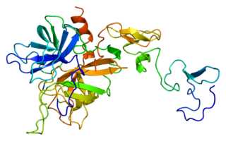
Protein C, also known as autoprothrombin IIA and blood coagulation factor XIV, is a zymogen, that is, an inactive enzyme. The activated form plays an important role in regulating anticoagulation, inflammation, and cell death and maintaining the permeability of blood vessel walls in humans and other animals. Activated protein C (APC) performs these operations primarily by proteolytically inactivating proteins Factor Va and Factor VIIIa. APC is classified as a serine protease since it contains a residue of serine in its active site. In humans, protein C is encoded by the PROC gene, which is found on chromosome 2.

<span class="mw-page-title-main">Thrombophilia</span> Abnormality of blood coagulation

Thrombophilia is an abnormality of blood coagulation that increases the risk of thrombosis. Such abnormalities can be identified in 50% of people who have an episode of thrombosis that was not provoked by other causes. A significant proportion of the population has a detectable thrombophilic abnormality, but most of these develop thrombosis only in the presence of an additional risk factor.

<span class="mw-page-title-main">Cryoglobulinemia</span> Medical condition

Cryoglobulinemia is a medical condition in which the blood contains large amounts of pathological cold sensitive antibodies called cryoglobulins – proteins that become insoluble at reduced temperatures. This should be contrasted with cold agglutinins, which cause agglutination of red blood cells.

<span class="mw-page-title-main">Schistocyte</span> Fragmented portion of a red blood cell

A schistocyte or schizocyte is a fragmented part of a red blood cell. Schistocytes are typically irregularly shaped, jagged, and have two pointed ends.

<span class="mw-page-title-main">Fresh frozen plasma</span> Liquid portion of whole blood

Fresh frozen plasma (FFP) is a blood product made from the liquid portion of whole blood. It is used to treat conditions in which there are low blood clotting factors or low levels of other blood proteins. It may also be used as the replacement fluid in plasma exchange. Using ABO compatible plasma, while not required, may be recommended. Use as a volume expander is not recommended. It is given by slow injection into a vein.

<span class="mw-page-title-main">Hypoprothrombinemia</span> Medical condition

Hypoprothrombinemia is a rare blood disorder in which a deficiency in immunoreactive prothrombin, produced in the liver, results in an impaired blood clotting reaction, leading to an increased physiological risk for spontaneous bleeding. This condition can be observed in the gastrointestinal system, cranial vault, and superficial integumentary system, affecting both the male and female population. Prothrombin is a critical protein that is involved in the process of hemostasis, as well as illustrating procoagulant activities. This condition is characterized as an autosomal recessive inheritance congenital coagulation disorder affecting 1 per 2,000,000 of the population, worldwide, but is also attributed as acquired.

<span class="mw-page-title-main">Warfarin necrosis</span> Medical condition

Warfarin-induced skin necrosis is a condition in which skin and subcutaneous tissue necrosis occurs due to acquired protein C deficiency following treatment with anti-vitamin K anticoagulants.

<span class="mw-page-title-main">Thrombotic microangiopathy</span> Medical condition

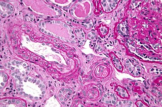
Thrombotic microangiopathy (TMA) is a pathology that results in thrombosis in capillaries and arterioles, due to an endothelial injury. It may be seen in association with thrombocytopenia, anemia, purpura and kidney failure.

<span class="mw-page-title-main">Protein C deficiency</span> Medical condition

Protein C deficiency is a rare genetic trait that predisposes to thrombotic disease. It was first described in 1981. The disease belongs to a group of genetic disorders known as thrombophilias. Protein C deficiency is associated with an increased incidence of venous thromboembolism, whereas no association with arterial thrombotic disease has been found.

<span class="mw-page-title-main">Calciphylaxis</span> Medical condition

Calciphylaxis, also known as calcific uremic arteriolopathy (CUA) or “Grey Scale”, is a rare syndrome characterized by painful skin lesions. The pathogenesis of calciphylaxis is unclear but believed to involve calcification of the small blood vessels located within the fatty tissue and deeper layers of the skin, blood clots, and eventual death of skin cells due to lack of blood flow. It is seen mostly in people with end-stage kidney disease but can occur in the earlier stages of chronic kidney disease and rarely in people with normally functioning kidneys. Calciphylaxis is a rare but serious disease, believed to affect 1-4% of all dialysis patients. It results in chronic non-healing wounds and indicates poor prognosis, with typical life expectancy of less than one year.

<span class="mw-page-title-main">Heparin cofactor II</span> Protein-coding gene in the species Homo sapiens

Heparin cofactor II (HCII), a protein encoded by the SERPIND1 gene, is a coagulation factor that inhibits IIa, and is a cofactor for heparin and dermatan sulfate.

<span class="mw-page-title-main">Upshaw–Schulman syndrome</span> Medical condition

Upshaw–Schulman syndrome (USS) is the recessively inherited form of thrombotic thrombocytopenic purpura (TTP), a rare and complex blood coagulation disease. USS is caused by the absence of the ADAMTS13 protease resulting in the persistence of ultra large von Willebrand factor multimers (ULVWF), causing episodes of acute thrombotic microangiopathy with disseminated multiple small vessel obstructions. These obstructions deprive downstream tissues from blood and oxygen, which can result in tissue damage and death. The presentation of an acute USS episode is variable but usually associated with thrombocytopenia, microangiopathic hemolytic anemia (MAHA) with schistocytes on the peripheral blood smear, fever and signs of ischemic organ damage in the brain, kidney and heart.

Factor XII deficiency is a deficiency in the production of factor XII (FXII), a plasma glycoprotein and clotting factor that participates in the coagulation cascade and activates factor XI. FXII appears to be not essential for blood clotting, as individuals with this condition are usually asymptomatic and form blood clots in vivo. FXII deficiency tends to be identified during presurgical laboratory screening for bleeding disorders.

Retiform purpura is the result of total vascular blockage and damage to the skin's blood vessels. These lesions can appear due to intravascular issues where clots, proteins, or emboli block skin vessels. They can also result from direct harm to the vessel walls, as seen in conditions like vasculitis, calciphylaxis, and certain severe opportunistic infections.

References

  1. James, William D.; Berger, Timothy G.; et al. (2006). Andrews' Diseases of the Skin: clinical Dermatology. Saunders Elsevier. ISBN   0-7216-2921-0.
  2. 1 2 3 4 5 6 7 8 9 10 11 12 13 14 15 16 17 18 19 20 21 Chalmers E, Cooper P, Forman K, Grimley C, Khair K, Minford A, Morgan M, Mumford AD (2011). "Purpura fulminans: recognition, diagnosis and management". Arch Dis Child. 96 (11): 1066–1071. doi:10.1136/adc.2010.199919. PMID   21233082. S2CID   206846385.
  3. 1 2 Ghosh SK, Bandyopadhyay D, Dutta A (2009). "Purpura fulminans: a cutaneous marker of disseminated intravascular coagulation". West J Emerg Med. 10 (1): 41. PMC   2672288 . PMID   19561767.
  4. 1 2 3 Estelles A, Garcia-Plaza I, Dasi A, Aznar J, Duart M, Sanz G, Perez-Requejo JL, Espana F, Jimenez C, Abeledo G (1984). "Severe inherited "homozygous" protein C deficiency in a newborn infant". Thromb Haemost. 52 (1): 53–56. doi:10.1055/s-0038-1661136. PMID   6548587. S2CID   1025282.
  5. D'Ursi P, Marino F, Caprera A, Milanesi L, Faioni EM, Rovida E (2007). "ProCMD: a database and 3D web resource for protein C mutants". BMC Bioinformatics. 8 (Suppl 1): S11. doi: 10.1186/1471-2105-8-s1-s11 . PMC   1885840 . PMID   17430555.
  6. Ursitti JA, Petrich BG, Lee PC, Resneck WG, Ye X, Yang J, Randall WR, Bloch RJ, Wang Y (2007). "Role of an alternatively spliced form of alphaII-spectrin in localization of connexin 43 in cardiomyocytes and regulation by stress-activated protein kinase". J Mol Cell Cardiol. 42 (3): 572–581. doi:10.1016/j.yjmcc.2006.11.018. PMC   1983066 . PMID   17276456.
  7. 1 2 3 4 5 6 7 8 9 10 11 Goldenberg NA, Manco-Johnson MJ (2008). "Protein C deficiency". Haemophilia. 14 (6): 1214–1221. doi: 10.1111/j.1365-2516.2008.01838.x . PMID   19141162. S2CID   2979452.
  8. Gurgey A. (1999). "Clinical manifestations in thrombotic children with factor V Leiden mutation". Pediatr Hematol Oncol. 16 (3): 233–237. doi:10.1080/088800199277281. PMID   10326221.
  9. 1 2 Levin M, Eley BS, Louis J, Cohen H, Young L, Heyderman RS (1995). "Postinfectious purpura fulminans caused by an autoantibody directed against protein S". J Pediatr. 127 (3): 355–63. doi:10.1016/s0022-3476(95)70063-3. PMID   7658262.
  10. Adcock DM, Brozna J, Marlar RA (1990). "Proposed classification and pathologic mechanisms of purpura fulminans and skin necrosis". Semin Thromb Hemost. 16 (4): 333–340. doi:10.1055/s-2007-1002686. PMID   2281322.
  11. Mosnier LO, Zlokovic BV, Griffin JH (2007). "The cytoprotective protein C pathway". Blood. 109 (8): 3161–3172. doi: 10.1182/blood-2006-09-003004 . PMID   17110453.
  12. Kondaveeti S, Hibberd ML, Booy R, Nadel S, Levin M (1999). "Effect of the Factor V Leiden mutation on the severity of meningococcal disease". Pediatr Infect Dis J. 18 (10): 893–896. doi:10.1097/00006454-199910000-00011. PMID   10530586.
  13. Adcock DM, Hicks MJ (1990). "Dermatopathology of skin necrosis associated with purpura fulminans". Semin Thromb Hemost. 16 (4): 283–292. doi:10.1055/s-2007-1002681. PMID   2281318.
  14. Fourrier F, Lestavel P, Chopin C, Marey A, Goudemand J, Rime A, Mangalaboyi J (1990). "Meningococcemia and purpura fulminans in adults: acute deficiencies of proteins C and S and early treatment with antithrombin III concentrates". Intensive Care Med. 16 (2): 121–124. doi:10.1007/bf01699858. PMID   2139671. S2CID   10997888.
  15. Paramo JA, Perez JL, Serrano M, Rocha E (1990). "Types 1 and 2 plasminogen activator inhibitor and tumor necrosis factor alpha in patients with sepsis". Thromb Haemost. 64 (1): 3–6. doi:10.1055/s-0038-1647143. PMID   2274926. S2CID   22756182.
  16. 1 2 Francis RB (1990). "Acquired purpura fulminans". Semin Thromb Hemost. 16 (4): 310–325. doi:10.1055/s-2007-1002684. PMID   2281320.
  17. Marlar RA, Montgomery RR, Broekmans AW (1989). "Diagnosis and treatment of homozygous protein C deficiency. Report of the Working Party on Homozygous Protein C Deficiency of the Subcommittee on Protein C and Protein S, International Committee on Thrombosis and Haemostasis". J Pediatr. 114 (4): 528–534. doi:10.1016/s0022-3476(89)80688-2. PMID   2647943.
  18. Dreyfus M, Magny JF, Bridey F, Schwarz HP, Planche C, Dehan M, Tchernia G (1991). "Treatment of homozygous protein C deficiency and neonatal purpura fulminans with a purified protein C concentrate". N Engl J Med. 325 (22): 1565–1568. doi: 10.1056/nejm199111283252207 . PMID   1944440.
  19. Dreyfus M, Masterson M, David M, Rivard GE, Muller FM, Kreuz W, Beeg T, Minford A, Allgrove J, Cohen JD, et al. (1995). "Replacement therapy with a monoclonal antibody purified protein C concentrate in newborns with severe congenital protein C deficiency". Semin Thromb Hemost. 21 (4): 371–381. doi:10.1055/s-2007-1000658. PMID   8747700.
  20. "Human protein C: new preparations. Effective replacement therapy for some clotting disorders". Prescrire Int. 12 (63): 11–13. 2003. PMID   12602374.
  21. Hartman KR, Manco-Johnson M, Rawlings JS, Bower DJ, Marlar RA (1989). "Homozygous protein C deficiency: early treatment with warfarin". Am J Pediatr Hematol Oncol. 11 (4): 395–401. PMID   2618972.
  22. 1 2 Monagle P, Andrew M, Halton J, Marlar R, Jardine L, Vegh P, Johnston M, Webber C, Massicotte MP (1998). "Homozygous protein C deficiency: description of a new mutation and successful treatment with low molecular weight heparin". Thromb Haemost. 79 (4): 756–761. doi:10.1055/s-0037-1615060. PMID   9569188. S2CID   3610434.
  23. Gladson CL, Groncy P, Griffin JH (1987). "Coumarin necrosis, neonatal purpura fulminans, and protein C deficiency". Arch Dermatol. 123 (12): 1701a–1706a. doi:10.1001/archderm.1987.01660360157029. PMID   2961308.
  24. Tuddenham, EG, Takase T, Thomas, AE, Awidi AS, Madanat FF, Abu Hajir MM, Kernoff PB Hoffbrand AV (1989). "Homozygous protein C deficiency with delayed onset of symptoms at 7 to 10 months". Thromb Res. 53 (5): 475–84. doi:10.1016/0049-3848(89)90202-8. PMID   2660320.{{cite journal}}: CS1 maint: multiple names: authors list (link)
  25. Lerolle N, Carlotti A, Melican K, Aubey F, Pierrot M, Diehl JL, Caille V, Hekimian G, Gandrille S, Mandet C, Bruneval P, Dumenil G, Borgel D (2013). "Assessment of the interplay between blood and skin vascular abnormalities in adult purpura fulminans". Am J Respir Crit Care Med. 188 (6): 684–692. doi:10.1164/rccm.201302-0228oc. PMID   23924269. S2CID   31204154.
  26. Wong VK, Hitchcock W, Mason WH (1989). "Meningococcal infections in children: a review of 100 cases". Pediatr Infect Dis J. 8 (4): 224–227. PMID   2654860.
  27. Manco-Johnson MJ, Abshire TC, Jacobson LJ, Marlar RA (1991). "Severe neonatal protein C deficiency: prevalence and thrombotic risk". J Pediatr. 119 (5): 793–798. doi:10.1016/s0022-3476(05)80305-1. PMID   1834822.
  28. Guelliot A (1884). "Note sur trois cas de purpusa infectieux foudroyant". Un Med Sci Nord-Est. 8: 25.