Quantitative susceptibility mapping

Last updated
A volume rendered brain QSM acquired at 3 Tesla and reconstructed with morphology enabled dipole inversion (MEDI). 3D VR Image 200.jpg
A volume rendered brain QSM acquired at 3 Tesla and reconstructed with morphology enabled dipole inversion (MEDI).

Quantitative susceptibility mapping (QSM) provides a novel contrast mechanism in magnetic resonance imaging (MRI) different from traditional susceptibility weighted imaging. [1] [2] [3] [4] [5]

Contents

The voxel intensity in QSM is linearly proportional to the underlying tissue apparent magnetic susceptibility, which is useful for chemical identification and quantification of specific biomarkers including iron, calcium, gadolinium, and super paramagnetic iron oxide (SPIO) nano-particles. QSM utilizes phase images, solves the magnetic field to susceptibility source inverse problem, and generates a three-dimensional susceptibility distribution. Due to its quantitative nature and sensitivity to certain kinds of material, potential QSM applications include standardized quantitative stratification of cerebral microbleeds and neurodegenerative disease, accurate gadolinium quantification in contrast enhanced MRI, and direct monitoring of targeted theranostic drug biodistribution in nanomedicine.

Background

A visualization of the cone in Fourier domain. Zero cone.jpg
A visualization of the cone in Fourier domain.

In MRI, the local field induced by non-ferromagnetic biomaterial susceptibility along the main polarization B0 field is the convolution of the volume susceptibility distribution with the dipole kernel : . This spatial convolution can be expressed as a point-wise multiplication in Fourier domain: [6] [7] . This Fourier expression provides an efficient way to predict the field perturbation when the susceptibility distribution is known. However, the field to source inverse problem involves division by zero at a pair of cone surfaces at the magic angle with respect to B0 in the Fourier domain. Consequently, susceptibility is underdetermined at the spatial frequencies on the cone surface, which often leads to severe streaking artifacts in the reconstructed QSM.

Techniques

Data acquisition

In principle, any 3D gradient echo sequence can be used for data acquisition. In practice, high resolution imaging with a moderately long echo time is preferred to obtain sufficient susceptibility effects, although the optimal imaging parameters depend on the specific applications and the field strength. A multi-echo acquisition is beneficial for accurate B0 field measurement without the contribution from B1 inhomogeneity. Flow compensation may further improve the accuracy of susceptibility measurement in venous blood, but there are certain technical difficulties to devise a fully flow compensated multi-echo sequence.

Background field removal

Estimated local field maps using left) high-pass filtering method, right) projection onto dipole fields (PDF) method. Local field 400.jpg
Estimated local field maps using left) high-pass filtering method, right) projection onto dipole fields (PDF) method.

In human brain quantitative susceptibility mapping, only the local susceptibility sources inside the brain are of interest. However, the magnetic field induced by the local sources is inevitably contaminated by the field induced by other sources such as main field inhomogeneity (imperfect shimming) and the air-tissue interface, whose susceptibility difference is orders of magnitudes stronger than that of the local sources. Therefore, the non-biological background field needs to be removed for clear visualization on phase images and precise quantification on QSM.

Ideally, the background field can be directly measured with a separate reference scan, where the sample of interest is replaced by a uniform phantom with the same shape while keeping the scanner shimming identical. However, for clinical application, such an approach is impossible and post-processing based methods are preferred. Traditional heuristic methods, including high-pass filtering, are useful for the background field removal, although they also tamper with the local field and degrade the quantitative accuracy.

More recent background field removal methods directly or indirectly exploit the fact that the background field is a harmonic function. Two recent methods based on physical principles, projection onto dipole fields (PDF) [8] and sophisticated harmonic artifact reduction on phase data (SHARP), [9] demonstrated improved contrast and higher precision on the estimated local field. Both methods model the background field as a magnetic field generated by an unknown background susceptibility distribution, and differentiate it from the local field using either the approximate orthogonality or the harmonic property. The background field can also be directly computed by solving the Laplace's equation with simplified boundary values, as demonstrated in the Laplacian boundary value (LBV) method. [10]

Field-to-source inversion

The field-to-source inverse problem can be solved by several methods with various associated advantages and limitations.

Calculation of susceptibility through multiple orientation sampling (COSMOS)

The first QSM image reconstructed using COSMOS to quantify gadolinium concentrations in vials. a) magnitude image; b) field map; c) QSM; d) linear regression. Gd cosmos.jpg
The first QSM image reconstructed using COSMOS to quantify gadolinium concentrations in vials. a) magnitude image; b) field map; c) QSM; d) linear regression.

COSMOS solves the inverse problem by oversampling from multiple orientations. [11] [12] COSMOS utilizes the fact that the zero cone surface in the Fourier domain is fixed at the magic angle with respect to the B0 field. Therefore, if an object is rotated with respect to the B0 field, then in the object's frame, the B0 field is rotated and thus the cone. Consequently, data that cannot be calculated due to the cone becomes available at the new orientations.

COSMOS assumes a model-free susceptibility distribution and keeps full fidelity to the measured data. This method has been validated extensively in in vitro , ex vivo and phantom experiments. Quantitative susceptibility maps obtained from in vivo human brain imaging also showed high degree of agreement with previous knowledge about brain anatomy. Three orientations are generally required for COSMOS, limiting the practicality for clinical applications. However, it may serve as a reference standard when available for calibrating other techniques.

Morphology enabled dipole inversion (MEDI)

A unique advantage of MRI is that it provides not only the phase image but also the magnitude image. In principle, the contrast change, or equivalently the edge, on a magnitude image arises from the underlying change of tissue type, which is the same cause for the change of susceptibility. This observation is translated into mathematics in MEDI, [13] where edges in a QSM which do not exist in the corresponding magnitude image are sparsified by solving a weighted norm minimization problem. [14]

MEDI has also been validated extensively in phantom, in vitro and ex vivo experiments. In an in vivo human brain, MEDI calculated QSM showed similar results compared to COSMOS without statistically significant difference. [15] MEDI only requires a single angle acquisition, so it is a more practical solution to QSM.

Thresholded K-space division (TKD)

The underdetermined data in Fourier domain is only at the location of the cone and its immediate vicinity. For this region in k-space, spatial-frequencies of the dipole kernel are set to a predetermined non-zero value for the division. Investigation of more advanced strategies for recovering data in this k-space region is also a topic of ongoing research. [16]

Thresholded k-space division [12] [17] only requires a single angle acquisition, and benefits from the ease of implementation as well as the fast calculation speed. However, streaking artifacts are frequently present in the QSM and the susceptibility value is underestimated compared to COSMOS calculated QSM.

Potential clinical applications

Differentiating calcification from iron

Differentiation between calcification and iron. From left to right are magnitude, phase and QSM. Iron calc 400.jpg
Differentiation between calcification and iron. From left to right are magnitude, phase and QSM.

It has been confirmed in in vivo and phantom experiments that cortical bones, whose major composition is calcification, are diamagnetic compared to water. [11] [18] Therefore, it is possible to use this diamagnetism to differentiate calcifications from iron deposits that usually demonstrate strong paramagnetism. [19] This may allow QSM to serve as a problem solving tool for the diagnosis of confounding hypointense findings on T2* weighted images.

Quantification of contrast agent

For exogenous susceptibility sources, the susceptibility value is theoretically linearly proportional to the concentration of the contrast agent. This provides a new way for in vivo quantification of gadolinium or SPIO concentrations. [20]

Related Research Articles

<span class="mw-page-title-main">Magnetic resonance imaging</span> Medical imaging technique

Magnetic resonance imaging (MRI) is a medical imaging technique used in radiology to form pictures of the anatomy and the physiological processes inside the body. MRI scanners use strong magnetic fields, magnetic field gradients, and radio waves to generate images of the organs in the body. MRI does not involve X-rays or the use of ionizing radiation, which distinguishes it from computed tomography (CT) and positron emission tomography (PET) scans. MRI is a medical application of nuclear magnetic resonance (NMR) which can also be used for imaging in other NMR applications, such as NMR spectroscopy.

Blood-oxygen-level-dependent imaging, or BOLD-contrast imaging, is a method used in functional magnetic resonance imaging (fMRI) to observe different areas of the brain or other organs, which are found to be active at any given time.

Magnetic resonance elastography (MRE) is a form of elastography that specifically leverages MRI to quantify and subsequently map the mechanical properties of soft tissue. First developed and described at Mayo Clinic by Muthupillai et al. in 1995, MRE has emerged as a powerful, non-invasive diagnostic tool, namely as an alternative to biopsy and serum tests for staging liver fibrosis.

During nuclear magnetic resonance observations, spin–lattice relaxation is the mechanism by which the longitudinal component of the total nuclear magnetic moment vector (parallel to the constant magnetic field) exponentially relaxes from a higher energy, non-equilibrium state to thermodynamic equilibrium with its surroundings (the "lattice"). It is characterized by the spin–lattice relaxation time, a time constant known as T1.

Kenneth Kin Man Kwong is a Hong Kong-born American nuclear physicist. He is a pioneer in human brain imaging. He received his bachelor's degree in Political Science in 1972 from the University of California, Berkeley. He went on to receive his Ph.D. in physics from the University of California, Riverside studying photon-photon collision interactions.

In vivo magnetic resonance spectroscopy (MRS) is a specialized technique associated with magnetic resonance imaging (MRI).

Signal enhancement by extravascular water protons, or SEEP, is a contrast mechanism for functional magnetic resonance imaging (fMRI), which is an alternative to the more commonly employed BOLD contrast. This mechanism for image contrast changes corresponding to changes in neuronal activity was first proposed by Dr. Patrick Stroman in 2001. SEEP contrast is based on changes in tissue water content which arise from the increased production of extracellular fluid and swelling of neurons and glial cells at sites of neuronal activity. Because the dominant sources of MRI signal in biological tissues are water and lipids, an increase in tissue water content is reflected by a local increase in MR signal intensity. A correspondence between BOLD and SEEP signal changes, and sites of activity, has been observed in the brain and appears to arise from the common dependence on changes in local blood flow to cause a change in blood oxygenation or to produce extracellular fluid. The advantage of SEEP contrast is that it can be detected with MR imaging methods which are relatively insensitive to magnetic susceptibility differences between air, tissues, blood, and bone. Such susceptibility differences can give rise to spatial image distortions and areas of low signal, and magnetic susceptibility changes in blood give rise to the BOLD contrast for fMRI. The primary application of SEEP to date has been fMRI of the spinal cord because the bone/tissue interfaces around the spinal cord cause poor image quality with conventional fMRI methods. The disadvantages of SEEP compared to BOLD contrast are that it reveals more localized areas of activity, and in the brain the signal intensity changes are typically lower, and it can therefore be more difficult to detect.

Functional magnetic resonance imaging (fMRI) of the spinal cord is an adaptation of the fMRI method that has been developed for use in the brain. Although the basic principles underlying the methods are the same, spinal fMRI requires a number of specific adaptations to accommodate the periodic motion of the spinal cord, the small cross-sectional dimensions and length of the spinal cord, and the fact that the magnetic field that is used for MRI varies with position in the spinal cord because of magnetic susceptibility differences between bone and tissues. Spinal fMRI has been used to produce maps of neuronal activity at most levels of the spinal cord in response to various stimuli, such as touch, vibration, and thermal changes, and with motor tasks. Research applications of spinal fMRI to date include studies of normal sensory and motor function, and studies of the effects of trauma and multiple sclerosis on the spinal cord.

<span class="mw-page-title-main">Susceptibility weighted imaging</span>

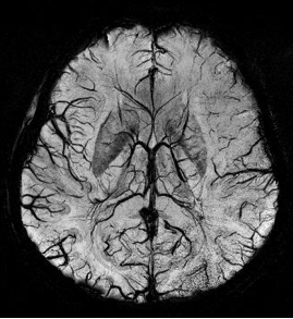
Susceptibility weighted imaging (SWI), originally called BOLD venographic imaging, is an MRI sequence that is exquisitely sensitive to venous blood, hemorrhage and iron storage. SWI uses a fully flow compensated, long echo, gradient recalled echo (GRE) pulse sequence to acquire images. This method exploits the susceptibility differences between tissues and uses the phase image to detect these differences. The magnitude and phase data are combined to produce an enhanced contrast magnitude image. The imaging of venous blood with SWI is a blood-oxygen-level dependent (BOLD) technique which is why it was referred to as BOLD venography. Due to its sensitivity to venous blood SWI is commonly used in traumatic brain injuries (TBI) and for high resolution brain venographies but has many other clinical applications. SWI is offered as a clinical package by Philips and Siemens but can be run on any manufacturer's machine at field strengths of 1.0 T, 1.5 T, 3.0 T and higher.

<span class="mw-page-title-main">Physics of magnetic resonance imaging</span> Overview article

Magnetic resonance imaging (MRI) is a medical imaging technique mostly used in radiology and nuclear medicine in order to investigate the anatomy and physiology of the body, and to detect pathologies including tumors, inflammation, neurological conditions such as stroke, disorders of muscles and joints, and abnormalities in the heart and blood vessels among others. Contrast agents may be injected intravenously or into a joint to enhance the image and facilitate diagnosis. Unlike CT and X-ray, MRI uses no ionizing radiation and is, therefore, a safe procedure suitable for diagnosis in children and repeated runs. Patients with specific non-ferromagnetic metal implants, cochlear implants, and cardiac pacemakers nowadays may also have an MRI in spite of effects of the strong magnetic fields. This does not apply on older devices, and details for medical professionals are provided by the device's manufacturer.

<span class="mw-page-title-main">Real-time MRI</span> Type of MRI

Real-time magnetic resonance imaging (RT-MRI) refers to the continuous monitoring ("filming") of moving objects in real time. Because MRI is based on time-consuming scanning of k-space, real-time MRI was possible only with low image quality or low temporal resolution. Using an iterative reconstruction algorithm these limitations have recently been removed: a new method for real-time MRI achieves a temporal resolution of 20 to 30 milliseconds for images with an in-plane resolution of 1.5 to 2.0 mm. Real-time MRI promises to add important information about diseases of the joints and the heart. In many cases MRI examinations may become easier and more comfortable for patients.

<span class="mw-page-title-main">Magnetic resonance imaging of the brain</span>

Magnetic resonance imaging of the brain uses magnetic resonance imaging (MRI) to produce high quality two-dimensional or three-dimensional images of the brain and brainstem as well as the cerebellum without the use of ionizing radiation (X-rays) or radioactive tracers.

<span class="mw-page-title-main">Intravoxel incoherent motion</span> Concept and a method initially introduced and developed by Le Bihan et al

Intravoxel incoherent motion (IVIM) imaging is a concept and a method initially introduced and developed by Le Bihan et al. to quantitatively assess all the microscopic translational motions that could contribute to the signal acquired with diffusion MRI. In this model, biological tissue contains two distinct environments: molecular diffusion of water in the tissue, and microcirculation of blood in the capillary network (perfusion). The concept introduced by D. Le Bihan is that water flowing in capillaries mimics a random walk (Fig.1), as long as the assumption that all directions are represented in the capillaries is satisfied.

Synthetic MRI is a simulation method in Magnetic Resonance Imaging (MRI), for generating contrast weighted images based on measurement of tissue properties. The synthetic (simulated) images are generated after an MR study, from parametric maps of tissue properties. It is thereby possible to generate several contrast weightings from the same acquisition. This is different from conventional MRI, where the signal acquired from the tissue is used to generate an image directly, often generating only one contrast weighting per acquisition. The synthetic images are similar in appearance to those normally acquired with an MRI scanner.

The history of magnetic resonance imaging (MRI) includes the work of many researchers who contributed to the discovery of nuclear magnetic resonance (NMR) and described the underlying physics of magnetic resonance imaging, starting early in the twentieth century. One researcher was American physicist Isidor Isaac Rabi who won the Nobel Prize in Physics in 1944 for his discovery of nuclear magnetic resonance, which is used in magnetic resonance imaging. MR imaging was invented by Paul C. Lauterbur who developed a mechanism to encode spatial information into an NMR signal using magnetic field gradients in September 1971; he published the theory behind it in March 1973.

<span class="mw-page-title-main">MRI pulse sequence</span>

An MRI pulse sequence in magnetic resonance imaging (MRI) is a particular setting of pulse sequences and pulsed field gradients, resulting in a particular image appearance.

An MRI artifact is a visual artifact in magnetic resonance imaging (MRI). It is a feature appearing in an image that is not present in the original object. Many different artifacts can occur during MRI, some affecting the diagnostic quality, while others may be confused with pathology. Artifacts can be classified as patient-related, signal processing-dependent and hardware (machine)-related.

Amplified magnetic resonance imaging (aMRI) is an MRI method that is coupled with video magnification processing methods to amplify the subtle spatial variations in MRI scans and to enable better visualization of tissue motion. aMRI can enable better visualization of tissue motion to aid the in vivo assessment of the biomechanical response in pathology. It is thought to have potential for helping with diagnosing and monitoring a range of clinical implications in the brain and other organs, including in Chiari Malformation, brain injury, hydrocephalus, other conditions associated with abnormal intracranial pressure, cerebrovascular, and neurodegenerative disease.

<span class="mw-page-title-main">Rolf Gruetter</span> Swiss physicist specialized in magnetic resonance

Rolf Gruetter is a Swiss physicist and neurobiologist specialized in magnetic resonance, biomedical imaging and brain metabolism. He is a professor of physics at EPFL and the head of the Laboratory Functional and Metabolic Imaging at the School of Basic Sciences.

Magnetic resonance fingerprinting (MRF) is methodology in quantitative magnetic resonance imaging (MRI) characterized by a pseudo-randomized acquisition strategy. It involves creating unique signal patterns or 'fingerprints' for different materials or tissues after which a pattern recognition algorithm matches these fingerprints with a predefined dictionary of expected signal patterns. This process translates the data into quantitative maps, revealing information about the magnetic properties being investigated.

References

  1. 1st International Workshop on MRI Phase Contrast and Quantitative Susceptibility Mapping, Jena (2011)
  2. 2nd International Workshop on MRI Phase Contrast & Quantitative Susceptibility Mapping, Cornell (2013)
  3. 3rd International Workshop on MRI Phase Contrast & Quantitative Susceptibility Mapping, Duke (2014)
  4. 4th International Workshop on MRI Phase Contrast & Quantitative Susceptibility Mapping, Graz (2016)
  5. Wang, Yi (2014). "Quantitative susceptibility mapping (QSM): Decoding MRI data for a tissue magnetic biomarker". Magnetic Resonance in Medicine. 73 (1): 82–101. doi:10.1002/mrm.25358. PMC   4297605 . PMID   25044035.
  6. Salomir, Rares; De Senneville, Baudouin Denis; Moonen, Chrit TW (2003). "A fast calculation method for magnetic field inhomogeneity due to an arbitrary distribution of bulk susceptibility". Concepts in Magnetic Resonance. 19B: 26–34. doi:10.1002/cmr.b.10083.
  7. Marques, J.P.; Bowtell, R. (2005). "Application of a Fourier-based method for rapid calculation of field inhomogeneity due to spatial variation of magnetic susceptibility". Concepts in Magnetic Resonance Part B: Magnetic Resonance Engineering. 25B: 65–78. doi: 10.1002/cmr.b.20034 . hdl: 2066/195319 .
  8. Liu, Tian; Khalidov, Ildar; de Rochefort, Ludovic; Spincemaille, Pascal; et al. (2011). "A novel background field removal method for MRI using projection onto dipole fields". NMR in Biomedicine . 24 (9): 1129–36. doi:10.1002/nbm.1670. PMC   3628923 . PMID   21387445.
  9. Schweser, Ferdinand; Deistung, Andreas; Lehr, Berengar Wendel; Reichenbach, Jürgen Rainer (2011). "Quantitative imaging of intrinsic magnetic tissue properties using MRI signal phase: an approach to in vivo brain iron metabolism?". NeuroImage. 54 (4): 2789–2807. doi:10.1016/j.neuroimage.2010.10.070. PMID   21040794. S2CID   19593094.
  10. Zhou, Dong; Liu, Tian; Spincemaille, Pascal; Wang, Yi (2014). "Background field removal by solving the Laplacian boundary value problem". NMR in Biomedicine. 27 (3): 312–9. doi:10.1002/nbm.3064. PMID   24395595. S2CID   1815936.
  11. 1 2 Liu, Tian; Spincemaille, Pascal; De Rochefort, Ludovic; Kressler, Bryan; et al. (2009). "Calculation of susceptibility through multiple orientation sampling (COSMOS): A method for conditioning the inverse problem from measured magnetic field map to susceptibility source image in MRI". Magnetic Resonance in Medicine. 61 (1): 196–204. doi: 10.1002/mrm.21828 . PMID   19097205. S2CID   41444063.
  12. 1 2 Wharton, Sam; Schäfer, Andreas; Bowtell, Richard (2010). "Susceptibility mapping in the human brain using threshold-based k-space division". Magnetic Resonance in Medicine. 63 (5): 1292–304. doi: 10.1002/mrm.22334 . PMID   20432300. S2CID   206278637.
  13. De Rochefort, Ludovic; Liu, Tian; Kressler, Bryan; Liu, Jing; et al. (2009). "Quantitative susceptibility map reconstruction from MR phase data using bayesian regularization: Validation and application to brain imaging". Magnetic Resonance in Medicine. 63 (1): 194–206. doi: 10.1002/mrm.22187 . PMID   19953507.
  14. Liu, J; Liu, T; de Rochefort, L; Khalidov, I (2010). "Quantitative susceptibility mapping by regulating the field to source inverse problem with a sparse prior derived from the Maxwell Equation: validation and application to brain" (PDF). Proc. Intl. Soc. Mag. Reson. Med. 18: 4996. Archived from the original (PDF) on October 16, 2015. Retrieved January 26, 2011.
  15. Liu, Tian; Liu, Jing; de Rochefort, Ludovic; Spincemaille, Pascal; et al. (September 2011). "Morphology enabled dipole inversion (MEDI) from a single-angle acquisition: Comparison with COSMOS in human brain imaging". Magnetic Resonance in Medicine. 66 (3): 777–83. doi: 10.1002/mrm.22816 . PMID   21465541.
  16. Li, Wei; Wu, Bing; Liu, Chunlei (2011). "Quantitative susceptibility mapping of human brain reflects spatial variation in tissue composition". NeuroImage. 55 (4): 1645–56. doi:10.1016/j.neuroimage.2010.11.088. PMC   3062654 . PMID   21224002.
  17. Shmueli, Karin; De Zwart, Jacco A.; Van Gelderen, Peter; Li, Tie-Qiang; et al. (2009). "Magnetic susceptibility mapping of brain tissue in vivo using MRI phase data". Magnetic Resonance in Medicine. 62 (6): 1510–22. doi:10.1002/mrm.22135. PMC   4275127 . PMID   19859937.
  18. De Rochefort, Ludovic; Brown, Ryan; Prince, Martin R.; Wang, Yi (2008). "Quantitative MR susceptibility mapping using piece-wise constant regularized inversion of the magnetic field". Magnetic Resonance in Medicine. 60 (4): 1003–9. doi: 10.1002/mrm.21710 . PMID   18816834.
  19. Schweser, Ferdinand; Deistung, Andreas; Lehr, Berengar W.; Reichenbach, JüRgen R. (2010). "Differentiation between diamagnetic and paramagnetic cerebral lesions based on magnetic susceptibility mapping". Medical Physics. 37 (10): 5165–78. Bibcode:2010MedPh..37.5165S. doi:10.1118/1.3481505. PMID   21089750.
  20. De Rochefort, Ludovic; Nguyen, Thanh; Brown, Ryan; Spincemaille, Pascal; et al. (2008). "In vivo quantification of contrast agent concentration using the induced magnetic field for time-resolved arterial input function measurement with MRI". Medical Physics. 35 (12): 5328–39. Bibcode:2008MedPh..35.5328D. doi:10.1118/1.3002309. PMID   19175092.