Related Research Articles

Functional magnetic resonance imaging or functional MRI (fMRI) measures brain activity by detecting changes associated with blood flow. This technique relies on the fact that cerebral blood flow and neuronal activation are coupled. When an area of the brain is in use, blood flow to that region also increases.

Functional neuroimaging is the use of neuroimaging technology to measure an aspect of brain function, often with a view to understanding the relationship between activity in certain brain areas and specific mental functions. It is primarily used as a research tool in cognitive neuroscience, cognitive psychology, neuropsychology, and social neuroscience.

Neuroimaging is the use of quantitative (computational) techniques to study the structure and function of the central nervous system, developed as an objective way of scientifically studying the healthy human brain in a non-invasive manner. Increasingly it is also being used for quantitative research studies of brain disease and psychiatric illness. Neuroimaging is highly multidisciplinary involving neuroscience, computer science, psychology and statistics, and is not a medical specialty. Neuroimaging is sometimes confused with neuroradiology.
Kenneth Kin Man Kwong is a Hong Kong-born American nuclear physicist. He is a pioneer in human brain imaging. He received his bachelor's degree in Political Science in 1972 from the University of California, Berkeley. He went on to receive his Ph.D. in physics from the University of California, Riverside studying photon-photon collision interactions.

Dr. Christopher deCharms is a neuroscientist, author, and inventor. Currently, Dr. deCharms is the founder and CEO of Brainful, a life-sciences companies focused on neurotechnology, including technology based on imaging methods that allow people to watch the activation of their own brains 'live' using functional magnetic resonance imaging (fMRI).
The Human Connectome Project (HCP) is a five-year project sponsored by sixteen components of the National Institutes of Health, split between two consortia of research institutions. The project was launched in July 2009 as the first of three Grand Challenges of the NIH's Blueprint for Neuroscience Research. On September 15, 2010, the NIH announced that it would award two grants: $30 million over five years to a consortium led by Washington University in St. Louis and the University of Minnesota, with strong contributions from University of Oxford (FMRIB) and $8.5 million over three years to a consortium led by Harvard University, Massachusetts General Hospital and the University of California Los Angeles.

Magnetic resonance imaging of the brain uses magnetic resonance imaging (MRI) to produce high quality two-dimensional or three-dimensional images of the brain and brainstem as well as the cerebellum without the use of ionizing radiation (X-rays) or radioactive tracers.
Mark Steven Cohen is an American neuroscientist and early pioneer of functional brain imaging using magnetic resonance imaging. He currently is a Professor of Psychiatry, Neurology, Radiology, Psychology, Biomedical Physics and Biomedical Engineering at the Semel Institute for Neuroscience and Human Behavior and the Staglin Center for Cognitive Neuroscience. He is also a performing musician.
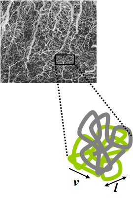
Intravoxel incoherent motion (IVIM) imaging is a concept and a method initially introduced and developed by Le Bihan et al. to quantitatively assess all the microscopic translational motions that could contribute to the signal acquired with diffusion MRI. In this model, biological tissue contains two distinct environments: molecular diffusion of water in the tissue, and microcirculation of blood in the capillary network (perfusion). The concept introduced by D. Le Bihan is that water flowing in capillaries mimics a random walk (Fig.1), as long as the assumption that all directions are represented in the capillaries is satisfied.
Arno Villringer is a Director at the Department of Neurology at the Max Planck Institute for Human Cognitive and Brain Sciences in Leipzig, Germany; Director of the Department of Cognitive Neurology at University of Leipzig Medical Center; and Academic Director of the Berlin School of Mind and Brain and the Mind&Brain Institute, Berlin. He holds a full professorship at University of Leipzig and an honorary professorship at Charité, Humboldt-Universität zu Berlin. From July 2022 to June 2025 he is the Chairperson of the Human Sciences Section of the Max Planck Society.

Resting state fMRI is a method of functional magnetic resonance imaging (fMRI) that is used in brain mapping to evaluate regional interactions that occur in a resting or task-negative state, when an explicit task is not being performed. A number of resting-state brain networks have been identified, one of which is the default mode network. These brain networks are observed through changes in blood flow in the brain which creates what is referred to as a blood-oxygen-level dependent (BOLD) signal that can be measured using fMRI.
Functional magnetic resonance spectroscopy of the brain (fMRS) uses magnetic resonance imaging (MRI) to study brain metabolism during brain activation. The data generated by fMRS usually shows spectra of resonances, instead of a brain image, as with MRI. The area under peaks in the spectrum represents relative concentrations of metabolites.
The following outline is provided as an overview of and topical guide to brain mapping:
Dynamic functional connectivity (DFC) refers to the observed phenomenon that functional connectivity changes over a short time. Dynamic functional connectivity is a recent expansion on traditional functional connectivity analysis which typically assumes that functional networks are static in time. DFC is related to a variety of different neurological disorders, and has been suggested to be a more accurate representation of functional brain networks. The primary tool for analyzing DFC is fMRI, but DFC has also been observed with several other mediums. DFC is a recent development within the field of functional neuroimaging whose discovery was motivated by the observation of temporal variability in the rising field of steady state connectivity research.
Bruce Rosen is an American physicist and radiologist and a leading expert in the area of functional neuroimaging. His research for the past 30 years has focused on the development and application of physiological and functional nuclear magnetic resonance techniques, as well as new approaches to combine functional magnetic resonance imaging (fMRI) data with information from other modalities such as positron emission tomography (PET), magnetoencephalography (MEG) and noninvasive optical imaging. The techniques his group has developed to measure physiological and metabolic changes associated with brain activation and cerebrovascular insult are used by research centers and hospitals throughout the world.
James S. Hyde was an American biophysicist. He held the James S. Hyde chair in Biophysics at the Medical College of Wisconsin (MCW) where he specialized in magnetic resonance instrumentation and methodology development in two distinct areas: electron paramagnetic resonance (EPR) spectroscopy and magnetic resonance imaging (MRI). He is senior author of the widely cited 1995 paper by B.B. Biswal et al. reporting the discovery of resting state functional connectivity (fcMRI) in the human brain. He also served as Director of the National Biomedical EPR Center, a Research Resource supported by the National Institutes of Health. He was author of more than 400 peer-reviewed papers and review articles and held 35 U.S. Patents. He was recognized by Festschrifts in both EPR and fcMRI.
Prof. Dr. med. Wolfgang Grodd is a German neuroradiologist and professor emeritus of the University hospital at the University of Tübingen. He is known for his scientific works on the development and application of structural and functional magnetic resonance imaging in metabolic diseases, sensorimotor representation, language production, and cognitive processing, cerebellum, thalamus, and basal ganglia. Currently, Wolfgang Grodd is a research scientist at the Department of the High-Field MR at the Max Planck Institute for Biological Cybernetics.
The history of magnetic resonance imaging (MRI) includes the work of many researchers who contributed to the discovery of nuclear magnetic resonance (NMR) and described the underlying physics of magnetic resonance imaging, starting early in the twentieth century. MR imaging was invented by Paul C. Lauterbur who developed a mechanism to encode spatial information into an NMR signal using magnetic field gradients in September 1971; he published the theory behind it in March 1973. The factors leading to image contrast had been described nearly 20 years earlier by physician and scientist Erik Odeblad and Gunnar Lindström. Among many other researchers in the late 1970s and 1980s, Peter Mansfield further refined the techniques used in MR image acquisition and processing, and in 2003 he and Lauterbur were awarded the Nobel Prize in Physiology or Medicine for their contributions to the development of MRI. The first clinical MRI scanners were installed in the early 1980s and significant development of the technology followed in the decades since, leading to its widespread use in medicine today.
Denis Le Bihan is a medical doctor, physicist, member of the Institut de France, member of the French Academy of Technologies and director since 2007 of NeuroSpin, an institution of the Atomic Energy and Alternative Energy Commission (CEA) in Saclay, dedicated to the study of the brain by magnetic resonance imaging (MRI) with a very high magnetic field. Denis Le Bihan has received international recognition for his outstanding work, introducing new imaging methods, particularly for the study of the human brain, as evidenced by the many international awards he has received, such as the Gold Medal of the International Society of Magnetic Resonance in Medicine (2001), the coveted Lounsbery Prize, the Louis D. Prize from the Institut de France, the prestigious Honda Prize (2012), the Louis-Jeantet Prize (2014), the Rhein Foundation Award (2021). His work has focused on the introduction, development and application of highly innovative methods, notably diffusion MRI.
References
- ↑ Sir Peter Mansfield – Autobiography. Nobelprize.org. Retrieved on 22 October 2011.
- ↑ News Item. Physics.sfu.ca (9 December 2009). Retrieved on 22 October 2011.
- ↑ Buxton, R.B. (2009) Introduction to functional magnetic resonance imaging: Principles and techniques (2nd ed). Cambridge, Cambridge University Press.
- ↑ Turner, R. Schmitt, F., & Stehling, M. K. (1998) The historical development of echo-planar magnetic resonance imaging. In, F. Schmitt, M. K. Stehling & R. Turner (Eds), Echo-planar imaging: Theory, technique and application. Berlin, Springer Verlag.
- ↑ http://nobelprize.org/nobel_prizes/medicine/laureates/2003/mansfield-autobio.html Nobelprize.org
- ↑ Stehling, M.K., & Liu, L. (1999). Echo planar imaging's impact on modern diagnostic MR-imaging: general principles and historic facts. Magnetic Resonance Materials in Physics, Biology and Medicine 9, 125–133
- ↑ Huettel, S.A., Song, A.W., & McCarthy, G (2004). Functional magnetic resonance imaging. Sunderland, MA. Sinauer Associates, Inc.
- ↑ Turner, R., Le Bihan, D., Maier, J., Vavrek, R., Hedges, L.K, & Pekar, J. (1990). Echo-planar imaging of intravoxel incoherent motion. Radiology 177: 407–414.
- ↑ Le Bihan, D., Mangin, J. F., Poupon, C., Clark, C. A., Pappata, S., Molko, N., & Chabriat, H. (2001). Diffusion tensor imaging: concepts and applications. Journal of Magnetic Resonance Imaging. 13(4):534–46.
- ↑ Cohen, M. S. & Bookheimer, S. Y. (1994) Localization of brain function with magnetic resonance imaging. Trends in Neurosciences 1 (7): 268–277.
- ↑ Turner, R., Le Bihan, D., Moonen, C. T., Despres, D., & Frank, J. (1991). Echo-planar time course of cat brain oxygenation changes. Magnetic Resonance in Medicine 22 (1): 159–66.
- ↑ Science (1993), vol. 261, 30 July 1993
- ↑ Turner, R. & Jones, T. (2003). Techniques for imaging neuroscience. British Medical Bulletin 65:3–20.
- ↑ Kwong, K. K., Belliveau, J. W., Chesler, D. A., Goldberg, I. E., Weisskoff, R. M., Poncelet, B. P., Kennedy, D. N., Hoppel, B. E., Cohen, M. S., Turner, R., Cheng, H., Brady, T .J., & Rosen B. R. (1992). Dynamic magnetic resonance imaging of human brain activity during primary sensory stimulation. PNAS 89(12):5675–5679
- ↑ Ogawa, S., Tank, D. W., Menon, R., Ellermann, J. M., Kim, S. G, Merkle, H., & Ugurbil, K. (1992). Intrinsic signal changes accompanying sensory stimulation: functional brain mapping with magnetic resonance imaging. PNAS 89(13):5951–55
- ↑ Buxton, R. B. (2009). Introduction to functional magnetic resonance imaging: Principles and techniques (2nd Edition). Cambridge. Cambridge University Press.
- ↑ United States Patent: 5492122. Patft.uspto.gov. Retrieved on 22 October 2011.
- ↑ United States Patent: 5185576. Patft.uspto.gov. Retrieved on 22 October 2011.
- ↑ United States Patent: 5077524. Patft.uspto.gov. Retrieved on 22 October 2011.
- ↑ United States Patent: 4896129. Patft.uspto.gov. Retrieved on 22 October 2011.
- ↑ United States Patent: 5666054. Patft.uspto.gov. Retrieved on 22 October 2011.
- ↑ esp@cenet original document. Gb.espacenet.com. Retrieved on 22 October 2011.
- ↑ esp@cenet document view. Gb.espacenet.com (8 April 1987). Retrieved on 22 October 2011.
- ↑ esp@cenet document view. Gb.espacenet.com (3 February 1988). Retrieved on 22 October 2011.
- ↑ esp@cenet document view. Gb.espacenet.com (16 August 1989). Retrieved on 22 October 2011.
- ↑ "ISMRM Gold Medal Award Winners". ISMRM. Retrieved 3 October 2020.