Thapsigargin

Last updated
Thapsigargin
Thapsigargin.png
Names
IUPAC name
(11S)-7,11-Dihydroxy-12-oxo-6β,12-epoxy-1β,7α,10α-guai-4-ene-2β,3α,8α,10-tetrayl 10-acetate 8-butanoate 3-[(2Z)-2-methylbut-2-enoate] 2-octanoate
Systematic IUPAC name
(3S,3aR,4S,6S,6aR,7S,8S,9bS)-3,3a-Dihydroxy-3,6,9-trimethyl-2-oxo-2,3,3a,4,5,6,6a,7,8,9b-decahydroazuleno[4,5-b]furan-4,6,7,8-tetrayl 6-acetate 4-butanoate 8-[(2Z)-2-methylbut-2-enoate] 7-octanoate
Identifiers
3D model (JSmol)
ChEBI
ChEMBL
ChemSpider
ECHA InfoCard 100.116.539 OOjs UI icon edit-ltr-progressive.svg
PubChem CID
UNII
  • InChI=1S/C34H50O12/c1-9-12-13-14-15-17-24(37)43-28-26-25(20(5)27(28)44-30(38)19(4)11-3)29-34(41,33(8,40)31(39)45-29)22(42-23(36)16-10-2)18-32(26,7)46-21(6)35/h11,22,26-29,40-41H,9-10,12-18H2,1-8H3/b19-11-/t22-,26+,27-,28-,29-,32-,33+,34+/m0/s1 Yes check.svgY
    Key: IXFPJGBNCFXKPI-FSIHEZPISA-N Yes check.svgY
  • InChI=1/C34H50O12/c1-9-12-13-14-15-17-24(37)43-28-26-25(20(5)27(28)44-30(38)19(4)11-3)29-34(41,33(8,40)31(39)45-29)22(42-23(36)16-10-2)18-32(26,7)46-21(6)35/h11,22,26-29,40-41H,9-10,12-18H2,1-8H3/b19-11-/t22-,26+,27-,28-,29-,32-,33+,34+/m0/s1
    Key: IXFPJGBNCFXKPI-FSIHEZPIBR
  • O=C3O[C@H]2C\1=C(\[C@H](OC(=O)\C(=C/C)C)[C@@H](OC(=O)CCCCCCC)[C@@H]/1[C@@](OC(=O)C)(C[C@H](OC(=O)CCC)[C@]2(O)[C@@]3(O)C)C)C
Properties
C34H50O12
Molar mass 650.762 g·mol−1
Except where otherwise noted, data are given for materials in their standard state (at 25 °C [77 °F], 100 kPa).
X mark.svgN  verify  (what is  Yes check.svgYX mark.svgN ?)

Thapsigargin is a non-competitive inhibitor of the sarco/endoplasmic reticulum Ca2+ ATPase (SERCA). [1] Structurally, thapsigargin is classified as a guaianolide, and is extracted from a plant, Thapsia garganica . [2] It is a tumor promoter in mammalian cells. [3]

Contents

Thapsigargin raises cytosolic (intracellular) calcium concentration by blocking the ability of the cell to pump calcium into the sarcoplasmic and endoplasmic reticula. Store-depletion can secondarily activate plasma membrane calcium channels, allowing an influx of calcium into the cytosol. Depletion of ER calcium stores leads to ER stress and activation of the unfolded protein response. [4] Non-resolved ER stress can cumulatively lead to cell death. [5] [6] Prolonged store depletion can protect against ferroptosis via remodeling of ER-synthesized phospholipids. [7]

Thapsigargin treatment and the resulting ER calcium depletion inhibits autophagy independent of the UPR. [8] [9]

Thapsigargin is useful in experimentation examining the impacts of increasing cytosolic calcium concentrations and ER calcium depletion.

A study from the University of Nottingham showed promising results for its use against Covid-19 and other coronavirus.

Biosynthesis

The complete biosynthesis of thapsigargin has yet to be elucidated. A proposed biosynthesis starts with the farnesyl pyrophosphate. The first step is controlled by the enzyme germacrene B synthase. In the second step, the C(8) position is easily activated for an allylic oxidation due to the position of the double bond. The next step is the addition of the acyloxy moiety by a P450 acetyltransferase; which is a well known reaction for the synthesis of the diterpene, taxol. In the third step, the lactone ring is formed by a cytochrome P450 enzyme using NADP+. With the butyloxy group on the C(8), the formation will only generate the 6,12-lactone ring. The fourth step is an epoxidation that initiates the last step of the base guaianolide formation. In the fifth step, a P450 enzyme closes the 5 + 7 guaianolide structure. The ring closing is important, because it will proceed via 1,10 - epoxidation in order to retain the 4,5 - double bond needed in thapsigargin. It is not known whether the secondary modifications to the guaianolide occur before, or after the formation of thapsigargin, but will need to be considered when elucidating the true biosynthesis. It should also be noted, that several of these enzymes are P450s, therefore oxygen and NADPH are likely crucial to this biosynthesis as well as other cofactors such as Mg2+ and Mn2+ may be needed. [10]

Research

Since inhibition of SERCA is a mechanism of action that has been used to target solid tumors, thapsigargin has attracted research interest. A prodrug of thapsigargin, mipsagargin, is currently undergoing clinical trials for the treatment of glioblastoma. [11] [12] [13] [14]

The biological activity has also attracted research into the laboratory synthesis of thapsigargin. To date, three distinct syntheses have been reported: one by Steven V. Ley, [15] one by Phil Baran., [16] and one by P. Andrew Evans. [17]

Preclinical studies demonstrated that other effects of thapsigargin include suppression of nicotinic acetylcholine receptors activity in neurons of the guinea-pig ileum submucous plexus [18] and rat superior cervical ganglion. [19]

Laboratory studies at the University of Nottingham, using in vitro cell cultures, indicates possible potential as a broad spectrum antiviral, with activity against the COVID-19 virus (SARS-CoV-2), a common cold virus, respiratory syncytial virus (RSV), and the influenza A virus. [20]

See also

Related Research Articles

<span class="mw-page-title-main">Endoplasmic reticulum</span> Cell organelle that synthesizes, folds and processes proteins

The endoplasmic reticulum (ER) is, in essence, the transportation system of the eukaryotic cell, and has many other important functions such as protein folding. It is a type of organelle made up of two subunits – rough endoplasmic reticulum (RER), and smooth endoplasmic reticulum (SER). The endoplasmic reticulum is found in most eukaryotic cells and forms an interconnected network of flattened, membrane-enclosed sacs known as cisternae, and tubular structures in the SER. The membranes of the ER are continuous with the outer nuclear membrane. The endoplasmic reticulum is not found in red blood cells, or spermatozoa.

<span class="mw-page-title-main">Endomembrane system</span> Membranes in the cytoplasm of a eukaryotic cell

The endomembrane system is composed of the different membranes (endomembranes) that are suspended in the cytoplasm within a eukaryotic cell. These membranes divide the cell into functional and structural compartments, or organelles. In eukaryotes the organelles of the endomembrane system include: the nuclear membrane, the endoplasmic reticulum, the Golgi apparatus, lysosomes, vesicles, endosomes, and plasma (cell) membrane among others. The system is defined more accurately as the set of membranes that forms a single functional and developmental unit, either being connected directly, or exchanging material through vesicle transport. Importantly, the endomembrane system does not include the membranes of plastids or mitochondria, but might have evolved partially from the actions of the latter.

<span class="mw-page-title-main">Lysosome</span> Cell organelle

A lysosome is a membrane-bound organelle found in many animal cells. They are spherical vesicles that contain hydrolytic enzymes that can break down many kinds of biomolecules. A lysosome has a specific composition, of both its membrane proteins, and its lumenal proteins. The lumen's pH (~4.5–5.0) is optimal for the enzymes involved in hydrolysis, analogous to the activity of the stomach. Besides degradation of polymers, the lysosome is involved in various cell processes, including secretion, plasma membrane repair, apoptosis, cell signaling, and energy metabolism.

Calcium release-activated channels (CRAC) are specialized plasma membrane Ca2+ ion channels. When calcium ions (Ca2+) are depleted from the endoplasmic reticulum (a major store of Ca2+) of mammalian cells, the CRAC channel is activated to slowly replenish the level of calcium in the endoplasmic reticulum. The Ca2+ Release-activated Ca2+ (CRAC) Channel (CRAC-C) Family (TC# 1.A.52) is a member of the Cation Diffusion Facilitator (CDF) Superfamily. These proteins typically have between 4 and 6 transmembrane α-helical spanners (TMSs). The 4 TMS CRAC channels arose by loss of 2TMSs from 6TMS CDF carriers, an example of 'reverse' evolution'.

<span class="mw-page-title-main">Calcium signaling</span> Intracellular communication process

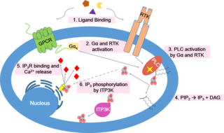
Calcium signaling is the use of calcium ions (Ca2+) to communicate and drive intracellular processes often as a step in signal transduction. Ca2+ is important for cellular signalling, for once it enters the cytosol of the cytoplasm it exerts allosteric regulatory effects on many enzymes and proteins. Ca2+ can act in signal transduction resulting from activation of ion channels or as a second messenger caused by indirect signal transduction pathways such as G protein-coupled receptors.

<span class="mw-page-title-main">Chelerythrine</span> Chemical compound

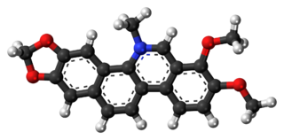
Chelerythrine is a benzophenanthridine alkaloid present in the plant Chelidonium majus. It is a potent, selective, and cell-permeable protein kinase C inhibitor in vitro. And an efficacious antagonist of G-protein-coupled CB1 receptors. This molecule also exhibits anticancer qualities and it has served as a base for many potential novel drugs against cancer. Structurally, this molecule has two distinct conformations, one being a positively charged iminium form, and the other being an uncharged form, a pseudo-base.

<span class="mw-page-title-main">Cyclopiazonic acid</span> Chemical compound

Cyclopiazonic acid (α-CPA), a mycotoxin and a fungal neurotoxin, is made by the molds Aspergillus and Penicillium. It is an indole-tetramic acid that serves as a toxin due to its ability to inhibit calcium-dependent ATPases found in the endoplasmic and sarcoplasmic reticulum. This inhibition disrupts the muscle contraction-relaxation cycle and the calcium gradient that is maintained for proper cellular activity in cells.

<span class="mw-page-title-main">Lipid signaling</span> Biological signaling using lipid molecules

Lipid signaling, broadly defined, refers to any biological signaling event involving a lipid messenger that binds a protein target, such as a receptor, kinase or phosphatase, which in turn mediate the effects of these lipids on specific cellular responses. Lipid signaling is thought to be qualitatively different from other classical signaling paradigms because lipids can freely diffuse through membranes. One consequence of this is that lipid messengers cannot be stored in vesicles prior to release and so are often biosynthesized "on demand" at their intended site of action. As such, many lipid signaling molecules cannot circulate freely in solution but, rather, exist bound to special carrier proteins in serum.

<span class="mw-page-title-main">Calcium ATPase</span> Class of enzymes

Ca2+ ATPase is a form of P-ATPase that transfers calcium after a muscle has contracted. The two kinds of calcium ATPase are:

<span class="mw-page-title-main">Nicotinic acid adenine dinucleotide phosphate</span> Chemical compound

Nicotinic acid adenine dinucleotide phosphate, (NAADP), is a Ca2+-mobilizing second messenger synthesised in response to extracellular stimuli. Like its mechanistic cousins, IP3 and cyclic adenosine diphosphoribose (Cyclic ADP-ribose), NAADP binds to and opens Ca2+ channels on intracellular organelles, thereby increasing the intracellular Ca2+ concentration which, in turn, modulates sundry cellular processes (see Calcium signalling). Structurally, it is a dinucleotide that only differs from the house-keeping enzyme cofactor, NADP by a hydroxyl group (replacing the nicotinamide amino group) and yet this minor modification converts it into the most potent Ca2+-mobilizing second messenger yet described. NAADP acts across phyla from plants to humans.

The unfolded protein response (UPR) is a cellular stress response related to the endoplasmic reticulum (ER) stress. It has been found to be conserved between mammalian species, as well as yeast and worm organisms.

<span class="mw-page-title-main">Farnesyl-diphosphate farnesyltransferase</span> Class of enzymes

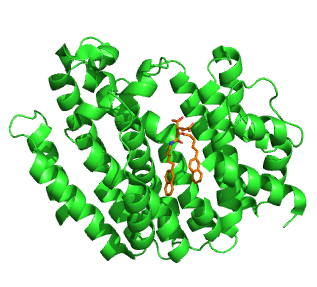
Squalene synthase (SQS) or farnesyl-diphosphate:farnesyl-diphosphate farnesyl transferase is an enzyme localized to the membrane of the endoplasmic reticulum. SQS participates in the isoprenoid biosynthetic pathway, catalyzing a two-step reaction in which two identical molecules of farnesyl pyrophosphate (FPP) are converted into squalene, with the consumption of NADPH. Catalysis by SQS is the first committed step in sterol synthesis, since the squalene produced is converted exclusively into various sterols, such as cholesterol, via a complex, multi-step pathway. SQS belongs to squalene/phytoene synthase family of proteins.

<span class="mw-page-title-main">ATP2A1</span> Protein-coding gene in the species Homo sapiens

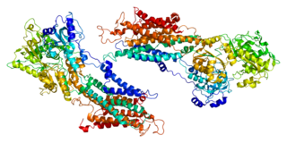
Sarcoplasmic/endoplasmic reticulum calcium ATPase 1 (SERCA1) is an enzyme that in humans is encoded by the ATP2A1 gene.

<span class="mw-page-title-main">ATP2A3</span> Protein-coding gene in the species Homo sapiens

Sarcoplasmic/endoplasmic reticulum calcium ATPase 3 is an enzyme that in humans is encoded by the ATP2A3 gene.

<span class="mw-page-title-main">STIM2</span> Protein-coding gene in the species Homo sapiens

Stromal interaction molecule 2 (STIM2) is a protein that in humans is encoded by the STIM2 gene.

<span class="mw-page-title-main">Pyocyanin</span> Chemical compound

Pyocyanin (PCN) is one of the many toxic compounds produced and secreted by the Gram negative bacterium Pseudomonas aeruginosa. Pyocyanin is a blue secondary metabolite, turning red below pH 4.9, with the ability to oxidise and reduce other molecules and therefore kill microbes competing against P. aeruginosa as well as mammalian cells of the lungs which P. aeruginosa has infected during cystic fibrosis. Since pyocyanin is a zwitterion at blood pH, it is easily able to cross the cell membrane. There are three different states in which pyocyanin can exist: oxidized (blue), monovalently reduced (colourless) or divalently reduced (red). Mitochondria play an important role in the cycling of pyocyanin between its redox states. Due to its redox-active properties, pyocyanin generates reactive oxygen species.

Membrane contact sites (MCS) are close appositions between two organelles. Ultrastructural studies typically reveal an intermembrane distance in the order of the size of a single protein, as small as 10 nm or wider, with no clear upper limit. These zones of apposition are highly conserved in evolution. These sites are thought to be important to facilitate signalling, and they promote the passage of small molecules, including ions, lipids and reactive oxygen species. MCS are important in the function of the endoplasmic reticulum (ER), since this is the major site of lipid synthesis within cells. The ER makes close contact with many organelles, including mitochondria, Golgi, endosomes, lysosomes, peroxisomes, chloroplasts and the plasma membrane. Both mitochondria and sorting endosomes undergo major rearrangements leading to fission where they contact the ER. Sites of close apposition can also form between most of these organelles most pairwise combinations. First mentions of these contact sites can be found in papers published in the late 1950s mainly visualized using electron microscopy (EM) techniques. Copeland and Dalton described them as “highly specialized tubular form of endoplasmic reticulum in association with the mitochondria and apparently in turn, with the vascular border of the cell”.

Beta cells are heavily engaged in the synthesis and secretion of insulin. They are therefore particularly sensitive to endoplasmic reticulum (ER) stress and the subsequent unfolded protein response (UPR). Severe or prolonged episodes of ER stress can lead to the death of beta cells, which can contribute to the development of both Type I and Type II diabetes.

<span class="mw-page-title-main">Istaroxime</span> Chemical compound

Istaroxime is an investigational drug under development for treatment of acute decompensated heart failure

<span class="mw-page-title-main">Ormdl sphingolipid biosynthesis regulator 3</span> Protein-coding gene in the species Homo sapiens

ORMDL sphingolipid biosynthesis regulator 3 is a protein that in humans is encoded by the ORMDL3 gene. This gene is associated with asthma in childhood. Transgenic mice which overexpress human ORMDL3 have increased levels of IgE. This correlated with increased numbers of macrophages, neutrophils, eosinophils, CD4+ and enhanced Th2 cytokine levels in the lung tissue.

References

  1. Rogers TB, Inesi G, Wade R, Lederer WJ (1995). "Use of thapsigargin to study Ca2+ homeostasis in cardiac cells". Biosci. Rep. 15 (5): 341–9. doi:10.1007/BF01788366. PMID   8825036. S2CID   29613387.
  2. Rasmussen U, Brøogger Christensen S, Sandberg F (1978). "Thapsigargine and thapsigargicine, two new histamine liberators from Thapsia garganica L.". Acta Pharm. Suec. 15 (2): 133–140. PMID   79299.{{cite journal}}: CS1 maint: multiple names: authors list (link)
  3. Hakii, H.; Fujiki, H.; Suganuma, M.; Nakayasu, M.; Tahira, T.; Sugimura, T.; Scheuer, P. J.; Christensen, S. B. (1986). "Thapsigargin, a histamine secretagogue, is a non-12-O-tetradecanolphorbol-13-acetate (TPA) type tumor promoter in two-stage mouse skin carcinogenesis". Journal of Cancer Research and Clinical Oncology. 111 (3): 177–181. doi:10.1007/BF00389230. PMID   2426275. S2CID   19093742.
  4. Malhotra, Jyoti D.; Kaufman, Randal J. (December 2007). "The endoplasmic reticulum and the unfolded protein response". Seminars in Cell & Developmental Biology. 18 (6): 716–731. doi:10.1016/j.semcdb.2007.09.003. PMC   2706143 . PMID   18023214.
  5. Hetz, Claudio; Papa, Feroz R. (January 2018). "The Unfolded Protein Response and Cell Fate Control". Molecular Cell. 69 (2): 169–181. doi: 10.1016/j.molcel.2017.06.017 . PMID   29107536.
  6. Sano, Renata; Reed, John C. (2013-12-01). "ER stress-induced cell death mechanisms". Biochimica et Biophysica Acta (BBA) - Molecular Cell Research. 1833 (12): 3460–3470. doi:10.1016/j.bbamcr.2013.06.028. ISSN   0167-4889. PMC   3834229 . PMID   23850759.
  7. Xin, Shan; Mueller, Constanze; Pfeiffer, Susanne; Kraft, Vanessa A. N.; Merl-Pham, Juliane; Bao, Xuanwen; Feederle, Regina; Jin, Xiang; Hauck, Stefanie M.; Schmitt-Kopplin, Philippe; Schick, Joel A. (2021-10-18). "MS4A15 drives ferroptosis resistance through calcium-restricted lipid remodeling". Cell Death & Differentiation. 29 (3): 670–686. doi:10.1038/s41418-021-00883-z. ISSN   1476-5403. PMC   8901757 . PMID   34663908. S2CID   239025829.
  8. Engedal, Nikolai; Torgersen, Maria L; Guldvik, Ingrid J; Barfeld, Stefan J; Bakula, Daniela; Sætre, Frank; Hagen, Linda K; Patterson, John B; Proikas-Cezanne, Tassula (2013-10-25). "Modulation of intracellular calcium homeostasis blocks autophagosome formation". Autophagy. 9 (10): 1475–1490. doi: 10.4161/auto.25900 . ISSN   1554-8627. PMID   23970164.
  9. Ganley, Ian G.; Wong, Pui-Mun; Gammoh, Noor; Jiang, Xuejun (2011). "Distinct Autophagosomal-Lysosomal Fusion Mechanism Revealed by Thapsigargin-Induced Autophagy Arrest" (PDF). Molecular Cell. 42 (6): 731–743. doi:10.1016/j.molcel.2011.04.024. PMC   3124681 . PMID   21700220.
  10. Drew, D.P.; Krichau, N.; Reichwald, K.; Simonsen, H.T. (2009). "Guaianolides in apiaceae: perspectives on pharmacology and biosynthesis". Phytochem. Rev. 8 (3): 581–599. doi:10.1007/s11101-009-9130-z. S2CID   37287410.
  11. Denmeade, S. R.; Mhaka, A. M.; Rosen, D. M.; Brennen, W. N.; Dalrymple, S; Dach, I; Olesen, C; Gurel, B; Demarzo, A. M.; Wilding, G; Carducci, M. A.; Dionne, C. A.; Møller, J. V.; Nissen, P; Christensen, S. B.; Isaacs, J. T. (2012). "Engineering a Prostate-Specific Membrane Antigen–Activated Tumor Endothelial Cell Prodrug for Cancer Therapy". Science Translational Medicine. 4 (140): 140ra86. doi:10.1126/scitranslmed.3003886. PMC   3715055 . PMID   22745436.
  12. Andersen, Trine; López, Carmen; Manczak, Tom; Martinez, Karen; Simonsen, Henrik (2015). "Thapsigargin—From Thapsia L. To Mipsagargin". Molecules. 20 (4): 6113–27. doi: 10.3390/molecules20046113 . PMC   6272310 . PMID   25856061.
  13. "Mipsagargin". NCI Drug Dictionary. National Cancer Institute. 2011-02-02.
  14. "Clinical Trials: Mipsagargin". National Cancer Institute. 17 August 2017.
  15. Ball, Matthew; Andrews, Stephen P.; Wierschem, Frank; Cleator, Ed; Smith, Martin D.; Ley, Steven V. (2007). "Total Synthesis of Thapsigargin, a Potent SERCA Pump Inhibitor". Organic Letters. 9 (4): 663–6. doi:10.1021/ol062947x. PMID   17256950.
  16. Chu, Hang; Smith, Joel M.; Felding, Jakob; Baran, Phil S. (2017). "Scalable Synthesis of (−)-Thapsigargin". ACS Central Science. 3 (1): 47–51. doi:10.1021/acscentsci.6b00313. PMC   5269647 . PMID   28149952.
  17. Chen, Dezhi; Evans, P Andrew. (2017). "A Concise, Efficient and Scalable Total Synthesis of Thapsigargin and Nortrilobolide from (R)-(-)-Carvone". J. Am. Chem. Soc. 139 (17): 6046–6049. doi:10.1021/jacs.7b01734. PMID   28422492.
  18. Glushakov, A. V.; Glushakova, H. Y.; Skok, V. I. (1999-01-15). "Modulation of nicotinic acetylcholine receptor activity in submucous neurons by intracellular messengers". Journal of the Autonomic Nervous System. 75 (1): 16–22. doi:10.1016/S0165-1838(98)00165-9. ISSN   0165-1838. PMID   9935265 via https://doi.org/10.1016/S0165-1838(98)00165-9.{{cite journal}}: External link in |via= (help)
  19. Voitenko, S. V.; Bobryshev, A. Yu; Skok, V. I. (2000-01-01). "Intracellular regulation of neuronal nicotinic cholinorceptors". Neuroscience and Behavioral Physiology. 30 (1): 19–25. doi:10.1007/BF02461388. ISSN   0097-0549. PMID   10768368. S2CID   10990124.
  20. "Powerful Antiviral Treatment for COVID-19 Discovered That Could Change How Epidemics Are Managed". 2 February 2021.

<15.Dey. S.; Bajaj, S. O "Promising anticancer drug thapsigargin: A perspective toward the total synthesis" Synthetic communication 2018, 48(1), 1-13/>

Further reading