Related Research Articles

Veins are blood vessels in the circulatory system of humans and most other animals that carry blood towards the heart. Most veins carry deoxygenated blood from the tissues back to the heart; exceptions are those of the pulmonary and fetal circulations which carry oxygenated blood to the heart. In the systemic circulation, arteries carry oxygenated blood away from the heart, and veins return deoxygenated blood to the heart, in the deep veins.

The femoral artery is a large artery in the thigh and the main arterial supply to the thigh and leg. The femoral artery gives off the deep femoral artery and descends along the anteromedial part of the thigh in the femoral triangle. It enters and passes through the adductor canal, and becomes the popliteal artery as it passes through the adductor hiatus in the adductor magnus near the junction of the middle and distal thirds of the thigh.

The great saphenous vein (GSV) or long saphenous vein is a large, subcutaneous, superficial vein of the leg. It is the longest vein in the body, running along the length of the lower limb, returning blood from the foot, leg and thigh to the deep femoral vein at the femoral triangle.

The inferior vena cava is a large vein that carries the deoxygenated blood from the lower and middle body into the right atrium of the heart. It is formed by the joining of the right and the left common iliac veins, usually at the level of the fifth lumbar vertebra.

The azygos vein is a vein running up the right side of the thoracic vertebral column draining itself towards the superior vena cava. It connects the systems of superior vena cava and inferior vena cava and can provide an alternative path for blood to the right atrium when either of the venae cavae is blocked.

In human anatomy, the abdominal aorta is the largest artery in the abdominal cavity. As part of the aorta, it is a direct continuation of the descending aorta.

Caput medusae is the appearance of distended and engorged superficial epigastric veins, which are seen radiating from the umbilicus across the abdomen. The name caput medusae originates from the apparent similarity to Medusa's head, which had venomous snakes in place of hair. It is also a sign of portal hypertension. It is caused by dilation of the paraumbilical veins, which carry oxygenated blood from mother to fetus in utero and normally close within one week of birth, becoming re-canalised due to portal hypertension caused by liver failure.

Inguinal lymph nodes are lymph nodes in the groin. They are situated in the femoral triangle of the inguinal region. They are subdivided into two groups: the superficial inguinal lymph nodes and deep inguinal lymph nodes.

An inferior vena cava filter is a medical device made of metal that is implanted by vascular surgeons or interventional radiologists into the inferior vena cava to prevent a life-threatening pulmonary embolism (PE) or venous thromboembolism (VTE).

In human anatomy, the inferior epigastric artery is an artery that arises from the external iliac artery. It is accompanied by the inferior epigastric vein; inferiorly, these two inferior epigastric vessels together travel within the lateral umbilical fold The inferior epigastric artery then traverses the arcuate line of rectus sheath to enter the rectus sheath, then anastomoses with the superior epigastric artery within the rectus sheath.

The round ligament of the liver, ligamentum teres or ligamentum teres hepatis is a ligament that forms part of the free edge of the falciform ligament of the liver. It connects the liver to the umbilicus. It is the remnant of the left umbilical vein. The round ligament divides the left part of the liver into medial and lateral sections.

The superficial epigastric artery arises from the front of the femoral artery about 1 cm below the inguinal ligament, and, passing through the femoral sheath and the fascia cribrosa, turns upward in front of the inguinal ligament, and ascends between the two layers of the superficial fascia of the abdominal wall nearly as far as the umbilicus.

The valve of the inferior vena cava is a venous valve that lies at the junction of the inferior vena cava and right atrium.
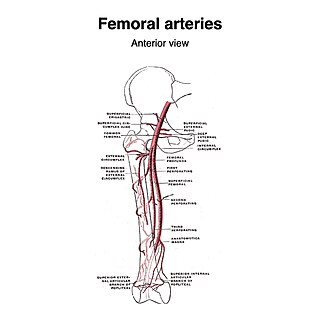
The superficial iliac circumflex artery, the smallest of the cutaneous branches of the femoral artery, arises close to the superficial epigastric artery, and, piercing the fascia lata, runs lateralward, parallel with the inguinal ligament, as far as the crest of the ilium.

The lumbar veins are four pairs of veins running along the inside of the posterior abdominal wall, and drain venous blood from parts of the abdominal wall. Each lumbar vein accompanies a single lumbar artery. The lower two pairs of lumbar veins all drain directly into the inferior vena cava, whereas the fate of the upper two pairs is more variable.
In the course of the round ligament of the liver, small paraumbilical veins are found which establish an anastomosis between the veins of the anterior abdominal wall and the portal vein, hypogastric, and iliac veins. These veins include Burrow's veins, and the veins of Sappey – superior veins of Sappey and the inferior veins of Sappey.
A portacaval anastomosis or portocaval anastomosis is a specific type of circulatory anastomosis that occurs between the veins of the portal circulation and the vena cava, thus forming one of the principal types of portasystemic anastomosis or portosystemic anastomosis, as it connects the portal circulation to the systemic circulation, providing an alternative pathway for the blood. When there is a blockage of the portal system, portocaval anastomosis enables the blood to still reach the systemic venous circulation. The inferior end of the esophagus and the superior part of the rectum are potential sites of a harmful portocaval anastomosis.
The lateral thoracic vein is a tributary of the axillary vein. It runs with the lateral thoracic artery and drains the Serratus anterior muscle and the Pectoralis major muscle.

In human anatomy, the liver is divided grossly into four parts or lobes: the right lobe, the left lobe, the caudate lobe, and the quadrate lobe. Seen from the front – the diaphragmatic surface – the liver is divided into two lobes: the right lobe and the left lobe. Viewed from the underside – the visceral surface – the other two smaller lobes, the caudate lobe and the quadrate lobe, are also visible. The two smaller lobes, the caudate lobe and the quadrate lobe, are known as superficial or accessory lobes, and both are located on the underside of the right lobe.
References
![]() This article incorporates text in the public domain from page 670 of the 20th edition of Gray's Anatomy (1918)
This article incorporates text in the public domain from page 670 of the 20th edition of Gray's Anatomy (1918)
| | This cardiovascular system article is a stub. You can help Wikipedia by expanding it. |