A tooth (pl.: teeth) is a hard, calcified structure found in the jaws (or mouths) of many vertebrates and used to break downfood. Some animals, particularly carnivores and omnivores, also use teeth to help with capturing or wounding prey, tearing food, for defensive purposes, to intimidate other animals often including their own, or to carry prey or their young. The roots of teeth are covered by gums. Teeth are not made of bone, but rather of multiple tissues of varying density and hardness that originate from the outermost embryonic germ layer, the ectoderm.
The general structure of teeth is similar across the vertebrates, although there is considerable variation in their form and position. The teeth of mammals have deep roots, and this pattern is also found in some fish, and in crocodilians. In most teleost fish, however, the teeth are attached to the outer surface of the bone, while in lizards they are attached to the inner surface of the jaw by one side. In cartilaginous fish, such as sharks, the teeth are attached by tough ligaments to the hoops of cartilage that form the jaw.[1]
Monophyodonts are animals that develop only one set of teeth, while diphyodonts grow an early set of deciduous teeth and a later set of permanent or "adult" teeth. Polyphyodonts grow many sets of teeth. For example, sharks, grow a new set of teeth every two weeks to replace worn teeth. Most extant mammals including humans are diphyodonts, but there are exceptions including elephants, kangaroos, and manatees, all of which are polyphyodonts.
Rodent incisors grow and wear away continually through gnawing, which helps maintain relatively constant length. The industry of the beaver is due in part to this qualification. Some rodents, such as voles and guinea pigs (but not mice), as well as lagomorpha (rabbits, hares and pikas), have continuously growing molars in addition to incisors.[2][3] Also, tusks (in tusked mammals) grow almost throughout life.[4]
Teeth are not always attached to the jaw, as they are in mammals. In many reptiles and fish, teeth are attached to the palate or to the floor of the mouth, forming additional rows inside those on the jaws proper. Some teleosts even have teeth in the pharynx. While not true teeth in the usual sense, the dermal denticles of sharks are almost identical in structure and are likely to have the same evolutionary origin. Indeed, teeth appear to have first evolved in sharks, and are not found in the more primitive jawless fish – while lampreys do have tooth-like structures on the tongue, these are in fact, composed of keratin, not of dentine or enamel, and bear no relationship to true teeth.[1] Though "modern" teeth-like structures with dentine and enamel have been found in late conodonts, they are now supposed to have evolved independently of later vertebrates' teeth.[5][6]
Living amphibians typically have small teeth, or none at all, since they commonly feed only on soft foods. In reptiles, teeth are generally simple and conical in shape, although there is some variation between species, most notably the venom-injecting fangs of snakes. The pattern of incisors, canines, premolars and molars is found only in mammals, and to varying extents, in their evolutionary ancestors. The numbers of these types of teeth vary greatly between species; zoologists use a standardised dental formula to describe the precise pattern in any given group.[1]
Etymology
The word tooth comes from Proto-Germanic*tanþs, derived from the Proto-Indo-European*h₁dent-, which was composed of the root *h₁ed-'to eat' plus the active participle suffix *-nt, therefore literally meaning 'that which eats'.[7]
The irregular plural form teeth is the result of Germanic umlaut whereby vowels immediately preceding a high vocalic in the following syllable were raised. As the nominative plural ending of the Proto-Germanic consonant stems (to which *tanþs belonged) was *-iz, the root vowel in the plural form *tanþiz (changed by this point to *tą̄þi via unrelated phonological processes) was raised to /œː/, and later unrounded to /eː/, resulting in the tōþ/tēþ alternation attested from Old English. Cf. also Old English bōc/bēċ'book/books' and 'mūs/mȳs''mouse/mice', from Proto-Germanic *bōks/bōkiz and *mūs/mūsiz respectively.
The genes governing tooth development in mammals are homologous to those involved in the development of fish scales.[9] Study of a tooth plate of a fossil of the extinct fish Romundina stellina showed that the teeth and scales were made of the same tissues, also found in mammal teeth, lending support to the theory that teeth evolved as a modification of scales.[10]
Teeth are among the most distinctive (and long-lasting) features of mammal species. Paleontologists use teeth to identify fossil species and determine their relationships. The shape of the animal's teeth are related to its diet. For example, plant matter is hard to digest, so herbivores have many molars for chewing and grinding. Carnivores, on the other hand, have canine teeth to kill prey and to tear meat.
Mammals, in general, are diphyodont, meaning that they develop two sets of teeth. In humans, the first set (the "baby", "milk", "primary" or "deciduous" set) normally starts to appear at about six months of age, although some babies are born with one or more visible teeth, known as neonatal teeth. Normal tooth eruption at about six months is known as teething and can be painful. Kangaroos, elephants, and manatees are unusual among mammals because they are polyphyodonts.
Aardvark
In aardvarks, teeth lack enamel and have many pulp tubules, hence the name of the order Tubulidentata.[11]
In dogs, the teeth are less likely than humans to form dental cavities because of the very high pH of dog saliva, which prevents enamel from demineralizing.[12] Sometimes called cuspids, these teeth are shaped like points (cusps) and are used for tearing and grasping food.[13]
Like human teeth, whale teeth have polyp-like protrusions located on the root surface of the tooth. These polyps are made of cementum in both species, but in human teeth, the protrusions are located on the outside of the root, while in whales the nodule is located on the inside of the pulp chamber. While the roots of human teeth are made of cementum on the outer surface, whales have cementum on the entire surface of the tooth with a very small layer of enamel at the tip. This small enamel layer is only seen in older whales where the cementum has been worn away to show the underlying enamel.[14]
The toothed whale is a suborder of the cetaceans characterized by having teeth. The teeth differ considerably among the species. They may be numerous, with some dolphins bearing over 100 teeth in their jaws. On the other hand, the narwhals have a giant unicorn-like tusk, which is a tooth containing millions of sensory pathways and used for sensing during feeding, navigation, and mating. It is the most neurologically complex tooth known. Beaked whales are almost toothless, with only bizarre teeth found in males. These teeth may be used for feeding but also for demonstrating aggression and showmanship.
In humans (and most other primates), there are usually 20 primary (also "baby" or "milk") teeth, and later up to 32 permanent teeth. Four of these 32 may be third molars or wisdom teeth, although these are not present in all adults, and may be removed surgically later in life.[15]
Among primary teeth, 10 of them are usually found in the maxilla (i.e. upper jaw) and the other 10 in the mandible (i.e. lower jaw). Among permanent teeth, 16 are found in the maxilla and the other 16 in the mandible. Most of the teeth have uniquely distinguishing features.
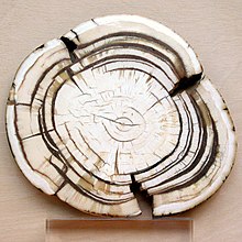
An adult horse has between 36 and 44 teeth. The enamel and dentin layers of horse teeth are intertwined.[16] All horses have 12 premolars, 12 molars, and 12 incisors.[17] Generally, all male equines also have four canine teeth (called tushes) between the molars and incisors. However, few female horses (less than 28%) have canines, and those that do usually have only one or two, which many times are only partially erupted.[18] A few horses have one to four wolf teeth, which are vestigial premolars, with most of those having only one or two. They are equally common in male and female horses and much more likely to be on the upper jaw. If present these can cause problems as they can interfere with the horse's bit contact. Therefore, wolf teeth are commonly removed.[17]
Horse teeth can be used to estimate the animal's age. Between birth and five years, age can be closely estimated by observing the eruption pattern on milk teeth and then permanent teeth. By age five, all permanent teeth have usually erupted. The horse is then said to have a "full" mouth. After the age of five, age can only be conjectured by studying the wear patterns on the incisors, shape, the angle at which the incisors meet, and other factors. The wear of teeth may also be affected by diet, natural abnormalities, and cribbing. Two horses of the same age may have different wear patterns.
A horse's incisors, premolars, and molars, once fully developed, continue to erupt as the grinding surface is worn down through chewing. A young adult horse will have teeth, which are 110–130mm (4.5–5 inches) long, with the majority of the crown remaining below the gumline in the dental socket. The rest of the tooth will slowly emerge from the jaw, erupting about 3mm (1⁄8in) each year, as the horse ages. When the animal reaches old age, the crowns of the teeth are very short and the teeth are often lost altogether. Very old horses, if lacking molars, may need to have their fodder ground up and soaked in water to create a soft mush for them to eat in order to obtain adequate nutrition.
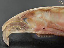
Elephants' tusks are specialized incisors for digging food up and fighting. Some elephant teeth are similar to those in manatees, and elephants are believed to have undergone an aquatic phase in their evolution.
At birth, elephants have a total of 28 molar plate-like grinding teeth not including the tusks. These are organized into four sets of seven successively larger teeth which the elephant will slowly wear through during its lifetime of chewing rough plant material. Only four teeth are used for chewing at a given time, and as each tooth wears out, another tooth moves forward to take its place in a process similar to a conveyor belt. The last and largest of these teeth usually becomes exposed when the animal is around 40 years of age, and will often last for an additional 20 years. When the last of these teeth has fallen out, regardless of the elephant's age, the animal will no longer be able to chew food and will die of starvation.[19][20]
Rabbit
Rabbits and other lagomorphs usually shed their deciduous teeth before (or very shortly after) their birth, and are usually born with their permanent teeth.[21] The teeth of rabbits complement their diet, which consists of a wide range of vegetation. Since many of the foods are abrasive enough to cause attrition, rabbit teeth grow continuously throughout life.[22] Rabbits have a total of six incisors, three upper premolars, three upper molars, two lower premolars, and two lower molars on each side. There are no canines. Dental formula is 2.0.3.31.0.2.3 = 28. Three to four millimeters of the tooth is worn away by incisors every week, whereas the cheek teeth require a month to wear away the same amount.[23]
The incisors and cheek teeth of rabbits are called aradicular hypsodont teeth. This is sometimes referred to as an elodent dentition. These teeth grow or erupt continuously. The growth or eruption is held in balance by dental abrasion from chewing a diet high in fiber.
Buccal view of top incisor from Rattus rattus. Top incisor outlined in yellow. Molars circled in blue.Buccal view of the lower incisor from the right dentary of a Rattus rattusLingual view of the lower incisor from the right dentary of a Rattus rattusMidsagittal view of top incisor from Rattus rattus. Top incisor outlined in yellow. Molars circled in blue.
Rodents
Rodents have upper and lower hypselodont incisors that can continuously grow enamel throughout its life without having properly formed roots.[24] These teeth are also known as aradicular teeth, and unlike humans whose ameloblasts die after tooth development, rodents continually produce enamel, they must wear down their teeth by gnawing on various materials.[25] Enamel and dentin are produced by the enamel organ, and growth is dependent on the presence of stem cells, cellular amplification, and cellular maturation structures in the odontogenic region.[26] Rodent incisors are used for cutting wood, biting through the skin of fruit, or for defense. This allows for the rate of wear and tooth growth to be at equilibrium.[24] The microstructure of rodent incisor enamel has shown to be useful in studying the phylogeny and systematics of rodents because of its independent evolution from the other dental traits. The enamel on rodent incisors are composed of two layers: the inner portio interna (PI) with Hunter-Schreger bands (HSB) and an outer portio externa (PE) with radial enamel (RE).[27] It usually involves the differential regulation of the epithelialstem cell niche in the tooth of two rodent species, such as guinea pigs.[28][29]
Lingual view of top incisor from Rattus rattus. Top incisor outlined in yellow. Molars circled in blue.
The teeth have enamel on the outside and exposed dentin on the inside, so they self-sharpen during gnawing. On the other hand, continually growing molars are found in some rodent species, such as the sibling vole and the guinea pig.[28][29] There is variation in the dentition of the rodents, but generally, rodents lack canines and premolars, and have a space between their incisors and molars, called the diastema region.
Manatee
Manatees are polyphyodont with mandibular molars developing separately from the jaw and are encased in a bony shell separated by soft tissue.[30][31]
Fish, such as sharks, may go through many teeth in their lifetime. The replacement of multiple teeth is known as polyphyodontia.
A class of prehistoric shark are called cladodonts for their strange forked teeth.
Unlike the continuous shedding of functional teeth seen in modern sharks,[33][34] the majority of stemchondrichthyan lineages retained all tooth generations developed throughout the life of the animal.[35] This replacement mechanism is exemplified by the tooth whorl-based dentitions of acanthodians,[36] which include the oldest known toothed vertebrate, Qianodusduplicis[37].
Amphibians
All amphibians have pedicellate teeth, which are modified to be flexible due to connective tissue and uncalcified dentine that separates the crown from the base of the tooth.[38]
Most amphibians exhibit teeth that have a slight attachment to the jaw or acrodont teeth. Acrodont teeth exhibit limited connection to the dentary and have little enervation.[39] This is ideal for organisms who mostly use their teeth for grasping, but not for crushing and allows for rapid regeneration of teeth at a low energy cost. Teeth are usually lost in the course of feeding if the prey is struggling. Additionally, amphibians that undergo a metamorphosis develop bicuspid shaped teeth.[40]
Reptiles
The teeth of reptiles are replaced constantly throughout their lives. Crocodilian juveniles replace teeth with larger ones at a rate as high as one new tooth per socket every month. Once mature, tooth replacement rates can slow to two years and even longer. Overall, crocodilians may use 3,000 teeth from birth to death. New teeth are created within old teeth.[41]
A skull of Ichthyornis discovered in 2014 suggests that the beak of birds may have evolved from teeth to allow chicks to escape their shells earlier, and thus avoid predators and also to penetrate protective covers such as hard earth to access underlying food.[42][43]
Invertebrates
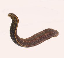
The European medicinal leech has three jaws with numerous sharp teeth which function like little saws for incising a host.
True teeth are unique to vertebrates,[44] although many invertebrates have analogous structures often referred to as teeth. The organisms with the simplest genome bearing such tooth-like structures are perhaps the parasitic worms of the family Ancylostomatidae.[45] For example, the hookworm Necator americanus has two dorsal and two ventral cutting plates or teeth around the anterior margin of the buccal capsule. It also has a pair of subdorsal and a pair of subventral teeth located close to the rear.[46]
Historically, the European medicinal leech, another invertebrate parasite, has been used in medicine to remove blood from patients.[47] They have three jaws (tripartite) that resemble saws in both appearance and function, and on them are about 100 sharp teeth used to incise the host. The incision leaves a mark that is an inverted Y inside of a circle. After piercing the skin and injecting anticoagulants (hirudin) and anaesthetics, they suck out blood, consuming up to ten times their body weight in a single meal.[48]
In some species of Bryozoa, the first part of the stomach forms a muscular gizzard lined with chitinous teeth that crush armoured prey such as diatoms. Wave-like peristaltic contractions then move the food through the stomach for digestion.[49]
The limpet rasps algae from rocks using teeth with the strongest known tensile strength of any biological material.
Molluscs have a structure called a radula, which bears a ribbon of chitinous teeth. However, these teeth are histologically and developmentally different from vertebrate teeth and are unlikely to be homologous. For example, vertebrate teeth develop from a neural crestmesenchyme-derived dental papilla, and the neural crest is specific to vertebrates, as are tissues such as enamel.[44]
The radula is used by molluscs for feeding and is sometimes compared rather inaccurately to a tongue. It is a minutely toothed, chitinous ribbon, typically used for scraping or cutting food before the food enters the oesophagus. The radula is unique to molluscs, and is found in every class of mollusc apart from bivalves.
Within the gastropods, the radula is used in feeding by both herbivorous and carnivorous snails and slugs. The arrangement of teeth (also known as denticles) on the radula ribbon varies considerably from one group to another as shown in the diagram on the left.
Predatory marine snails such as the Naticidae use the radula plus an acidic secretion to bore through the shell of other molluscs. Other predatory marine snails, such as the Conidae, use a specialized radula tooth as a poisoned harpoon. Predatory pulmonate land slugs, such as the ghost slug, use elongated razor-sharp teeth on the radula to seize and devour earthworms. Predatory cephalopods, such as squid, use the radula for cutting prey.
In most of the more ancient lineages of gastropods, the radula is used to graze by scraping diatoms and other microscopic algae off rock surfaces and other substrates. Limpets scrape algae from rocks using radula equipped with exceptionally hard rasping teeth.[50] These teeth have the strongest known tensile strength of any biological material, outperforming spider silk.[50] The mineral protein of the limpet teeth can withstand a tensile stress of 4.9GPa, compared to 4GPa of spider silk and 0.5GPa of human teeth.[51]
Fossilization and taphonomy
Because teeth are very resistant, often preserved when bones are not,[52] and reflect the diet of the host organism, they are very valuable to archaeologists and palaeontologists.[53] Early fish such as the thelodonts had scales composed of dentine and an enamel-like compound, suggesting that the origin of teeth was from scales which were retained in the mouth. Fish as early as the late Cambrian had dentine in their exoskeletons, which may have functioned in defense or for sensing their environments.[54] Dentine can be as hard as the rest of teeth and is composed of collagen fibres, reinforced with hydroxyapatite.[54]
Though teeth are very resistant, they also can be brittle and highly susceptible to cracking.[55] However, cracking of the tooth can be used as a diagnostic tool for predicting bite force. Additionally, enamel fractures can also give valuable insight into the diet and behaviour of archaeological and fossil samples.
Decalcification removes the enamel from teeth and leaves only the organic interior intact, which comprises dentine and cementine.[56] Enamel is quickly decalcified in acids,[57] perhaps by dissolution by plant acids or via diagenetic solutions, or in the stomachs of vertebrate predators.[56] Enamel can be lost by abrasion or spalling,[56] and is lost before dentine or bone are destroyed by the fossilisation process.[57] In such a case, the 'skeleton' of the teeth would consist of the dentine, with a hollow pulp cavity.[56] The organic part of dentine, conversely, is destroyed by alkalis.[57]
Horse teeth refers to the dentition of equine species, including horses and donkeys. Equines are both heterodontous and diphyodontous, which means that they have teeth in more than one shape, and have two successive sets of teeth, the deciduous and permanent sets.
Human teeth function to mechanically break down items of food by cutting and crushing them in preparation for swallowing and digesting. As such, they are considered part of the human digestive system. Humans have four types of teeth: incisors, canines, premolars, and molars, which each have a specific function. The incisors cut the food, the canines tear the food and the molars and premolars crush the food. The roots of teeth are embedded in the maxilla or the mandible and are covered by gums. Teeth are made of multiple tissues of varying density and hardness.
Dentition pertains to the development of teeth and their arrangement in the mouth. In particular, it is the characteristic arrangement, kind, and number of teeth in a given species at a given age. That is, the number, type, and morpho-physiology of the teeth of an animal.
The molars or molar teeth are large, flat teeth at the back of the mouth. They are more developed in mammals. They are used primarily to grind food during chewing. The name molar derives from Latin, molaris dens, meaning "millstone tooth", from mola, millstone and dens, tooth. Molars show a great deal of diversity in size and shape across the mammal groups. The third molar of humans is sometimes vestigial.
Tusks are elongated, continuously growing front teeth that protrude well beyond the mouth of certain mammal species. They are most commonly canine teeth, as with narwhals, chevrotains, musk deer, water deer, muntjac, pigs, peccaries, hippopotamuses and walruses, or, in the case of elephants, elongated incisors. Tusks share common features such as extra-oral position, growth pattern, composition and structure, and lack of contribution to ingestion. Tusks are thought to have adapted to the extra-oral environments, like dry or aquatic or arctic. In most tusked species both the males and the females have tusks although the males' are larger. Most mammals with tusks have a pair of them growing out from either side of the mouth. Tusks are generally curved and have a smooth, continuous surface. The male narwhal's straight single helical tusk, which usually grows out from the left of the mouth, is an exception to the typical features of tusks described above. Continuous growth of tusks is enabled by formative tissues in the apical openings of the roots of the teeth.
Incisors are the front teeth present in most mammals. They are located in the premaxilla above and on the mandible below. Humans have a total of eight. Opossums have 18, whereas armadillos have none.
Gigantopithecus is an extinct genus of ape that lived in southern China from 2 million to approximately 300-200,000 years ago during the Early to Middle Pleistocene, represented by one species, Gigantopithecus blacki. Potential identifications have also been made in Thailand, Vietnam, and Indonesia. The first remains of Gigantopithecus, two third molar teeth, were identified in a drugstore by anthropologist Ralph von Koenigswald in 1935, who subsequently described the ape. In 1956, the first mandible and more than 1,000 teeth were found in Liucheng, and numerous more remains have since been found in at least 16 sites. Only teeth and four mandibles are known currently, and other skeletal elements were likely consumed by porcupines before they could fossilise. Gigantopithecus was once argued to be a hominin, a member of the human line, but it is now thought to be closely allied with orangutans, classified in the subfamily Ponginae.
Hypodontia is defined as the developmental absence of one or more teeth excluding the third molars. It is one of the most common dental anomalies, and can have a negative impact on function, and also appearance. It rarely occurs in primary teeth and the most commonly affected are the adult second premolars and the upper lateral incisors. It usually occurs as part of a syndrome that involves other abnormalities and requires multidisciplinary treatment.
Tooth development or odontogenesis is the complex process by which teeth form from embryonic cells, grow, and erupt into the mouth. For human teeth to have a healthy oral environment, all parts of the tooth must develop during appropriate stages of fetal development. Primary (baby) teeth start to form between the sixth and eighth week of prenatal development, and permanent teeth begin to form in the twentieth week. If teeth do not start to develop at or near these times, they will not develop at all, resulting in hypodontia or anodontia.
A polyphyodont is any animal whose teeth are continually replaced. In contrast, diphyodonts are characterized by having only two successive sets of teeth.
Tooth development or odontogenesis is the process in which teeth develop and grow into the mouth. Tooth development varies among species.
Dental anatomy is a field of anatomy dedicated to the study of human tooth structures. The development, appearance, and classification of teeth fall within its purview. Tooth formation begins before birth, and the teeth's eventual morphology is dictated during this time. Dental anatomy is also a taxonomical science: it is concerned with the naming of teeth and the structures of which they are made, this information serving a practical purpose in dental treatment.
A cusp is a pointed, projecting, or elevated feature. In animals, it is usually used to refer to raised points on the crowns of teeth. The concept is also used with regard to the leaflets of the four heart valves. The mitral valve, which has two cusps, is also known as the bicuspid valve, and the tricuspid valve has three cusps.
Post-canine megadontia is a relative enlargement of the molars and premolars compared to the size of the incisors and canines. This phenomenon is seen in some early hominid ancestors such as Paranthropus aethiopicus.
Eritherium is an extinct genus of early Proboscidea found in the Ouled Abdoun basin, Morocco. It lived about 60 million years ago. It was first named by Emmanuel Gheerbrant in 2009 and the type species is Eritherium azzouzorum. Eritherium is the oldest, smallest and most primitive known elephant relative.
Teeth are common to most vertebrates, but mammalian teeth are distinctive in having a variety of shapes and functions. This feature first arose among early therapsids during the Permian, and has continued to the present day. All therapsid groups with the exception of the mammals are now extinct, but each of these groups possessed different tooth patterns, which aids with the classification of fossils.
The infundibulum of a tooth is the funnel-like center that is filled with cementum. The funnel is widest at the top (crown) which is the grinding (occlusal) surface. The infundibulum is also known as the dental cup. Simple tooth infundibula occur most notably in the incisors of horses and other equids, but they also occur in the premolars and molars of ruminants and camelids. The infundibula found in ruminants can get quite complex some with two funneling centers, and with multiple folding in the sides of the cup. These folds produce greater amounts of enamel in vertical curtains that substantially increase the durability of the tooth. The cheek teeth of elephants express this in a slightly different form with the vertical curtains of enamel coming in from the sides and meeting in the middle.
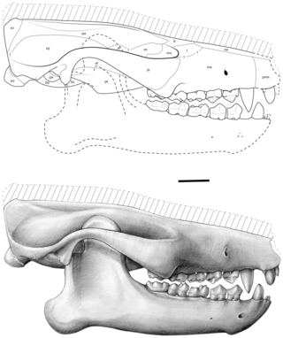
Ocepeia is an extinct genus of afrotherian mammal that lived in present-day Morocco during the middle Paleocene epoch, approximately 60 million years ago. First named and described in 2001, the type species is O. daouiensis from the Selandian stage of Morocco's Ouled Abdoun Basin. A second, larger species, O. grandis, is known from the Thanetian, a slightly younger stage in the same area. In life, the two species are estimated to have weighed about 3.5 kg (7.7 lb) and 10 kg (22 lb), respectively, and are believed to have been specialized leaf-eaters. The fossil skulls of Ocepeia are the oldest known afrotherian skulls, and the best-known of any Paleocene mammal in Africa.
Changes to the dental morphology and jaw are major elements of hominid evolution. These changes were driven by the types and processing of food eaten. The evolution of the jaw is thought to have facilitated encephalization, speech, and the formation of the uniquely human chin.
Azygonyx was a small tillodont mammal, likely the size of a cat to raccoon, that lived in North America during the Paleocene and Eocene in the early part of the Cenozoic Era. The only fossils that have been recovered are from the Willwood and Fort Union Formations in the Bighorn Basin of Wyoming, United States, and date to the Clarkforkian to Wasatchian, about 56 to 50 million years ago. Fifty-six collections that have been recovered thus far include the remains of Azygonyx. Azygonyx survived the Paleocene Eocene Thermal Maximum along with other mammals like Phenacodus and Ectocion, both of which were ground-dwelling mammals. Azygonyx probably was a generalist terrestrial mammal that may have roamed around the ground, but was also capable of climbing trees.
References
1 2 3 Romer, Alfred Sherwood; Parsons, Thomas S. (1977). The Vertebrate Body. Philadelphia, PA: Holt-Saunders International. pp.300–310. ISBN978-0-03-910284-5.
1 2 Cox, Philip; Hautier, Lionel (2015). Evolution of the Rodents: Advances in Phylogeny, Functional Morphology and Development. Cambridge University Press. p.482. ISBN9781107044333.
↑ Caceci, Thomas. Veterinary Histology with subtitle "Digestive System: Oral Cavity" found hereArchived 2006-04-30 at the Wayback Machine .
1 2 Tummers M and Thesleff I. Root or crown: a developmental choice orchestrated by the differential regulation of the epithelial stem cell niche in the tooth of two rodent species. Development (2003). 130(6):1049-57.
1 2 AM Hunt. A description of the molar teeth and investing tissues of normal guinea pigs. J Dent Res. (1959) 38(2):216-31.
↑ Shoshani, J., ed. (2000). Elephants: Majestic Creatures of the Wild. Checkmark Books. ISBN0-87596-143-6.
↑ Kardong, Kenneth (1995). Vertebrate: Comparative Anatomy, Function, Evolution. New York: McGraw-HIll. pp. 215–225. ISBN9780078023026.
↑ Xiong, Jianli (2014). "Comparison of vomerine tooth rows in juvenile and adult Hynobius guabangshanensis". Vertebrate Zoology. 64: 215–220.
↑ Poole, D. F. G. (January 1961). "Notes on Tooth Replacement in the Nile Crocodile Crocodilus niloticus". Proceedings of the Zoological Society of London. 136 (1): 131–140. doi:10.1111/j.1469-7998.1961.tb06083.x.
↑ Roberts, Larry S., and John Janovy, Jr. Foundations of Parasitology. Seventh ed. Singapore: McGraw-Hill, 2006.
↑ Brian Payton (1981). Kenneth Muller; John Nicholls; Gunther Stent (eds.). Neurobiology of the Leech. New York: Cold Spring Harbor Laboratory. pp.27–34. ISBN978-0-87969-146-2.
1 2 Asa H. Barber; Dun Lu; Nicola M. Pugno (18 February 2015), "Extreme strength observed in limpet teeth", Journal of the Royal Society Interface, 12 (105): 20141326, doi:10.1098/rsif.2014.1326, PMC4387522, PMID25694539
1 2 Teaford, Mark F and Smith, Moya Meredith, 2007. Development, Function and Evolution of Teeth, Cambridge University Press. ISBN978-0-521-03372-5, Chapter 5.
1 2 3 4 Fisher, Daniel C (1981). "Taphonomic Interpretation of Enamel-Less Teeth in the Shotgun Local Fauna (Paleocene, Wyoming)". Museum of Paleontology Contributions, the University of Michigan. 25 (13): 259–275. hdl:2027.42/48503.
Shoshani, Jeheskel (2002). "Tubulidentata". In Robertson, Sarah (ed.). Encyclopedia of Life Sciences. Vol.18: Svedberg, Theodor to Two-hybrid and Related Systems. London, UK: Nature Publishing Group. ISBN978-1-56159-274-6.
This page is based on this Wikipedia article Text is available under the CC BY-SA 4.0 license; additional terms may apply. Images, videos and audio are available under their respective licenses.