Tunicamycin

Last updated
Tunicamycin
Tunicamycin.svg
Names
IUPAC name
(E)-N-[(2S,3R,4R,5R,6R)-2-[(2R,3R,4R,5S,6R)-

3-acetamido-4,5-dihydroxy-6-(hydroxymethyl)oxan-2-yl]oxy- 6-[2-[(2R,3S,4R,5R)-5-(2,4-dioxopyrimidin-1-yl)- 3,4-dihydroxyoxolan-2-yl]-2-hydroxyethyl]-4,5-dihydroxyoxan-

3-yl]-5-methylhex-2-enamide

Contents

Other names
NSC 177382
Identifiers
3D model (JSmol)
ChEMBL
ChemSpider
ECHA InfoCard 100.115.295 OOjs UI icon edit-ltr-progressive.svg
EC Number
  • 601-012-4
MeSH Tunicamycin
PubChem CID
UNII
  • InChI=1S/C30H46N4O16/c1-11(2)5-4-6-16(38)32-19-23(43)20(40)14(47-29(19)50-28-18(31-12(3)36)22(42)21(41)15(10-35)48-28)9-13(37)26-24(44)25(45)27(49-26)34-8-7-17(39)33-30(34)46/h4,6-8,11,13-15,18-29,35,37,40-45H,5,9-10H2,1-3H3,(H,31,36)(H,32,38)(H,33,39,46)/b6-4+/t13?,14-,15-,18-,19-,20+,21-,22-,23-,24+,25-,26-,27-,28-,29+/m1/s1
    Key: ZHSGGJXRNHWHRS-VIDYELAYSA-N
  • CC(C)C\C=C\C(=O)N[C@@H]1[C@H]([C@H]([C@H](O[C@H]1O[C@@H]2[C@@H]([C@H]([C@@H]([C@H](O2)CO)O)O)NC(=O)C)CC([C@@H]3[C@H]([C@H]([C@@H](O3)N4C=CC(=O)NC4=O)O)O)O)O)O
Properties
C39H64N4O16
Molar mass N/A
Hazards
GHS labelling: [1]
GHS-pictogram-skull.svg
Danger
H300
Except where otherwise noted, data are given for materials in their standard state (at 25 °C [77 °F], 100 kPa).
X mark.svgN  verify  (what is  Yes check.svgYX mark.svgN ?)

Tunicamycin is a mixture of homologous nucleoside antibiotics that inhibits the UDP-HexNAc: polyprenol-P HexNAc-1-P family of enzymes. In eukaryotes, this includes the enzyme GlcNAc phosphotransferase (GPT), which catalyzes the transfer of N-acetylglucosamine-1-phosphate from UDP-N-acetylglucosamine to dolichol phosphate in the first step of glycoprotein synthesis. Tunicamycin blocks N-linked glycosylation (N-glycans) and treatment of cultured human cells with tunicamycin causes cell cycle arrest in G1 phase. It is used as an experimental tool in biology, e.g. to induce unfolded protein response. [2] Tunicamycin is produced by several bacteria, including Streptomyces clavuligerus and Streptomyces lysosuperificus .

Tunicamycin homologues have varying molecular weights owing to the variability in fatty acid side chain conjugates. [3]

Biosynthesis

The biosynthesis of tunicamycins was studied in Streptomyces chartreusis and a proposed biosynthetic pathway was characterized. The bacteria utilize the enzymes in the tun gene cluster (TunA-N) to make tunicamycins. [4]

TunA uses the starter unit uridine diphosphate-N-acetyl-glucosamine (UDP-GlcNAc) and catalyzes the dehydration of the 6’ hydroxyl group. First, a Tyr residue in TunA abstracts a proton from the 4’ hydroxyl group, forming a ketone at that position. A hydride is subsequently abstracted from the 4’ carbon by NAD+, forming NADH. The ketone is stabilized by hydrogen bonding from the Tyr residue, and a nearby Thr residue. A glutamate residue then abstracts a proton from the 5’ carbon, pushing the electrons up to form a double bond between the 5’ and 6’ carbon. A nearby cysteine donates a proton to the hydroxyl group as it leaves as water. NADH donates a hydride to the 4’ carbon, reforming a hydroxide in that position and forming UDP-6’-deoxy-5-6-ene-GlcNAc. TunF then catalyzes the epimerization of the intermediate to UDP-6’-deoxy-5-6-ene-GalNAc, changing the 4’ hydroxyl from the equatorial to axial position. [5]

The other starter unit for tunicamycin is uridine, which is produced from uridine triphosphate (UTP). TunN is a nucleotide diphosphatase, and catalyzes the removal of pyrophosphate from UTP to form uridine monophosphate. The last phosphate is removed by the putative monophosphatase, TunG.

Once uridine and UDP-6’-deoxy-5-6-ene-GalNAc are produced, TunB catalyzes their linkage at the 6’ carbon of UDP-6’-deoxy-5-6-ene-GalNAc. TunB uses S-adenyslmethionine (SAM) to form a radical on the 5’ carbon of the ribose on uracil. TunM is thought to catalyze the formation of a new bond between the 5’ carbon of uridine and the 6’ carbon of UDP-6’-deoxy-5-6-ene-GalNAc using the electron from the uridine radical and one of the electrons from the double bond of UDP-6’-deoxy-5-6-ene-GalNAc. The radical on UDP-6’-deoxy-5-6-ene-GalNAc is then quenched by abstracting a hydrogen from SAM. [6] The resulting molecule is UDP-N-acetyl-tunicamine. TunH then catalyzes the hydrolysis of UDP from UDP-N-acetyl-tunicamine. Another molecule of UDP-GlcNAc is introduced, and a β-1,1 glycosidic bond is subsequently formed, catalyzed by TunD. The resulting molecule is deacetylated by TunE. TunL and a fatty acyl-ACP ligase are used to load metabolic fatty acids onto the acyl carrier protein, TunK. TunC then attaches the fatty acid to the free amine, producing tunicamycin.

See also

Related Research Articles

Peptidoglycan or murein is a unique large macromolecule, a polysaccharide, consisting of sugars and amino acids that forms a mesh-like peptidoglycan layer (sacculus) that surrounds the bacterial cytoplasmic membrane. The sugar component consists of alternating residues of β-(1,4) linked N-acetylglucosamine (NAG) and N-acetylmuramic acid (NAM). Attached to the N-acetylmuramic acid is an oligopeptide chain made of three to five amino acids. The peptide chain can be cross-linked to the peptide chain of another strand forming the 3D mesh-like layer. Peptidoglycan serves a structural role in the bacterial cell wall, giving structural strength, as well as counteracting the osmotic pressure of the cytoplasm. This repetitive linking results in a dense peptidoglycan layer which is critical for maintaining cell form and withstanding high osmotic pressures, and it is regularly replaced by peptidoglycan production. Peptidoglycan hydrolysis and synthesis are two processes that must occur in order for cells to grow and multiply, a technique carried out in three stages: clipping of current material, insertion of new material, and re-crosslinking of existing material to new material.

In enzymology, an UDP-N-acetylglucosamine 6-dehydrogenase (EC 1.1.1.136) is an enzyme that catalyzes the chemical reaction

<span class="mw-page-title-main">UDP-N-acetylmuramate dehydrogenase</span> Class of enzymes

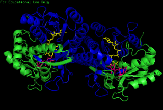
In enzymology, an UDP-N-acetylmuramate dehydrogenase (EC 1.3.1.98) is an enzyme that catalyzes the chemical reaction

Uridine diphosphate <i>N</i>-acetylglucosamine Chemical compound

Uridine diphosphate N-acetylglucosamine or UDP-GlcNAc is a nucleotide sugar and a coenzyme in metabolism. It is used by glycosyltransferases to transfer N-acetylglucosamine residues to substrates. D-Glucosamine is made naturally in the form of glucosamine-6-phosphate, and is the biochemical precursor of all nitrogen-containing sugars. To be specific, glucosamine-6-phosphate is synthesized from fructose 6-phosphate and glutamine as the first step of the hexosamine biosynthesis pathway. The end-product of this pathway is UDP-GlcNAc, which is then used for making glycosaminoglycans, proteoglycans, and glycolipids.

<span class="mw-page-title-main">UDP-glucose 4-epimerase</span> Class of enzymes

The enzyme UDP-glucose 4-epimerase, also known as UDP-galactose 4-epimerase or GALE, is a homodimeric epimerase found in bacterial, fungal, plant, and mammalian cells. This enzyme performs the final step in the Leloir pathway of galactose metabolism, catalyzing the reversible conversion of UDP-galactose to UDP-glucose. GALE tightly binds nicotinamide adenine dinucleotide (NAD+), a co-factor required for catalytic activity.

<span class="mw-page-title-main">UDP-N-acetylglucosamine 2-epimerase</span> Class of enzymes

In enzymology, an UDP-N-acetylglucosamine 2-epimerase is an enzyme that catalyzes the chemical reaction

Nucleotide sugars are the activated forms of monosaccharides. Nucleotide sugars act as glycosyl donors in glycosylation reactions. Those reactions are catalyzed by a group of enzymes called glycosyltransferases.

<span class="mw-page-title-main">N-acetylglucosamine-6-phosphate deacetylase</span>

In enzymology, N-acetylglucosamine-6-phosphate deacetylase (EC 3.5.1.25), also known as GlcNAc-6-phosphate deacetylase or NagA, is an enzyme that catalyzes the deacetylation of N-acetylglucosamine-6-phosphate (GlcNAc-6-P) to glucosamine-6-phosphate (GlcN-6-P):

<span class="mw-page-title-main">UDP-N-acetylglucosamine 1-carboxyvinyltransferase</span> Class of enzymes

In enzymology, an UDP-N-acetylglucosamine 1-carboxyvinyltransferase is an enzyme that catalyzes the first committed step in peptidoglycan biosynthesis of bacteria:

In enzymology, a N-acetyllactosaminide 3-alpha-galactosyltransferase is an enzyme that catalyzes the chemical reaction

In enzymology, a N-acetyllactosaminide beta-1,3-N-acetylglucosaminyltransferase is an enzyme that catalyzes the chemical reaction

In enzymology, a protein N-acetylglucosaminyltransferase is an enzyme that catalyzes the chemical reaction

In enzymology, an UDP-N-acetylglucosamine—dolichyl-phosphate N-acetylglucosaminephosphotransferase is an enzyme that catalyzes the chemical reaction

<span class="mw-page-title-main">UDP-N-acetylglucosamine diphosphorylase</span> Class of enzymes

In enzymology, an UDP-N-acetylglucosamine diphosphorylase is an enzyme that catalyzes the chemical reaction

<span class="mw-page-title-main">GALNT3</span> Protein-coding gene in the species Homo sapiens

Polypeptide N-acetylgalactosaminyltransferase 3 is an enzyme that in humans is encoded by the GALNT3 gene.

<span class="mw-page-title-main">DPAGT1</span> Protein-coding gene in the species Homo sapiens

UDP-N-acetylglucosamine—dolichyl-phosphate N-acetylglucosaminephosphotransferase is an enzyme that in humans is encoded by the DPAGT1 gene.

<span class="mw-page-title-main">GALNT2</span> Protein-coding gene in the species Homo sapiens

Polypeptide N-acetylgalactosaminyltransferase 2 is an enzyme that in humans is encoded by the GALNT2 gene.

<span class="mw-page-title-main">Ribostamycin</span> Aminoglycoside antibiotic

Ribostamycin is an aminoglycoside-aminocyclitol antibiotic isolated from a streptomycete, Streptomyces ribosidificus, originally identified in a soil sample from Tsu City of Mie Prefecture in Japan. It is made up of 3 ring subunits: 2-deoxystreptamine (DOS), neosamine C, and ribose. Ribostamycin, along with other aminoglycosides with the DOS subunit, is an important broad-spectrum antibiotic with important use against human immunodeficiency virus and is considered a critically important antimicrobial by the World Health Organization., Resistance against aminoglycoside antibiotics, such as ribostamycin, is a growing concern. The resistant bacteria contain enzymes that modify the structure through phosphorylation, adenylation, and acetylation and prevent the antibiotic from being able to interact with the bacterial ribosomal RNAs.

O-linked glycosylation is the attachment of a sugar molecule to the oxygen atom of serine (Ser) or threonine (Thr) residues in a protein. O-glycosylation is a post-translational modification that occurs after the protein has been synthesised. In eukaryotes, it occurs in the endoplasmic reticulum, Golgi apparatus and occasionally in the cytoplasm; in prokaryotes, it occurs in the cytoplasm. Several different sugars can be added to the serine or threonine, and they affect the protein in different ways by changing protein stability and regulating protein activity. O-glycans, which are the sugars added to the serine or threonine, have numerous functions throughout the body, including trafficking of cells in the immune system, allowing recognition of foreign material, controlling cell metabolism and providing cartilage and tendon flexibility. Because of the many functions they have, changes in O-glycosylation are important in many diseases including cancer, diabetes and Alzheimer's. O-glycosylation occurs in all domains of life, including eukaryotes, archaea and a number of pathogenic bacteria including Burkholderia cenocepacia, Neisseria gonorrhoeae and Acinetobacter baumannii.

Protein <i>O</i>-GlcNAc transferase Protein-coding gene in the species Homo sapiens

Protein O-GlcNAc transferase also known as OGT or O-linked N-acetylglucosaminyltransferase is an enzyme that in humans is encoded by the OGT gene. OGT catalyzes the addition of the O-GlcNAc post-translational modification to proteins.

References

  1. "C&L Inventory". echa.europa.eu.
  2. Chan SW, Egan PA (2005). "Hepatitis C virus envelope proteins regulate CHOP via induction of the unfolded protein response". The FASEB Journal. 19 (11): 1510–1512. doi: 10.1096/fj.04-3455fje . PMID   16006626.
  3. Tunicamycin product details]
  4. Wyszynski F, Hesketh A, Bibb M, Davis B (2010). "Dissecting tunicamycin biosynthesis by genome mining: cloning and heterologous expression of a minimal gene cluster". Chemical Science. 1 (5): 581. doi:10.1039/c0sc00325e.
  5. Wyszynski F, Lee S, Yabe T, Wang H, Gomez-Escribano JP, Bibb M (July 2012). "Biosynthesis of the tunicamycin antibiotics proceeds via unique exo-glycal intermediates". Nature Chemistry. 4 (7): 539–546. Bibcode:2012NatCh...4..539W. doi:10.1038/nchem.1351. PMID   22717438.
  6. Giese B (August 1989). "The Stereoselectivity of Intermolecular Free Radical Reactions [New Synthetic Methods (78)]". Angewandte Chemie International Edition in English. 28 (8): 969–980. doi:10.1002/anie.198909693.