Unique hues

Last updated
A concept of four unique hues of psychologist Charles Hubbard Judd (1917) Opponent color circle 1917.png
A concept of four unique hues of psychologist Charles Hubbard Judd (1917)

Unique hue is a term used in perceptual psychology of color vision and generally applied to the purest hues of blue, green, yellow and red. The proponents of the opponent process theory believe that these hues cannot be described as a mixture of other hues, and are therefore pure, whereas all other hues are composite. [1] The neural correlate of the unique hues are approximated by the extremes of the opponent channels in opponent process theory. [2] In this context, unique hues are sometimes described as "psychological primaries" as they can be considered analogous to the primary colors of trichromatic color theory. [3] [4]

Contents

Opponent Process Theory

Approximations within the sRGB gamut to the "aim colors" of the Natural Color System, a model based on the opponent process theory of color vision. Opponent colors.svg
Approximations within the sRGB gamut to the "aim colors" of the Natural Color System, a model based on the opponent process theory of color vision.

The concept of certain hues as 'unique' came with the introduction of opponent process theory, which Ewald Hering introduced in 1878. [5] [6] Hering first proposed the idea that red, green, blue, and yellow were unique hues ("Urfarben"), based on the concept that these colors could not be simultaneously perceived. These hues represented the extremes of two perpendicular axes of color: a red-green axis and a blue-yellow axis. While this theory with 4 unique hues was initially considered contradictory to the Young-Helmholtz trichromatic theory's three primary colors, the two theories were reconciled theoretically by Erwin Schrödinger [7] and the later discovery of color-opponent cells in the retina and lateral geniculate nucleus (LGN) related the two theories physiologically. [8]

Physiology

Diagram of the opponent process Diagram of the opponent process.png
Diagram of the opponent process
Signal path from the eyes to the LGN. Gray722.png
Signal path from the eyes to the LGN.

A physiological pathway from the cones in the retina to a neural correlate for the psychological unique hues has been elusive. [9] Mollon and Jordan state: “...the nature of the unique hues remains mysterious and we do not know whether they tell us anything about the neural organisation of the visual system.” [10] The first transformation of light to a neuronal signal (visual phototransduction) yields 3 channels, each proportional to the quantal catch of one cone type (L-, M- and S-), estimated by the LMS color space. The second transformation occurs in the color-opponent cells and produces the opponent process channels: L+M (luminance), L-M (red-green), and S-(L+M) (blue-yellow), the latter of which form the cardinal axes. [9]

Hering and researchers until the mid 20th century expected that the cardinal axes would correspond to the unique hues, i.e. the unique hues would exist when one opponent channel is maximally stimulated and the other opponent channel is in equilibrium. [5] [4] However, subsequent psychophysical tests demonstrated that while unique red lies on the extreme of the L-M axis, the other unique hues do not lie on the extremes of either opponent channel (L-M and S-(L+M) axes). [11] [12] Therefore, the cardinal axes are not a direct correlate of our experience of unique hues and a further (third) transformation must be applied to identify correlates, i.e. each unique hue is a synthesis of the opponent process channels. One theory suggests a conversion at a point later than the LGN, and that this produces non-linear combinations resulting in our experience of color being non-linear to the cardinal axes. [13] However, while opponent-cells have been found in the LGN that respond to cone combinations other than those of the cardinal axes, such as M-S, [14] there is no physiological understanding of this third transformation. An opposing theory therefore suggests that hues are learned based on variations in the visual environment; that unique hues represent an adaptation away from the cardinal axes and unique hues cannot be explained by relative numbers of excited L- and M-cones or their sensitivities. [10] [15]

There is mixed evidence as to whether unique hues are perceptually privileged compared to other colors. Some research suggests that there is no greater sensitivity for unique hues compared to other colors, [16] but other evidence suggests there is greater sensitivity for yellows and blues, which may be due to them coinciding with the daylight locus. [17] There is no direct evidence that larger populations of neurons are dedicated to unique hues compared to other colors, but some EEG research suggests that the latency of some EEG components may be shorter for unique hues compared to non-unique hues, [18] and that colors can be decoded with a higher accuracy from EEG signals when they are unique hues. [19]

Measurement

Unique hues are typically quantified as wavelength of monochromatic light, [20] Munsell color, [21] or hue degree derived from a RGB color space. [22] [23] [12] The subject is asked to determine the hue that is not contaminated by neighboring unique hues, either by the method of adjustment, where the subject freely adjusts the color until they reach the unique hue, or two-alternative forced choice (2AFC) staircases. In the latter, the subject iteratively chooses which of two spectral color options is more pure. The unchosen color is replaced with a color on the opposite side of the chosen color. When the same color is chosen twice in a row, this constitutes a reversal, and the step size decreases. After a certain number of reversals, the wavelength/hue of the unique hue is determined. [20]

Variability

The unique hues have been experimentally determined to represent average hue angles of 353° (carmine-red), 128° (cobalt green), 228° (cobalt blue), 58° (yellow). [22] However, the values have large inter-subject [22] and slight intra-subject variability, depending on the state of adaptation of the visual system. [10] For example, the wavelength attributed to unique green varies by up to 70 nm between subjects. [12] The variance greatly exceeds the variance that would be expected from differing L:M cone ratios or spectral sensitivities, but the source of this variance has not been identified. [12]

Unique hues are a useful tool in measuring intra-subject variability in color perception. [24] Neitz et al (2002) show that unique yellow shifts towards longer wavelengths following multi-day adaptation to red environments, and is also shifted for deuteranomalous colorblind observers. [20] The researchers interpret these results as suggesting a long-term normalisation mechanism which can change the weighting of cone inputs to compensate for global changes in illumination, allowing color vision to remain optimal in a changing chromatic environment. Unique hues have also been shown to change over the course of the year as a result of adaptation to differences in the color spectrum of the environment in summer compared to winter, [25] and have been shown to change after surgery to remove cataracts. [26]

Unique hues have played an important role in understanding linguistic relativity or the idea that language has a significant influence on thought. The way in which language and culture affects color naming is debated and not yet fully understood. The Universalist side of the debate argues that unique color terms are biologically tied to the human visual system and the visual environment and are the same regardless of language and culture. The Relativist side argues that language contextualizes thought and therefore perception, the idea being that having a different environment and culture causes the perception of the individual to be different. [27]

In CVD

Unique hues have different meaning in subjects with color vision deficiency. Unique yellow was determined to skew to higher wavelengths for anomalous trichromats (deuteranomaly), approaching 700 nm for strong deutans. [20] Dichromats, who possess a single chromatic opponent channel, thereby have unique hues at the extremes of their visible spectrum, where each cone is excited independently, which renders unique hues an ineffective tool for quantifying dichromatic color vision. However, it is common to use similar techniques for defining the wavelength corresponding to "unique white" (achromatic point) of dichromats as means for quantifying their color vision. [28] While imbalance in the L:M cone ratio is linked to mild red-green CVD, there is no dependence of unique yellow on the L:M ratio. [20] Likewise, there is no change to unique yellow for carriers of dichromacy. [10]

See also

Related Research Articles

<span class="mw-page-title-main">Color</span> Visual perception of the light spectrum

Color or colour is the visual perception based on the electromagnetic spectrum. Though color is not an inherent property of matter, color perception is related to an object's light absorption, reflection, emission spectra and interference. For most humans, colors are perceived in the visible light spectrum with three types of cone cells (trichromacy). Other animals may have a different number of cone cell types or have eyes sensitive to different wavelength, such as bees that can distinguish ultraviolet, and thus have a different color sensitivity range. Animal perception of color originates from different light wavelength or spectral sensitivity in cone cell types, which is then processed by the brain.

<span class="mw-page-title-main">Color blindness</span> Decreased ability to see color or color differences

Color blindness or color vision deficiency (CVD) is the decreased ability to see color or differences in color. The severity of color blindness ranges from mostly unnoticeable to full absence of color perception. Color blindness is usually an inherited problem or variation in the functionality of one or more of the three classes of cone cells in the retina, which mediate color vision. The most common form is caused by a genetic condition called congenital red–green color blindness, which affects up to 1 in 12 males (8%) and 1 in 200 females (0.5%). The condition is more prevalent in males, because the opsin genes responsible are located on the X chromosome. Rarer genetic conditions causing color blindness include congenital blue–yellow color blindness, blue cone monochromacy, and achromatopsia. Color blindness can also result from physical or chemical damage to the eye, the optic nerve, parts of the brain, or from medication toxicity. Color vision also naturally degrades in old age.

<span class="mw-page-title-main">Primary color</span> Sets of colors that can be mixed to produce gamut of colors

A set of primary colors or primary colours consists of colorants or colored lights that can be mixed in varying amounts to produce a gamut of colors. This is the essential method used to create the perception of a broad range of colors in, e.g., electronic displays, color printing, and paintings. Perceptions associated with a given combination of primary colors can be predicted by an appropriate mixing model that reflects the physics of how light interacts with physical media, and ultimately the retina.

<span class="mw-page-title-main">Hue</span> Property of a color

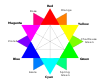
In color theory, hue is one of the main properties of a color, defined technically in the CIECAM02 model as "the degree to which a stimulus can be described as similar to or different from stimuli that are described as red, orange, yellow, green, blue, violet," within certain theories of color vision.

<span class="mw-page-title-main">Natural Color System</span> Proprietary color model

The Natural Color System (NCS) is a proprietary perceptual color model. It is based on the color opponency hypothesis of color vision, first proposed by German physiologist Ewald Hering. The current version of the NCS was developed by the Swedish Colour Centre Foundation, from 1964 onwards. The research team consisted of Anders Hård, Lars Sivik and Gunnar Tonnquist, who in 1997 received the AIC Judd award for their work. The system is based entirely on the phenomenology of human perception and not on color mixing. It is illustrated by a color atlas, marketed by NCS Colour AB in Stockholm.

<span class="mw-page-title-main">Color constancy</span> How humans perceive color

Color constancy is an example of subjective constancy and a feature of the human color perception system which ensures that the perceived color of objects remains relatively constant under varying illumination conditions. A green apple for instance looks green to us at midday, when the main illumination is white sunlight, and also at sunset, when the main illumination is red. This helps us identify objects.

<span class="mw-page-title-main">Color vision</span> Ability to perceive differences in light frequency

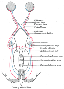
Color vision, a feature of visual perception, is an ability to perceive differences between light composed of different frequencies independently of light intensity.

<span class="mw-page-title-main">Tetrachromacy</span> Type of color vision with four types of cone cells

Tetrachromacy is the condition of possessing four independent channels for conveying color information, or possessing four types of cone cell in the eye. Organisms with tetrachromacy are called tetrachromats.

<span class="mw-page-title-main">Afterimage</span> Image that continues to appear in the eyes after a period of exposure to the original image

An afterimage is an image that continues to appear in the eyes after a period of exposure to the original image. An afterimage may be a normal phenomenon or may be pathological (palinopsia). Illusory palinopsia may be a pathological exaggeration of physiological afterimages. Afterimages occur because photochemical activity in the retina continues even when the eyes are no longer experiencing the original stimulus.

<span class="mw-page-title-main">Trichromacy</span> Possessing of three independent channels for conveying color information

Trichromacy or trichromatism is the possession of three independent channels for conveying color information, derived from the three different types of cone cells in the eye. Organisms with trichromacy are called trichromats.

Dichromacy is the state of having two types of functioning photoreceptors, called cone cells, in the eyes. Organisms with dichromacy are called dichromats. Dichromats require only two primary colors to be able to represent their visible gamut. By comparison, trichromats need three primary colors, and tetrachromats need four. Likewise, every color in a dichromat's gamut can be evoked with monochromatic light. By comparison, every color in a trichromat's gamut can be evoked with a combination of monochromatic light and white light.

The opponent process is a color theory that states that the human visual system interprets information about color by processing signals from photoreceptor cells in an antagonistic manner. The opponent-process theory suggests that there are three opponent channels, each comprising an opposing color pair: red versus green, blue versus yellow, and black versus white (luminance). The theory was first proposed in 1892 by the German physiologist Ewald Hering.

A color model is an abstract mathematical model describing the way colors can be represented as tuples of numbers, typically as three or four values or color components. When this model is associated with a precise description of how the components are to be interpreted, taking account of visual perception, the resulting set of colors is called "color space."

<span class="mw-page-title-main">Abney effect</span> Perceived hue shift when white light is added to a monochromatic light source

The Abney effect or the purity-on-hue effect describes the perceived hue shift that occurs when white light is added to a monochromatic light source.

<span class="mw-page-title-main">Infant visual development</span>

Infant vision concerns the development of visual ability in human infants from birth through the first years of life. The aspects of human vision which develop following birth include visual acuity, tracking, color perception, depth perception, and object recognition.

<span class="mw-page-title-main">Chromostereopsis</span> Visual illusion whereby the impression of depth is conveyed in two-dimensional color images

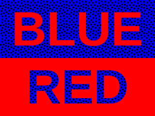
Chromostereopsis is a visual illusion whereby the impression of depth is conveyed in two-dimensional color images, usually of red–blue or red–green colors, but can also be perceived with red–grey or blue–grey images. Such illusions have been reported for over a century and have generally been attributed to some form of chromatic aberration.

<span class="mw-page-title-main">Impossible color</span> Color that cannot be perceived under ordinary viewing conditions

Impossible colors are colors that do not appear in ordinary visual functioning. Different color theories suggest different hypothetical colors that humans are incapable of perceiving for one reason or another, and fictional colors are routinely created in popular culture. While some such colors have no basis in reality, phenomena such as cone cell fatigue enable colors to be perceived in certain circumstances that would not be otherwise.

<span class="mw-page-title-main">Parasol cell</span>

A parasol cell, sometimes called an M cell or M ganglion cell, is one type of retinal ganglion cell (RGC) located in the ganglion cell layer of the retina. These cells project to magnocellular cells in the lateral geniculate nucleus (LGN) as part of the magnocellular pathway in the visual system. They have large cell bodies as well as extensive branching dendrite networks and as such have large receptive fields. Relative to other RGCs, they have fast conduction velocities. While they do show clear center-surround antagonism, they receive no information about color. Parasol ganglion cells contribute information about the motion and depth of objects to the visual system.

<span class="mw-page-title-main">Russell L. De Valois</span>

Russell L. De Valois was an American scientist recognized for his pioneering research on spatial and color vision.

<span class="mw-page-title-main">Memory color effect</span>

The memory color effect is the phenomenon that the canonical hue of a type of object acquired through experience can directly modulate the appearance of the actual colors of objects.

References

  1. Allen, Keith (2010). "Locating the Unique Hues". Rivista di estetica. 43 (43): 13–28. doi: 10.4000/estetica.1786 .
  2. Valberg, Arne (2001). "Unique hues: an old problem for a new generation". Vision Research. 41 (13): 1645–1657. doi: 10.1016/S0042-6989(01)00041-4 . PMID   11348647.
  3. Wright, Wayne (2013). "Psychologically Pure Colors". Encyclopedia of Color Science and Technology. pp. 1–4. doi:10.1007/978-3-642-27851-8_78-8. ISBN   978-3-642-27851-8.
  4. 1 2 Hurvich, Leo (1955). "Some quantitative aspects of an opponent-colors theory. II. Brightness, saturation, and hue in normal and dichromatic vision". JOSA. 45 (8): 602–616. doi:10.1364/JOSA.45.000602. PMID   13243163.
  5. 1 2 Hering, Ewald (1878). Zur Lehre von Lichtsinne.
  6. Hering, Ewald (1964). Outlines of a Theory of the Light Sense. Harvard.
  7. Niall, Keith K. (1988). "On the trichromatic and opponent-process theories: An article by E. Schrödinger". Spatial Vision. 3 (2): 79–95. doi:10.1163/156856888x00050. PMID   3153667.
  8. Svaetichin G, Macnichol EF (November 1959). "Retinal mechanisms for chromatic and achromatic vision". Annals of the New York Academy of Sciences. 74 (2): 385–404. Bibcode:1959NYASA..74..385S. doi:10.1111/j.1749-6632.1958.tb39560.x. PMID   13627867. S2CID   27130943.
  9. 1 2 Wuerger, Sophie M.; Atkinson, Philip; Cropper, Simon (1 November 2005). "The cone inputs to the unique-hue mechanisms". Vision Research. 45 (25): 3210–3223. doi:10.1016/j.visres.2005.06.016. PMID   16087209. S2CID   5778387.
  10. 1 2 3 4 Mollon, J. D.; Jordan, Gabriele (1997). "On the Nature of Unique Hues". In Dickinson, C.; Murray, I.; Carden, D. (eds.). John Dalton's Colour Vision Legacy. Taylor and Francis. pp. 381–392.
  11. Krauskopf, John; Williams, David R.; Heeley, David W. (January 1982). "Cardinal directions of color space". Vision Research. 22 (9): 1123–1131. doi:10.1016/0042-6989(82)90077-3. PMID   7147723. S2CID   10480870.
  12. 1 2 3 4 Webster, Michael A.; Miyahara, Eriko; Malkoc, Gokhan; Raker, Vincent E. (1 September 2000). "Variations in normal color vision II Unique hues". Journal of the Optical Society of America A. 17 (9): 1545. Bibcode:2000JOSAA..17.1545W. doi:10.1364/josaa.17.001545.
  13. De Valois (1993). "A multi-stage color model". Vision Research. 33 (8): 1053–1065. doi: 10.1016/0042-6989(93)90240-W . PMID   8506645.
  14. Valberg, Arne (1986). "Neurones with strong inhibitory S-cone inputs in the macaque lateral geniculate nucleus". Vision Research. 26 (7): 1061–1064. doi: 10.1016/0042-6989(86)90040-4 . PMID   3798743.
  15. Mollon, J.D. (1982). "Color vision". Annual Review of Psychology. 33: 41–85. doi:10.1146/annurev.ps.33.020182.000353. PMID   6977310.
  16. Krauskopf, John; Karl, Gegenfurtner (1992). "Color discrimination and adaptation". Vision Research. 32 (11): 2165–2175. doi: 10.1016/0042-6989(92)90077-v . ISSN   0042-6989. PMID   1304093.
  17. Danilova, M. V.; Mollon, J. D. (2014-02-12). "Symmetries and asymmetries in chromatic discrimination". Journal of the Optical Society of America A. 31 (4): A247-53. Bibcode:2014JOSAA..31A.247D. doi:10.1364/josaa.31.00a247. ISSN   1084-7529. PMID   24695178.
  18. Forder, Lewis; Bosten, Jenny; He, Xun; Franklin, Anna (2017-02-10). "A neural signature of the unique hues". Scientific Reports. 7 (1): 42364. Bibcode:2017NatSR...742364F. doi:10.1038/srep42364. ISSN   2045-2322. PMC   5301231 . PMID   28186142.
  19. Chauhan, Tushar; Jakovljev, Ivana; Thompson, Lindsay N.; Wuerger, Sophie M.; Martinovic, Jasna (2021-06-17). "Decoding of EEG signals reveals non-uniformities in the neural geometry of colour". bioRxiv   10.1101/2021.06.17.448044 .
  20. 1 2 3 4 5 Neitz, Jay (2002). "Color perception is mediated by a plastic neural mechanism that is adjustable in adults". Neuron. 35 (4): 783–792. doi: 10.1016/S0896-6273(02)00818-8 .
  21. Kuehni, Rolf G. (2004). "Variability in unique hue selection: A surprising phenomenon" (PDF). Color Research & Application. 29 (2): 158–162. doi:10.1002/col.10237.
  22. 1 2 3 Miyahara, E (2003). "Focal colors and unique hues". Perceptual and Motor Skills. 97 (3_suppl): 1038–42. doi:10.2466/pms.2003.97.3f.1038. PMC   1404500 . PMID   15002843.
  23. De Valois, Russell L; De Valois, Karen K; Switkes, Eugene; Mahon, Luke (April 1997). "Hue Scaling of Isoluminant and Cone-specific Lights". Vision Research. 37 (7): 885–897. doi: 10.1016/s0042-6989(96)00234-9 . PMID   9156186.
  24. Tregillus, Katherine (2019). "Long-term adaptation to color". Current Opinion in Behavioral Sciences. 30: 116–121. doi: 10.1016/j.cobeha.2019.07.005 .
  25. Welbourne, Lauren (2015). "Human colour perception changes between seasons". Current Biology. 25 (15): R646-7. doi: 10.1016/j.cub.2015.06.030 . PMID   26241135.
  26. Delahunt, Peter (2004). "Long-term renormalization of chromatic mechanisms following cataract surgery". Visual Neuroscience. 21 (3): 301–307. doi:10.1017/S0952523804213025. PMC   2633455 . PMID   15518204.
  27. Kuehni, Rolf G. (2013). "Unique hues and their stimuli-state of the art". Color Research & Application. 39 (3): 279–287. doi:10.1002/col.21793.
  28. Massof, Robert W.; Bailey, James E. (1976). "Achromatic points in protanopes and deuteranopes". Vision Research. 16 (1): 53–58. doi:10.1016/0042-6989(76)90076-6. PMID   1083102. S2CID   9909092.