
The endoplasmic reticulum (ER) is a type of organelle made up of two subunits – rough endoplasmic reticulum (RER), and smooth endoplasmic reticulum (SER). The endoplasmic reticulum is found in most eukaryotic cells and forms an interconnected network of flattened, membrane-enclosed sacs known as cisternae, and tubular structures in the SER. The membranes of the ER are continuous with the outer nuclear membrane. The endoplasmic reticulum is not found in red blood cells, or spermatozoa.
Diastrophic dysplasia is an autosomal recessive dysplasia which affects cartilage and bone development. Diastrophic dysplasia is due to mutations in the SLC26A2 gene.

Atelosteogenesis, type II is a severe disorder of cartilage and bone development. It is rare, and infants with the disorder are usually stillborn; however, those who survive birth die soon after
Osteochondromas are the most common benign tumors of the bones. The tumors take the form of cartilage-capped bony projections or outgrowth on the surface of bones exostoses. It is characterized as a type of overgrowth that can occur in any bone where cartilage forms bone. Tumors most commonly affect long bones about the knee and in the forearm. Additionally, flat bones such as the pelvis and scapula may be affected. Hereditary multiple exostoses usually present during childhood. Yet, the vast majority of affected individuals become clinically manifest by the time they reach adolescence. Osteochondromas occur in 3% of the general population and represent 35% of all benign tumors and 8% of all bone tumors. The majority of these tumors are solitary non-hereditary lesions and approximately 15% of osteochondromas occur as hereditary multiple exostoses preferably known as hereditary multiple osteochondromas (HMOs). Osteochondromas do not result from injury and the exact cause remains unknown. Recent research has indicated that multiple osteochondromas is an autosomal dominant inherited disease. Germ line mutations in EXT1 and EXT2 genes located on chromosomes 8 and 11 have been associated with the cause of the disease. The treatment choice for osteochondroma is surgical removal of solitary lesion or partial excision of the outgrowth, when symptoms cause motion limitations or nerve and blood vessel impingements. In hereditary multiple exostoses the indications of surgery are based upon multiple factors that are taken collectively, namely: patient's age, tumor location and number, accompanying symptomatology, esthetic concerns, family history and underlying gene mutation. A variety of surgical procedures have been employed to remedy hereditary multiple exostoses such as osteochondroma excision, bone lengthening, corrective osteotomy and hemiepiphysiodesis. Sometimes a combination of the previous procedures is used. The indicators of surgical success in regard to disease and patient characteristics are greatly disputable. Because most studies of hereditary multiple exostoses are retrospective and of limited sample size with missing data, the best evidence for each of the currently practiced surgical procedures is lacking.
The type II and XI collagenopathies are a group of disorders that affect connective tissue, the tissue that supports the body's joints and organs. These disorders are caused by defects in type II or type XI collagen. Collagens are complex molecules that provide structure, strength, and elasticity to connective tissue. Type II and type XI collagen disorders are grouped together because both types of collagen are components of the cartilage found in joints and the spinal column, the inner ear, and the jelly-like substance that fills the eyeball. The type II and XI collagenopathies result in similar clinical features.
Hypochondrogenesis is a severe genetic disorder causing malformations of bone growth. The condition is characterized by a short body and limbs and abnormal bone formation in the spine and pelvis.
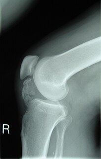
Achondrogenesis, type 2 results in short arms and legs, a small chest with short ribs, and underdeveloped lungs at birth. Achondrogenesis, type 2 is a subtype of collagenopathy, types II and XI. This condition is also associated with a lack of bone formation (ossification) in the spine and pelvis. Typical facial features include a prominent forehead, a small chin, and, in some cases, an opening in the roof of the mouth. The abdomen is enlarged, and affected infants often have a condition called hydrops fetalis in which excess fluid builds up in the body before birth. The skull bones may be soft, but they often appear normal on X-ray images. In contrast, bones in the spine (vertebrae) and pelvis do not harden.

Spondyloepiphyseal dysplasia congenita is a rare disorder of bone growth that results in dwarfism, characteristic skeletal abnormalities, and occasionally problems with vision and hearing. The name of the condition indicates that it affects the bones of the spine (spondylo-) and the ends of bones (epiphyses), and that it is present from birth (congenital). The signs and symptoms of spondyloepiphyseal dysplasia congenita are similar to, but milder than, the related skeletal disorders achondrogenesis type 2 and hypochondrogenesis. Spondyloepiphyseal dysplasia congenita is a subtype of collagenopathy, types II and XI.
Achondrogenesis, type 1B is a severe autosomal recessive skeletal disorder, invariably fatal in the perinatal period. It is characterized by extremely short limbs, a narrow chest and a prominent, rounded abdomen. The fingers and toes are short and the feet may be rotated inward. Affected infants frequently have a soft out-pouching around the belly-button or near the groin.

Collagen, type II, alpha 1 , also known as COL2A1, is a human gene that provides instructions for the production of the pro-alpha1(II) chain of type II collagen.

Cartilage associated protein is a protein that in humans is encoded by the CRTAP gene.
VPS13B also known as vacuolar protein sorting-associated protein 13B is a protein that in humans is encoded by the VPS13B gene. It is a giant protein associated with the Golgi apparatus that is believed to be involved in post Golgi apparatus sorting and trafficking. Mutations in the human VPS13B gene cause Cohen syndrome.

Ceroid-lipofuscinosis neuronal protein 6 is a protein that in humans is encoded by the CLN6 gene.

KDEL (Lys-Asp-Glu-Leu) endoplasmic reticulum protein retention receptor 1, also known as KDELR1, is a protein which in humans is encoded by the KDELR1 gene.
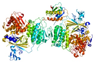
Sec23 homolog A , also known as SEC23A, is a protein which in humans is encoded by the SEC23A gene.

Boomerang dysplasia is a lethal form of osteochondrodysplasia known for a characteristic congenital feature in which bones of the arms and legs are malformed into the shape of a boomerang. Death usually occurs in early infancy due to complications arising from overwhelming systemic bone malformations.
Wrinkly skin syndrome(WSS) is a rare genetic condition characterized by sagging, wrinkled skin, low skin elasticity, and delayed fontanel closure along with a range of other symptoms. The disorder exhibits an autosomal recessive inheritance pattern with mutations in the ATP6V0A2 gene, leading to abnormal glycosylation events. There are only about 30 known cases of WSS as of 2010. Given its rarity and symptom overlap to other dermatological conditions, reaching an accurate diagnosis is difficult and requires specialized dermatological testing. Limited treatment options are available but long-term prognosis is variable from patient-to-patient, on the basis of individual case studies. Some skin symptoms recede with increasing age while progressive neurological advancement of the disorder causes seizures and mental deterioration later in life for some patients.
Cranio-lenticulo-sutural dysplasia is a neonatal/infancy disease caused by a disorder in the 14th chromosome. It is an autosomal recessive disorder, meaning that both recessive genes must be inherited from each parent in order for the disease to manifest itself. The disease causes a significant dilation of the endoplasmic reticulum in fibroblasts of the host with CLSD. Due to the distension of the endoplasmic reticulum, export of proteins from the cell is disrupted.

Bruck syndrome is characterized as the combination of arthrogryposis multiplex congenita and osteogenesis imperfecta. Both diseases are uncommon, but concurrence is extremely rare which makes Bruck syndrome very difficult to research. Bruck syndrome is thought to be an atypical variant of osteogenesis imperfecta most resembling type III, if not its own disease. Multiple gene mutations associated with osteogenesis imperfecta are not seen in Bruck syndrome. Many affected individuals are within the same family, and pedigree data supports that the disease is acquired through autosomal recessive inheritance. Bruck syndrome has features of congenital contractures, bone fragility, recurring bone fractures, flexion joint and limb deformities, pterygia, short body height, and progressive kyphoscoliosis. Individuals encounter restricted mobility and pulmonary function. A reduction in bone mineral content and larger hydroxyapatite crystals are also detectable Joint contractures are primarily bilateral and symmetrical, and most prone to ankles. Bruck syndrome has no effect on intelligence, vision, or hearing.

The Golgi matrix is a collection of proteins involved in the structure and function of the Golgi apparatus. The matrix was first isolated in 1994 as an amorphous collection of 12 proteins that remained associated together in the presence of detergent and 150 mM NaCl. Treatment with a protease enzyme removed the matrix, which confirmed the importance of proteins for the matrix structure. Modern freeze etch electron microscopy (EM) clearly shows a mesh connecting Golgi cisternae and associated vesicles. Further support for the existence of a matrix comes from EM images showing that ribosomes are excluded from regions between and near Golgi cisternae.
