Aggressive periodontitis

Last updated

Aggressive periodontitis describes a type of periodontal disease and includes two of the seven classifications of periodontitis as defined by the 1999 classification system: [1]

Contents

  1. Localized aggressive periodontitis (LAP)
  2. Generalized aggressive periodontitis (GAP)

LAP is localised to first molar or incisor interproximal attachment loss, whereas GAP is the interproximal attachment loss affecting at least three permanent teeth other than incisors and first molar. [2] The prevalence of LAP is less than 1% and that of GAP is 0.13%. [2] Approximately 0.1% of white Caucasians [3] (with 0.1% in northern and in central Europe, 0.5% in southern Europe, and 0.1-0.2% in North America [2] ) and 2.6% of black Africans may have LAP. [3] Estimates of the disease prevalence are 1-5% in the African population and in groups of African descent, 2.6% in African-Americans, 0.5-1.0% in Hispanics in North America, 0.3-2.0% in South America, and 0.2-1.0% in Asia. [2] On the other hand, in Asia, the prevalence rate of 1.2% for LAP and 0.6% for GAP in Baghdad and Iran population, and 0.47% in Japanese population. [2]

Therefore, the prevalence of LAP varies considerably between continents, and differences in race or ethnicity seem to be a major contributing factor. [2]

Aggressive periodontitis is much less common than chronic periodontitis and generally affects younger patients than does the chronic form. [4] [5] Around 1 in every 1000 patients experience more rapid loss of attachment. [6] Males seem to be at higher risk of GAP than females [2]

The localized and generalized forms are not merely different in extent; they differ in etiology and pathogenesis.

Etiology

Microbiology

Of the microflora characterised in aggressive periodontitis, approximately 65-75% of bacteria are Gram-negative bacilli, with few spirochaetes or motile rods present. [7] Aggressive periodontitis is often characterised by a rapid loss of periodontal attachment associated with highly pathogenic bacteria and an impaired immune response. Various studies have associated Aggregatibacter actinomycetemcomitans , formerly known as Actinobacillus actinomycetemcomitans, with aggressive periodontitis. An early study dating back to 1983 explains its prevalence and documents its role in localised aggressive periodontitis. [8]

Virulence factors are the attributes of microorganisms that enable it to colonise a particular niche in its host, overcome the host defences and initiate a disease process. [9] Fives Taylor et al. (2000) have categorised the virulence factors of Aggregatibacter actinomycetemcomitans as follows. [9]

Promote colonization and persistence in the oral cavity:Interfere with host defences:Destroy host tissues:Inhibit host repair of tissues:
AdhesinsLeukotoxinCytotoxinsInhibitors of fibroblast proliferation
InvasinsChemotactic inhibitorsCollagenase
BacteriocinsImmunosuppressive proteinsBone resorption agentsInhibitors of bone formation
Antibiotic resistanceFc-binding proteinsStimulators of inflammatory mediators

Samaranayake notes the evidence for the specific involvement of Aggregatibacter actinomycetemcomitans includes: an increased incidence of it found in subgingival plaque obtained from lesional sites, high level of its antibody which tends to fall following successful treatment, its possession of a wide range of potentially pathogenic products and its elimination with concordant disease regression, following treatment with successful periodontal therapy and adjunctive tetracycline. [7]

Porphyromonas gingivalis is a Gram-negative anaerobe associated with the pathogenicity of periodontal disease, [10] and aggressive periodontitis is no exception. Greater numbers of both Porphyromonas gingivalis and Aggregatibacter actinomycetemcomitans were found in active, destructive periodontal lesions in comparison to non-active sites. [10]

Capnocytophaga spp are implicated as prime periodontal pathogens, especially in localised aggressive periodontitis. [7] Both Capnocytophaga spp and Prevotella intermedia were the most frequently detected microorganisms in a study, [11] which also noted that Capnocytophaga spp was the most prominent bacteria in subgingival samples of patients with aggressive periodontitis. [11] [12]

An impaired ability of peripheral blood lymphocytes to react to chemotactic stimuli is found in the majority of patients with aggressive periodontitis. As well as Aggregatibacter actinomycetemcomitans being associated with this, the synergism of the disease also accounts for both Capnocytophaga spp and Porphyromonas gingivalis. [7]

Pathophysiology

Aggressive periodontitis is a multifactorial disease with many complex interactions including host factors, microbiology and genetics.

Host defences involve multiple factors; saliva, epithelium, inflammatory response, immune response and chemical mediators. The inflammatory exudate in the gingival tissues and gingival crevicular fluid is mostly polymorph neutrophils but also includes B cells and plasma cells. The neutrophils may show an intrinsic functional defect and respond abnormally when challenged by certain pathogens. [13] The plasma cells produce specific antibodies in response to the periodontal pathogens, which diffuse into the gingival crevicular fluid. They produce mainly IgG, with some IgA. [13] It has been suggested that these gingival crevicular fluid antibody levels could be potentially useful in the development of a vaccine. [14] Patients with localised aggressive periodontitis have large amount of Aggregatibacter actinomycetemcomitans specific IgG2. This is suggested to be protective against wider spread periodontal breakdown. However, patients with generalized aggressive periodontitis have decreased ability to mount high titres of IgG to Porphyromonas gingivalis and Aggregatibacter actinomycetemcomitans.

It has also been found that a low T-helper to T-suppressor ratio is found in aggressive periodontitis which may alter local immune regulation. Monocytes respond to bacterial and inflammatory stimuli with very high levels of local release inflammatory mediators and induce hyper-inflammatory reaction with activation of tissue degrading matrix-metalloproteinases. These is also evidence they produce increased amounts IL-1α and IL-1β which cause osteoclastic bone resorption. These amounts are greatly reduced following treatment. [13]

Studies of families, twins and sibling pairs have provided strong evidence for a genetic basis for aggressive periodontitis. [15] A person's genetic predisposition to the condition is determined by a single gene of major effect, inherited as an autosomal dominant trait. However, for the disease process to initiate the person must be exposed to the presence of periodontal pathogens and potentially also various environmental factors.

Smoking is a generalized risk factor for generalized forms of aggressive periodontitis. Studies found that smokers have more affected teeth than non-smokers and high levels of attachment loss. This is due to the suppression of serum IgG2 and antibody against Aggregatibacter actinomycetemcomitans found in smokers. [16]

Features

According to the 1999 International Workshop for the Classification of Periodontal Diseases, aggressive periodontitis was defined according to 3 primary features, [17] in contrast to chronic periodontitis. [18] These features are common for both localized and generalized form of disease. [19] [20]

Primary features

Patients do not have any underlying systemic disease that would contribute to aggressive periodontitis. [21] For instance, diabetes is proved to be associated with periodontitis- it is a major risk factor when glycaemic control is poor. [22]

Loss of attachment refers to the destruction of periodontium whereas the bone refers to the alveolar bone supporting the teeth. [23] The loss can be determined by using a calibrated periodontal probe and taking radiographs of the dentition. [24] Usually the loss of attachment is greater than 2mm per year.

Familial aggregation of aggressive periodontitis is often discovered by taking a thorough medical history of the patient. [25] The patient is said to have a high genetic susceptibility to aggressive periodontitis. Many studies have shown that genetic factors contribute to the pathogenesis of this disease. [26] In this case, the manifestation of aggressive periodontitis is believed to be the result of genetic mutation, combined with environmental factors. [26]

Secondary features

Secondary features are characteristics which are frequently seen but not always present in every patient diagnosed with aggressive periodontitis.

The amount of bacteria is often indicated by the level of dental plaque. [27] This feature implies that when aggressive periodontitis is present, loss of attachment and bone loss tend to occur even if the plaque level is low.

These gram-negative microbes are considered the chief aetiological agent of aggressive periodontitis. They are implicated in the development of aggressive periodontitis by triggering inflammatory response in periodontal tissue.

Phagocytes are essential in resolving inflammation. The impairment of their phagocytic activity results in persistent inflammation in periodontal tissues. [28]

Due to the increased responsiveness, the macrophages produce excessive levels of inflammatory mediator and cytokine, such as prostaglandin E2 (PGE2) and interleukin-1β (IL-1B). [19] Their hyperactivity is associated with periodontal tissue destruction and bone loss. [29]

In some patients, the disease may burnout without any cause-related therapy. [30] Caries levels have seen to be lower in cases of aggressive periodontitis. [31] [32]

Staging Cases of aggressive periodontitis have been staged into Stage I, II and III based on the severity of the cases. The staging index was proposed based on clinical features, radiological features and possible risk factors. [25] The proposed index was validated with 10 cases of aggressive periodontitis followed for 10 years.

Clinical and radiographic features

Localized aggressive periodontitis

Clinical features

LAP begins around the age of puberty where there is interproximal loss of attachment of the first molar, and or incisors. [2] on at least two permanent teeth (one which is a first molar) and no involvement of more than two teeth other than the first molars and incisors, [2] [33] lack of inflammation and evidence of deep periodontal pocket with advanced bone loss. [2] There is also a relatively fast progression of periodontal tissue loss. [33]

With an increase in the age of the patient, there may be progression of the disease involving the adjacent teeth and lead to the patient developing GAP. [34] [17] The periodontal tissue also exhibits minimal signs of inflammation clinically [35] and show a robust response with serum antibodies to pathogens. [33]

The amount of plaque present is inconsistent with the amount and severity of tissue destruction [2] [33] but with a high plaque pathogenicity due to the presence of increased levels of bacteria like Aggregatibacter actinomycetemcomitans (A.a) and Porphyromonas Gingivalis (P.g). [2]

Secondary features of LAP may also be present including; [2]

  • diastema formation with disto-labial migration of the incisors
  • increased mobility of the affected teeth, sensitivity due to exposed root,
  • deep dull pain that radiates to the jaw
  • periodontal abscess with lymph node enlargement

Radiographic features

Radiographically, the periodontal lesion often presents with alveolar bone loss in a horizontal pattern at the interproximal surface of the permanent first molars [2] [33] [34] and usually horizontal bone pattern of bone loss at the interproximal surface of the incisors as the bone is thinner than at the interproximal surface of the molars. [33]

The alveolar bone loss patterns are usually bilateral and similar on both sides and has been referred to as being a 'mirror-image' pattern. [34] [33]

In advanced cases the alveolar bone loss may be depicted as a horizontal bone loss pattern radiographically. [33] [34]

Generalized aggressive periodontitis

Clinical features

  • Mostly in individuals under 30 years old [36]
    • In GAP, the clinical appearance of the disease resembles chronic periodontitis. The difference is that individuals affected by GAP are much younger and the progression of disease appears more rapid. [35]
  • There is a poor serum response against infecting agents [36]
    • Destruction is present that is not in balance with the amount of local irritants present [35]
  • Generalized inter-proximal attachment loss on 3 or more permanent teeth, excluding the first molars or incisors [36]
    • The main distinction between the localized and generalized form of AgP lies in the number of teeth affected. GAP brings about attachment loss involving more than 30% of sites on teeth; [1] effectively being at least three permanent teeth other than the first molars or incisors. [20]
  • Episodic nature of attachment loss Two main tissue responses have been found in GAP cases: [36]
    • Tissue may have severe acute inflammation and often presents with an angry red appearance and ulceration. [37] There may be spontaneous bleeding or suppuration. This response is known to be present in the destructive phase, where there is presence of bone and attachment loss.
    • The other response is known as a period of quiescence, where gingival tissue may appear with no inflammation, pink appearance with some possible stippling. In addition to this mild appearance there may be deep pockets upon probing.

Radiographic features

  • The key diagnostic feature of AgP is vertical bone loss around teeth including the first molars and incisors. This tends to begin around puberty in otherwise healthy individuals. [36] There may be an appearance of "arc-shaped loss of alveolar bone extending from the distal surface of the second premolar to the mesial surface of the second molar". [38]
  • In GAP, generalized bone destruction is present that ranges from mild crestal bone resorption to severe alveolar bone destruction, depending on the severity of the disease. [38] There may be a combination of vertical and horizontal bone loss defects. [38]

Screening

Early diagnosis of aggressive periodontitis is important as it can cause rapid permanent destruction of the periodontal tissues. It is essential that all patients undergo a routine periodontal examination to screen for any form of periodontal disease during a dental checkup.

Clinical examination

At the start of the clinical examination of the gingival and periodontal tissues, the dental practitioner would look at the appearance of the gingiva first. A healthy periodontium in a Caucasian would appear stippled and pink with a knife edge margin where it abuts the tooth (pigmentation may differ in other races). [39] After that, gingival probing depths would be checked. This would normally be carried out using a basic periodontal probe (WHO CPI). [40] On probing, patients with AgP should have evidence of significant periodontal pocket depths and loss of attachment (LOA). Dental practitioners should also be aware of false pocketing around erupting/newly erupted teeth in the mixed dentition phase and also in the presence of gingival inflammation. [40] [41] The presence of bleeding on probing (BOP) should be noted which is an indicator of active disease.

Radiographs

Radiographic assessment should be carried out for patients with evidence of periodontitis to observe alveolar bone levels which can help to identify signs of AgP. [40] In healthy periodontal tissues, the distance from the amelocemental junction (ACJ) to the alveolar bone crest is typically in the order of 1mm in young people. [42] If the distance between the ACJ and alveolar bone crest is more than 2-3mm then there is a possible suggestion of AgP. In addition to that, presence of angular or vertical bone loss (especially at 6's) and arrowhead or furcation lesions are also a strong suggestion of AgP.

Strong family association

It is also important for a dental practitioner to check for family history of periodontal disease for each patient. This is because AgP may have an autosomal dominant inheritance pattern which suggests that up to 50% of siblings could be affected if one parent has the disease. [43] Careful interpretation of the history is required but it may provide vital evidence in diagnosing AgP. If a case of Agp is diagnosed, it is important to screen the patient's family members as well for AgP. [44] [45] Early detection of AgP allows intervention to be carried out before extensive periodontal destruction has taken place which further simplifies treatment.

Treatment

Following the initial assessment and diagnosis of AgP, a treatment plan is usually developed for each individual undergoing therapy. As the overall treatment concepts and goals for AgP are not significantly different from that of chronic periodontitis, the different treatment phases (cause related therapy; re-examination for response to therapy; definitive therapy; and maintenance) are similar for both types of periodontitis.

Nevertheless, the considerable amount of bone loss relative to the young age of the individual in AgP necessitates an often more aggressive treatment approach, to halt further periodontal destruction and regain as much periodontal attachment as possible. The objective of treatment is to create a conducive clinical condition for retaining as many teeth, for as long as possible. [46]

This stage involves discussion of the disease with the patient.

The periodontal therapy carried out at this stage is of a non-surgical approach, which is aimed at the removal of supra- and sub-gingival plaque and calculus deposits, to decrease the microbial load, bacteria biofilm, and calculus from the periodontally involved sites. [50]

Re-examination/Response to Therapy

This stage of treatment involves the reassessment of the individual's compliance (i.e. level of oral hygiene) and the tissue response to the treatment. This is carried out 10–12 weeks following RSD. If the disease is stabilised, the treatment progresses on to the maintenance stage. In the case where the disease is not stabilised, the cause of failure should be considered, and the treatment progresses on to the stage of definitive therapy, if the cause is correctable.

Definitive Therapy

Maintenance

Periodontal treatment may help to stabilise the disease, but it does not change one's susceptibility to the disease. Given the high susceptibility for disease progression of the individual with AgP, there is a higher risk of disease recurrence. [57] It is thus necessary to attend frequent review appointments at the dentist to ensure there is no relapse of the disease, and that the periodontal health is maintained after active periodontal therapy. [46]

Related Research Articles

<span class="mw-page-title-main">Periodontal disease</span> Medical condition

Periodontal disease, also known as gum disease, is a set of inflammatory conditions affecting the tissues surrounding the teeth. In its early stage, called gingivitis, the gums become swollen and red and may bleed. It is considered the main cause of tooth loss for adults worldwide. In its more serious form, called periodontitis, the gums can pull away from the tooth, bone can be lost, and the teeth may loosen or fall out. Bad breath may also occur.

<span class="mw-page-title-main">Gums</span> Mouth anatomy

The gums or gingiva consist of the mucosal tissue that lies over the mandible and maxilla inside the mouth. Gum health and disease can have an effect on general health.

<span class="mw-page-title-main">Periodontal fiber</span> Group of specialized connective tissue fibers

The periodontal ligament, commonly abbreviated as the PDL, is a group of specialized connective tissue fibers that essentially attach a tooth to the alveolar bone within which it sits. It inserts into root cementum on one side and onto alveolar bone on the other.

Periodontology or periodontics is the specialty of dentistry that studies supporting structures of teeth, as well as diseases and conditions that affect them. The supporting tissues are known as the periodontium, which includes the gingiva (gums), alveolar bone, cementum, and the periodontal ligament. A periodontist is a dentist that specializes in the prevention, diagnosis and treatment of periodontal disease and in the placement of dental implants.

<span class="mw-page-title-main">Veterinary dentistry</span> Branch of veterinary medicine

Veterinary dentistry is the field of dentistry applied to the care of animals. It is the art and science of prevention, diagnosis, and treatment of conditions, diseases, and disorders of the oral cavity, the maxillofacial region, and its associated structures as it relates to animals.

<span class="mw-page-title-main">Gingival sulcus</span> Space between tooth and gums

The gingival sulcus is an area of potential space between a tooth and the surrounding gingival tissue and is lined by sulcular epithelium. The depth of the sulcus is bounded by two entities: apically by the gingival fibers of the connective tissue attachment and coronally by the free gingival margin. A healthy sulcular depth is three millimeters or less, which is readily self-cleansable with a properly used toothbrush or the supplemental use of other oral hygiene aids.

<span class="mw-page-title-main">Gingival and periodontal pocket</span>

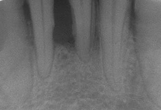
Gingival and periodontal pockets are dental terms indicating the presence of an abnormal depth of the gingival sulcus near the point at which the gingival tissue contacts the tooth.

<span class="mw-page-title-main">Scaling and root planing</span> Dental procedure

Scaling and root planing, also known as conventional periodontal therapy, non-surgical periodontal therapy or deep cleaning, is a procedure involving removal of dental plaque and calculus and then smoothing, or planing, of the (exposed) surfaces of the roots, removing cementum or dentine that is impregnated with calculus, toxins, or microorganisms, the agents that cause inflammation. It is a part of non-surgical periodontal therapy. This helps to establish a periodontium that is in remission of periodontal disease. Periodontal scalers and periodontal curettes are some of the tools involved.

Gingivectomy is a dental procedure in which a dentist or oral surgeon cuts away part of the gums in the mouth.

Laser-assisted new attachment procedure (LANAP) is a surgical therapy for the treatment of periodontitis, intended to work through regeneration rather than resection. This therapy and the laser used to perform it have been in use since 1994. It was developed by Robert H. Gregg II and Delwin McCarthy.

Guided bone regeneration (GBR) and guided tissue regeneration (GTR) are dental surgical procedures that use barrier membranes to direct the growth of new bone and gingival tissue at sites with insufficient volumes or dimensions of bone or gingiva for proper function, esthetics or prosthetic restoration. Guided bone regeneration typically refers to ridge augmentation or bone regenerative procedures; guided tissue regeneration typically refers to regeneration of periodontal attachment.

<span class="mw-page-title-main">Gingivitis</span> Inflammation of the gums

Gingivitis is a non-destructive disease that causes inflammation of the gums; ulitis is an alternative term. The most common form of gingivitis, and the most common form of periodontal disease overall, is in response to bacterial biofilms that are attached to tooth surfaces, termed plaque-induced gingivitis. Most forms of gingivitis are plaque-induced.

<span class="mw-page-title-main">Debridement (dental)</span> Removal of plaque and calculus from teeth

In dentistry, debridement refers to the removal by dental cleaning of accumulations of plaque and calculus (tartar) in order to maintain dental health. Debridement may be performed using ultrasonic instruments, which fracture the calculus, thereby facilitating its removal, as well as hand tools, including periodontal scaler and curettes, or through the use of chemicals such as hydrogen peroxide.

Epidemiology of periodontal disease is the study of patterns, causes, and effects of periodontal diseases. Periodontal disease is a disease affecting the tissue surrounding the teeth. This causes the gums and the teeth to separate making spaces that become infected. The immune system tries to fight the toxins breaking down the bone and tissue connecting to the teeth to the gums. The teeth will have to be removed. This is an advance stage of gum disease that has multiple definitions. Adult periodontitis affects less than 10 to 15% of the population in industrialized countries, mainly adults around the ages of 50 to 60. The disease is now declining world-wide.

Chronic periodontitis is one of the seven categories of periodontitis as defined by the American Academy of Periodontology 1999 classification system. Chronic periodontitis is a common disease of the oral cavity consisting of chronic inflammation of the periodontal tissues that is caused by the accumulation of profuse amounts of dental plaque. Periodontitis initially begins as gingivitis and can progress onto chronic and subsequent aggressive periodontitis according to the 1999 classification.

<span class="mw-page-title-main">Peri-implantitis</span> Inflammatory disease

Peri-implantitis is a destructive inflammatory process affecting the soft and hard tissues surrounding dental implants. The soft tissues become inflamed whereas the alveolar bone, which surrounds the implant for the purposes of retention, is lost over time.

In dentistry, numerous types of classification schemes have been developed to describe the teeth and gum tissue in a way that categorizes various defects. All of these classification schemes combine to provide the periodontal diagnosis of the aforementioned tissues in their various states of health and disease.

<span class="mw-page-title-main">Tooth mobility</span> Medical condition

Tooth mobility is the horizontal or vertical displacement of a tooth beyond its normal physiological boundaries around the gingival area, i.e. the medical term for a loose tooth.

Periodontal surgery is a form of dental surgery that prevents or corrects anatomical, traumatic, developmental, or plaque-induced defects in the bone, gingiva, or alveolar mucosa. The objectives of this surgery include accessibility of instruments to root surface, elimination of inflammation, creation of an oral environment for plaque control, periodontal diseases control, oral hygiene maintenance, maintain proper embrasure space, address gingiva-alveolar mucosa problems, and esthetic improvement. The surgical procedures include crown lengthening, frenectomy, and mucogingival flap surgery.

References

  1. 1 2 Armitage GC (December 1999). "Development of a classification system for periodontal diseases and conditions". Annals of Periodontology. 4 (1): 1–6. doi:10.1902/annals.1999.4.1.1. PMID   10863370.
  2. 1 2 3 4 5 6 7 8 9 10 11 12 13 14 Joshipura V, Yadalam U, Brahmavar B (2015-01-01). "Aggressive periodontitis: A review". Journal of the International Clinical Dental Research Organization. 7 (1): 11. doi: 10.4103/2231-0754.153489 .
  3. 1 2 Clerehugh V (2012). "Guidelines for periodontal screening and management of children and adolescents under 18 years of age" (PDF). British Society of Periodontology and The British Society of Paediatric Dentistry. Retrieved 6 Dec 2017.
  4. Albandar JM, Tinoco EM (2002). "Global epidemiology of periodontal diseases in children and young persons". Periodontol. 2000. 29: 153–76. doi:10.1034/j.1600-0757.2002.290108.x. PMID   12102707.[ verification needed ]
  5. Papapanou PN (November 1996). "Periodontal diseases: epidemiology". Ann. Periodontol. 1 (1): 1–36. doi:10.1902/annals.1996.1.1.1. PMID   9118256.[ verification needed ]
  6. Needleman I (2016). "The Good Practitioner's Guide to Periodontology" (PDF). British Society of Periodontology. Archived from the original (PDF) on 21 December 2019. Retrieved 6 Dec 2017.
  7. 1 2 3 4 Whiley RA (2006-11-25). "Essential microbiology for dentistry". British Dental Journal (3rd ed.). 201 (10): 679. doi: 10.1038/sj.bdj.4814299 .
  8. Zambon JJ, Christersson LA, Slots J (December 1983). "Actinobacillus actinomycetemcomitans in human periodontal disease. Prevalence in patient groups and distribution of biotypes and serotypes within families". Journal of Periodontology. 54 (12): 707–11. doi:10.1902/jop.1983.54.12.707. PMID   6358452. S2CID   27904962.
  9. 1 2 Fives-Taylor PM, Meyer DH, Mintz KP, Brissette C (June 1999). "Virulence factors of Actinobacillus actinomycetemcomitans". Periodontology 2000. 20: 136–67. doi:10.1111/j.1600-0757.1999.tb00161.x. PMID   10522226.
  10. 1 2 Thiha K, Takeuchi Y, Umeda M, Huang Y, Ohnishi M, Ishikawa I (June 2007). "Identification of periodontopathic bacteria in gingival tissue of Japanese periodontitis patients". Oral Microbiology and Immunology. 22 (3): 201–7. doi:10.1111/j.1399-302X.2007.00354.x. PMID   17488447.
  11. 1 2 Nonnenmacher C, Mutters R, de Jacoby LF (April 2001). "Microbiological characteristics of subgingival microbiota in adult periodontitis, localized juvenile periodontitis and rapidly progressive periodontitis subjects". Clinical Microbiology and Infection. 7 (4): 213–7. doi: 10.1046/j.1469-0691.2001.00210.x . PMID   11422244.
  12. Genco RJ, Zambon JJ, Christersson LA (November 1986). "Use and interpretation of microbiological assays in periodontal diseases". Oral Microbiology and Immunology. 1 (1): 73–81. doi:10.1111/j.1399-302X.1986.tb00324.x. PMID   3295682.
  13. 1 2 3 Wilson TG, Kornman KS (2003). fundamentals of periodontics (2nd ed.). Quintessence Publishing Company. ISBN   978-0-86715-405-4.[ page needed ]
  14. Garcia, Monique; Cappelli, David. "UTCAT2409, Found CAT view, CRITICALLY APPRAISED TOPICs". cats.uthscsa.edu. Retrieved 2017-12-07.
  15. Kinane DF, Hart TC (2003). "Genes and gene polymorphisms associated with periodontal disease". Critical Reviews in Oral Biology and Medicine. 14 (6): 430–49. doi: 10.1177/154411130301400605 . PMID   14656898.
  16. Schenkein HA, Gunsolley JC, Koertge TE, Schenkein JG, Tew JG (August 1995). "Smoking and its effects on early-onset periodontitis". Journal of the American Dental Association. 126 (8): 1107–13. doi:10.14219/jada.archive.1995.0327. PMID   7560567.
  17. 1 2 "1999 International International Workshop for a Classification of Periodontal Diseases and Conditions. Papers. Oak Brook, Illinois, October 30-November 2, 1999". Annals of Periodontology. 4 (1): i, 1–112. December 1999. doi:10.1902/annals.1999.4.1.i. PMID   10896458.
  18. Ramachandra, Srinivas Sulugodu; Gupta, Vivek Vijay; Mehta, Dhoom Singh; Gundavarapu, Kalyan C; Luigi, Nibali (2017). "Differential Diagnosis between Chronic versus Aggressive Periodontitis and Staging of Aggressive Periodontitis: A Cross-sectional Study". Contemporary Clinical Dentistry. 8 (4): 594–603. doi: 10.4103/ccd.ccd_623_17 . PMC   5754981 . PMID   29326511.
  19. 1 2 3 4 5 6 7 8 9 10 Armitage, Gary C. (February 2004). "Periodontal diagnoses and classification of periodontal diseases". Periodontology 2000. 34 (1): 9–21. doi:10.1046/j.0906-6713.2002.003421.x. PMID   14717852.
  20. 1 2 Lang N, Bartold PM, Cullinan M, Jeffcoat M, Mombelli A, Murakami S, et al. (December 1999). "Consensus Report: Aggressive Periodontitis". Annals of Periodontology. 4 (1): 53. doi:10.1902/annals.1999.4.1.53.
  21. Bronstein, Diana; Kravchenko, Dmitriy; Suzuki, Jon B. (September 2016). "Managing Aggressive Periodontitis". Decisions in Dentistry. 2 (9): 46–49.
  22. Preshaw PM, Alba AL, Herrera D, Jepsen S, Konstantinidis A, Makrilakis K, Taylor R (January 2012). "Periodontitis and diabetes: a two-way relationship". Diabetologia. 55 (1): 21–31. doi:10.1007/s00125-011-2342-y. PMC   3228943 . PMID   22057194.
  23. "Periodontitis, aggressive". Oxford Reference. Retrieved 2017-12-07.
  24. Armitage GC (2004). "The complete periodontal examination". Periodontology 2000. 34: 22–33. doi:10.1046/j.0906-6713.2002.003422.x. PMID   14717853.
  25. 1 2 Ramachandra, SS; Dopico, J; Donos, N; Nibali, L (1 September 2017). "Disease Staging Index for Aggressive Periodontitis". Oral Health and Preventive Dentistry. 15 (4): 371–378. doi:10.3290/j.ohpd.a38746. PMID   28831460.
  26. 1 2 Vieira AR, Albandar JM (June 2014). "Role of genetic factors in the pathogenesis of aggressive periodontitis". Periodontology 2000. 65 (1): 92–106. doi:10.1111/prd.12021. PMID   24738588.
  27. Schaeken MJ, Creugers TJ, Van der Hoeven JS (September 1987). "Relationship between dental plaque indices and bacteria in dental plaque and those in saliva". Journal of Dental Research. 66 (9): 1499–502. doi:10.1177/00220345870660091701. PMID   3476622. S2CID   38972315.
  28. Fredman G, Oh SF, Ayilavarapu S, Hasturk H, Serhan CN, Van Dyke TE (2011). "Impaired phagocytosis in localized aggressive periodontitis: rescue by Resolvin E1". PLOS ONE. 6 (9): e24422. Bibcode:2011PLoSO...624422F. doi: 10.1371/journal.pone.0024422 . PMC   3173372 . PMID   21935407.
  29. Shaddox L, Wiedey J, Bimstein E, Magnuson I, Clare-Salzler M, Aukhil I, Wallet SM (February 2010). "Hyper-responsive phenotype in localized aggressive periodontitis". Journal of Dental Research. 89 (2): 143–8. doi:10.1177/0022034509353397. PMC   3096871 . PMID   20042739.
  30. Asano, Masahiro; Asahara, Yoji; Kirino, Akinori; Ohishi, Mika; Akimaru, Noriko; Hama, Hideki; Sury, Yono; Shionoya, Akemi; Kido, Jun-ichi; Nagata, Toshihiko (2003). "思春期後に歯槽骨吸収が自然停止した早期発症型歯周炎患者の1症例" [Case Report of an Early-onset Periodontitis Patient Showing Self-Arrest of Alveolar Bone Loss after Puberty]. Nihon Shishubyo Gakkai Kaishi (Journal of the Japanese Society of Periodontology) (in Japanese). 45 (3): 279–288. doi: 10.2329/perio.45.279 .
  31. Fine, D. H.; Goldberg, D.; Karol, R. (April 1984). "Caries Levels in Patients With Juvenile Periodontitis". Journal of Periodontology. 55 (4): 242–246. doi:10.1902/jop.1984.55.4.242. PMID   6585543.
  32. Sulugodu Ramachandra, Srinivas (April 2014). "Low levels of caries in aggressive periodontitis: A literature review". The Saudi Dental Journal. 26 (2): 47–49. doi:10.1016/j.sdentj.2013.12.002. PMC   4229677 . PMID   25408595.
  33. 1 2 3 4 5 6 7 8 Albandar JM (June 2014). "Aggressive periodontitis: case definition and diagnostic criteria". Periodontology 2000. 65 (1): 13–26. doi:10.1111/prd.12014. PMID   24738584.
  34. 1 2 3 4 Albandar JM (June 2014). "Aggressive and acute periodontal diseases". Periodontology 2000. 65 (1): 7–12. doi:10.1111/prd.12013. PMID   24738583.
  35. 1 2 3 Armitage GC, Cullinan MP (June 2010). "Comparison of the clinical features of chronic and aggressive periodontitis". Periodontology 2000. 53: 12–27. doi:10.1111/j.1600-0757.2010.00353.x. PMID   20403102.
  36. 1 2 3 4 5 Kebschull, Moritz; Dommisch, Henrik (17 July 2018). "Aggressive Periodontitis". Newman and Carranza's Clinical Periodontology. Elsevier. pp. 352–360.e4. ISBN   978-0-323-52300-4.
  37. Ramachandra, Srinivas Sulugodu; Hegde, Manjunath; Prasad, Umesh Chandra (2 June 2012). "Gingival enlargement and mesiodens associated with generalized aggressive periodontitis: a case report". Dental Update. 39 (5): 364–369. doi:10.12968/denu.2012.39.5.364. PMID   22852514.
  38. 1 2 3 4 5 Roshna T, Nandakumar K (2012). "Generalized aggressive periodontitis and its treatment options: case reports and review of the literature". Case Reports in Medicine. 2012: 535321. doi: 10.1155/2012/535321 . PMC   3265097 . PMID   22291715.
  39. Highfield J (September 2009). "Diagnosis and classification of periodontal disease". Australian Dental Journal. 54 (Suppl 1): S11-26. doi: 10.1111/j.1834-7819.2009.01140.x . PMID   19737262.
  40. 1 2 3 Preshaw PM (2015-09-15). "Detection and diagnosis of periodontal conditions amenable to prevention". BMC Oral Health. 15 (Suppl 1): S5. doi: 10.1186/1472-6831-15-S1-S5 . PMC   4580822 . PMID   26390822.
  41. Clerehugh V, Kindelan S (2012). "Guidelines for Periodontal Screening and Management of Children and Adolescents Under 18 Years of Age" (PDF). British Society of Periodontology. Archived from the original (PDF) on 2020-07-25. Retrieved 2017-12-07.
  42. Jonasson G (July 2015). "Five-year alveolar bone level changes in women of varying skeletal bone mineral density and bone trabeculation". Oral Surgery, Oral Medicine, Oral Pathology and Oral Radiology. 120 (1): 86–93. doi:10.1016/j.oooo.2015.04.009. PMID   26093684.
  43. Melnick M, Shields ED, Bixler D (July 1976). "Periodontosis: a phenotypic and genetic analysis". Oral Surgery, Oral Medicine, and Oral Pathology. 42 (1): 32–41. doi:10.1016/0030-4220(76)90029-3. PMID   1065840.
  44. Nibali L, Donos N, Brett PM, Parkar M, Ellinas T, Llorente M, Griffiths GS (December 2008). "A familial analysis of aggressive periodontitis - clinical and genetic findings". Journal of Periodontal Research. 43 (6): 627–34. doi:10.1111/j.1600-0765.2007.01039.x. PMID   18752567.
  45. Llorente MA, Griffiths GS (February 2006). "Periodontal status among relatives of aggressive periodontitis patients and reliability of family history report". Journal of Clinical Periodontology. 33 (2): 121–5. doi:10.1111/j.1600-051X.2005.00887.x. PMID   16441736.
  46. 1 2 3 Teughels W, Dhondt R, Dekeyser C, Quirynen M (June 2014). "Treatment of aggressive periodontitis". Periodontology 2000. 65 (1): 107–33. doi:10.1111/prd.12020. PMID   24738589.
  47. Shahabuddin N, Boesze-Battaglia K, Lally ET (2016). "Trends in Susceptibility to Aggressive Periodontal Disease". International Journal of Dentistry and Oral Health. 2 (4). doi:10.16966/2378-7090.197. PMC   5172390 . PMID   28008419.
  48. Mullally BH, Breen B, Linden GJ (April 1999). "Smoking and patterns of bone loss in early-onset periodontitis". Journal of Periodontology. 70 (4): 394–401. doi:10.1902/jop.1999.70.4.394. PMID   10328651.
  49. (SDCEP), Scottish Dental Clinical Effectiveness Programme (June 2014). "Prevention and Treatment of Periodontal Diseases in Primary Care, Dental Clinical Guidance" (PDF). Archived from the original (PDF) on 27 October 2021. Retrieved 7 Dec 2017.
  50. Aimetti M (2014). "Nonsurgical periodontal treatment". The International Journal of Esthetic Dentistry. 9 (2): 251–67. PMID   24765632.
  51. Keestra JA, Grosjean I, Coucke W, Quirynen M, Teughels W (December 2015). "Non-surgical periodontal therapy with systemic antibiotics in patients with untreated aggressive periodontitis: a systematic review and meta-analysis". Journal of Periodontal Research. 50 (6): 689–706. doi:10.1111/jre.12252. PMID   25522248.
  52. (BSP), British Society of Periodontology (Mar 2016). "The Good Practitioner's Guide to Periodontology" (PDF). Archived from the original (PDF) on 21 December 2019. Retrieved 7 Dec 2017.
  53. Vohra F, Akram Z, Safii SH, Vaithilingam RD, Ghanem A, Sergis K, Javed F (March 2016). "Role of antimicrobial photodynamic therapy in the treatment of aggressive periodontitis: A systematic review". Photodiagnosis and Photodynamic Therapy. 13: 139–147. doi:10.1016/j.pdpdt.2015.06.010. PMID   26184762.
  54. Collins F. "Periodontal Treatment: The Delivery and Role of Locally Applied Therapeutics" (PDF). Retrieved 7 Dec 2017.
  55. Gupta, Vivek Vijay; Ramachandra, Srinivas Sulugodu (2019). "Aggressive periodontitis with a history of orthodontic treatment". Journal of Indian Society of Periodontology. 23 (4): 371–376. doi: 10.4103/jisp.jisp_654_18 . PMC   6628778 . PMID   31367137.
  56. Singh, R; Ramachandra, SS (August 2016). "Resective or Regenerative Periodontal Therapy: Considerations during Treatment Planning: A Case Report". The New York State Dental Journal. 82 (4): 46–49. PMID   30561962.
  57. Kamma JJ, Baehni PC (June 2003). "Five-year maintenance follow-up of early-onset periodontitis patients". Journal of Clinical Periodontology. 30 (6): 562–72. doi:10.1034/j.1600-051x.2003.00289.x. PMID   12795796.