Endodontics

Last updated
Endodontist
US Navy 040324-N-8213G-148 Lt. Cmdr. David Craig, of Brilliant, Ohio, performs a root canal on his patient.jpg
An endodontist operating on his patient.
Occupation
Occupation type
Specialty
Activity sectors
Dentistry
Description
Education required
Dental Degree, Specialty certificate in endodontics
Fields of
employment
Hospitals, Private Practices
Related jobs
Oral and Maxillofacial Surgery

Endodontics (from the Greek roots endo- "inside" and odont- "tooth") is the dental specialty concerned with the study and treatment of the dental pulp. [1]

Contents

Overview

Endodontics encompasses the study (practice) of the basic and clinical sciences of normal dental pulp, the etiology, diagnosis, prevention, and treatment of diseases and injuries of the dental pulp along with associated periradicular conditions.

In clinical terms, endodontics involves either preserving part, or all of the dental pulp in health, or removing all of the pulp in irreversible disease. This includes teeth with irreversibly inflamed and infected pulpal tissue. Not only does endodontics involve treatment when a dental pulp is present, but also includes preserving teeth which have failed to respond to non-surgical endodontic treatment, or for teeth that have developed new lesions, e.g., when root canal re-treatment is required, or periradicular surgery. [2]

Endodontic treatment is one of the most common procedures. If the dental pulp (containing nerves, arterioles, venules, lymphatic tissue, and fibrous tissue) becomes diseased or injured, endodontic treatment is required to save the tooth. [3]

Procedures

Root canal treatment

Root canal treatment is a dental procedure used to treat infected tooth pulp which would be otherwise extracted. The pulp is the soft tissue core of the tooth which contains nerves, blood supply and connective tissue necessary for tooth health. [4] This is usually caused when bacteria enter the pulp through a deep cavity or failed filling. [4]

Root canal treatment is required when the dental pulp is irreversibly damaged and involves both coronal and apical pulp. [5] Root canal treatment can also be carried out on teeth with doubtful pulpal state before placing post-retained crowns and overdentures. [6] Root canal therapy is not only performed when pain relief from an infected or inflamed pulp is required. It is also done to prevent adverse signs and symptoms from the surrounding sequelae and promote the healing and repair of the surrounding periradicular tissues. An example of which is if there is trauma to a front tooth which has caused it to be avulsed from the bony socket; endodontic treatment is required following re-implantation to preserve the aesthetics and function of the tooth, even though there may be no adverse symptoms of the dental pulp, or pain present at the time. [7] [8]

Prior to root canal treatment, clinical examination and radiographic examinations are carried out to diagnose and plan treatment. Local anaesthesia is delivered to make the procedure pain free. The tooth to be treated is then isolated using a rubber dam, which prevents saliva entering the tooth during treatment and protects the airway from the fine files and strong chemicals used. The root canal treatment procedure is often carried out over single [4] or multiple appointments. [5] Root canal treatment involves: [9]

Instrument fractures are common procedural mishaps in root canal treatment. It is essential to prevent instrument fractures. The success of removing of broken instrument depends on the location, direction and type of instrument. [10] A sodium hypochlorite accident can result in long-term functional and aesthetic complications. Extrusion of sodium hypochlorite irrigating solution during a root canal procedure can cause a severe inflammatory reaction and tissue damage. Treatment is provided based on the severity of the injury. [11] Tooth discolouration as a result of root canal treatment can occur if the pulpal tissue remnants are not completely removed or if a root canal sealer material containing silver is used. [10]

Periradicular surgery

Periradicular surgeries involve the root surface. These include apicoectomy (removal of a root end), root resection (removal of an entire root), repair of an injured root due to perforation or resorption, removal of broken fragments of the tooth or a filling material, and exploratory surgery to look for root fractures. [12] [13]

Apicoectomy

An apicoectomy is a surgical procedure through which the apex of a root is resected, and a root-end filling is placed, preventing bacterial leakage into the root canal system from the periradicular tissues. [14] A microsurgical technique is used to carry out apicectomy, which improves post-operative healing.

An apicoectomy can be carried out when a previous root canal treatment fails, and re-root canal treatment is not possible. [14] This may be as a result of anatomical features, such as root dilaceration, which can compromise the completion of cleaning and obturating the root canal system. Procedural errors including ledges or perforations, may also be indications for an apicectomy.

Local anaesthetic is utilised to achieve anaesthesia as well as haemostasis for improved visualisation. A flap in the gum is designed, and then raised to allow for exposure of the periapical lesion. [15] Bone removal (osteotomy) is carried out to enable access to root apex, and diseased tissue is removed at this point through curettage. [15] The root end resection is carried out, removing 3mm apically. The canal(s) is then obturated, and the flap is sutured. There are a number of root-end filling materials available, including zinc oxide eugenol cements, and mineral trioxide aggregate.

Complications that may arise include:

  • pain: anti-inflammatory agents or analgesics should be taken
  • swelling: intermittent ice will aid in eradicating this. Swelling resolves usually within 24–48 hours.
  • ecchymosis (discolouration): this will often occur distant from the surgical site
  • paraesthesia: usually transient as a result of inflammatory swelling, and sensation will return to normal in 4 weeks
  • serious infection is rare, but can be treated with antibiotics, which should be administered with caution to avoid bacterial resistance [16]
  • maxillary sinus perforation [17]

Other procedures

Other non-surgical endodontic procedures include pulp capping, pulpotomy, apexification, and pulpal regeneration. Hemisection, where a root and its overlying portion of the crown are separated from the rest of the tooth and optionally removed, is another (non-periradicular) endodontic surgery. [13]

Tools

Microsurgical endodontics, the use of magnification devices such as microscopes, and dental loupes, has been widely accepted among endodontists and practitioners; [18] its use is believed to increase accuracy and visualization in the operating field. [19] However, a Cochrane review in 2015 found no evidence to determine whether there is a difference in the outcome of a procedure done by magnification devices or a conventional procedure done with no magnification. [20] The American Association of Endodontists strongly encourages its members to pursue the use of an oral microscope to ensure the highest level of excellence. [21]

The use of a CBCT is also becoming the standard of care. [22] [23]

Training

Endodontists are specialist dentists with additional training, experience and formal qualifications in endodontic treatment, apicectomies, microsurgery, and dental emergency and trauma management. [18] Endodontics is recognized as a specialty by many national dental organizations including the Dental Board of Australia, British General Dental Council, American Dental Association, Royal College of Dentists of Canada, Indian Dental Association, and Royal Australasian College of Dental Surgeons.

Australia

In Australia, endodontics is recognized as one of the thirteen registered dental specialties. In addition to a dental degree, endodontists have an additional three years of postgraduate University training in the area of endodontics to be recognized and registered by the Dental Board of Australia. A general dentist is permitted to perform endodontic treatment, but must be competent in the skills required for the treatment and refer complex cases for specialist management.[ citation needed ]

United States

In the United States after finishing a dental degree, a dentist must undergo 2–3 additional years of postgraduate training to become an endodontist. American Dental Association (ADA) accredited programs are a minimum of two years in length. Following successful completion of this training, the dentist becomes eligible to sit for the American Board Of Endodontology examination. Successful completion of board certification results in Diplomate status in the American Board of Endodontics. [24]

Although general dentists can perform endodontic treatment, there are several things which set endodontists apart. Endodontists use microscopes during treatment to better treat the small internal anatomy of teeth without taking away too much tooth structure, or causing iatrogenic damage. Also, endodontists use CBCT (3D imaging) to assess case difficulty and for diagnosis and treatment planning of endodontic cases.

See also

Related Research Articles

<span class="mw-page-title-main">Dentist</span> Health care occupations caring for the mouth and teeth

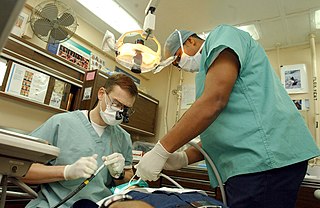
A dentist, also known as a dental surgeon, is a health care professional who specializes in dentistry, the branch of medicine focused on the teeth, gums, and mouth. The dentist's supporting team aids in providing oral health services. The dental team includes dental assistants, dental hygienists, dental technicians, and sometimes dental therapists.

<span class="mw-page-title-main">Root canal</span> Hollow part of the root of a tooth

A root canal is the naturally occurring anatomic space within the root of a tooth. It consists of the pulp chamber, the main canal(s), and more intricate anatomical branches that may connect the root canals to each other or to the surface of the root.

<span class="mw-page-title-main">Apicoectomy</span> Endodontic root end surgery

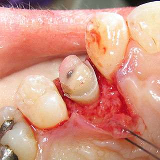
A root end surgery, also known as apicoectomy, apicectomy, retrograde root canal treatment or root-end filling, is an endodontic surgical procedure whereby a tooth's root tip is removed and a root end cavity is prepared and filled with a biocompatible material. It is an example of a periradicular surgery.

Dens invaginatus (DI), also known as tooth within a tooth, is a rare dental malformation and a developmental anomaly where there is an infolding of enamel into dentin. The prevalence of this condition is 0.3 - 10%, affecting males more frequently than females. The condition presents in two forms, coronal involving tooth crown and radicular involving tooth root, with the former being more common.

<span class="mw-page-title-main">Crown lengthening</span> Dental procedure

Crown lengthening is a surgical procedure performed by a dentist, or more frequently a periodontist, where more tooth is exposed by removing some of the gingival margin (gum) and supporting bone. Crown lengthening can also be achieved orthodontically by extruding the tooth.

<span class="mw-page-title-main">Root canal treatment</span> Dental treatment

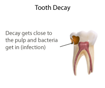
Root canal treatment is a treatment sequence for the infected pulp of a tooth which is intended to result in the elimination of infection and the protection of the decontaminated tooth from future microbial invasion. Root canals, and their associated pulp chamber, are the physical hollows within a tooth that are naturally inhabited by nerve tissue, blood vessels and other cellular entities. Together, these items constitute the dental pulp.

Mineral trioxide aggregate (MTA) was developed for use as a dental root repair material by Mahmoud Torabinejad. It is formulated from commercial Portland cement, combined with bismuth oxide powder for radio-opacity. MTA is used for creating apical plugs during apexification, repairing root perforations during root canal therapy, and treating internal root resorption. This can be used for root-end filling material and as pulp capping material. Originally, MTA was dark gray in color, but white versions have been on the market since 2002.

<span class="mw-page-title-main">Pulpotomy</span>

Pulpotomy is a minimally invasive procedure performed in children on a primary tooth with extensive caries but without evidence of root pathology. The minimally invasive endodontic techniques of vital pulp therapy (VPT) are based on improved understanding of the capacity of pulp (nerve) tissues to heal and regenerate plus the availability of advanced endodontic materials. During the caries removal, this results in a carious or mechanical pulp exposure (bleeding) from the cavity. During pulpotomy, the inflamed/diseased pulp tissue is removed from the coronal pulp chamber of the tooth leaving healthy pulp tissue which is dressed with a long-term clinically successful medicament that maintains the survival of the pulp and promotes repair. There are various types of medicament placed above the vital pulp such as Buckley's Solution of formocresol, ferric sulfate, calcium hydroxide or mineral trioxide aggregate (MTA). MTA is a more recent material used for pulpotomies with a high rate of success, better than formocresol or ferric sulfate. It is also recommended to be the preferred pulpotomy agent in the future. After the coronal pulp chamber is filled, the tooth is restored with a filling material that seals the tooth from microleakage, such as a stainless steel crown which is the most effective long-term restoration. However, if there is sufficient remaining supporting tooth structure, other filling materials such as amalgam or composite resin can provide a functional alternative when the primary tooth has a life span of two years or less. The medium- to long-term treatment outcomes of pulpotomy in symptomatic permanent teeth with caries, especially in young people, indicate that pulpotomy can be a potential alternative to root canal therapy (RCT).

Pulp necrosis is a clinical diagnostic category indicating the death of cells and tissues in the pulp chamber of a tooth with or without bacterial invasion. It is often the result of many cases of dental trauma, caries and irreversible pulpitis.

Dental pulpal testing is a clinical and diagnostic aid used in dentistry to help establish the health of the dental pulp within the pulp chamber and root canals of a tooth. Such investigations are important in aiding dentists in devising a treatment plan for the tooth being tested.

A phoenix abscess is an acute exacerbation of a chronic periapical lesion. It is a dental abscess that can occur immediately following root canal treatment. Another cause is due to untreated necrotic pulp. It is also the result of inadequate debridement during the endodontic procedure. Risk of occurrence of a phoenix abscess is minimised by correct identification and instrumentation of the entire root canal, ensuring no missed anatomy.

<span class="mw-page-title-main">Operating microscope</span> Optical microscope used in surgery

An operating microscope or surgical microscope is an optical microscope specifically designed to be used in a surgical setting, typically to perform microsurgery.

In dentistry, the smear layer is a layer found on root canal walls after root canal instrumentation. It consists of microcrystalline and organic particle debris. It was first described in 1975 and research has been performed since then to evaluate its importance in bacteria penetration into the dentinal tubules and its effects on endodontic treatment. More broadly, it is the organic layer found over all hard tooth surfaces.

<span class="mw-page-title-main">Regenerative endodontics</span> Dental specialty

Regenerative endodontic procedures is defined as biologically based procedures designed to replace damaged structures such as dentin, root structures, and cells of the pulp-dentin complex. This new treatment modality aims to promote normal function of the pulp. It has become an alternative to heal apical periodontitis. Regenerative endodontics is the extension of root canal therapy. Conventional root canal therapy cleans and fills the pulp chamber with biologically inert material after destruction of the pulp due to dental caries, congenital deformity or trauma. Regenerative endodontics instead seeks to replace live tissue in the pulp chamber. The ultimate goal of regenerative endodontic procedures is to regenerate the tissues and the normal function of the dentin-pulp complex.

<span class="mw-page-title-main">Pulp capping</span> Dental restoration technique

Pulp capping is a technique used in dental restorations to prevent the dental pulp from necrosis, after being exposed, or nearly exposed during a cavity preparation, from a traumatic injury, or by a deep cavity that reaches the center of the tooth causing the pulp to die. When dental caries is removed from a tooth, all or most of the infected and softened enamel and dentin are removed. This can lead to the pulp of the tooth either being exposed or nearly exposed which causes pulpitis (inflammation). Pulpitis, in turn, can become irreversible, leading to pain and pulp necrosis, and necessitating either root canal treatment or extraction. The ultimate goal of pulp capping or stepwise caries removal is to protect a healthy dental pulp and avoid the need for root canal therapy.

In the dental specialty of endodontics, periradicular surgery is surgery to the external root surface. Examples of periradicular surgery include apicoectomy, root resection, repair of root perforation or resorption defects, removal of broken fragments of the tooth or a filling material, and exploratory surgery to look for root fractures.

Periapical granuloma, also sometimes referred to as a radicular granuloma or apical granuloma, is an inflammation at the tip of a dead (nonvital) tooth. It is a lesion or mass that typically starts out as an epithelial lined cyst, and undergoes an inward curvature that results in inflammation of granulation tissue at the root tips of a dead tooth. This is usually due to dental caries or a bacterial infection of the dental pulp. Periapical granuloma is an infrequent disorder that has an occurrence rate between 9.3 to 87.1 percent. Periapical granuloma is not a true granuloma due to the fact that it does not contain granulomatous inflammation; however, periapical granuloma is a common term used.

Tooth replantation is a form of restorative dentistry in which an avulsed or luxated tooth is reinserted and secured into its socket through a combination of dental procedures. The purposes of tooth replantation is to resolve tooth loss and preserve the natural landscape of the teeth. Whilst variations of the procedure exist including, Allotransplantation, where a tooth is transferred from one individual to another individual of the same species. It is a largely defunct practice due to the improvements made within the field of dentistry and due to the risks and complications involved including the transmission of diseases such as syphilis, histocompatibility, as well as the low success rate of the procedure, has resulted in its practice being largely abandoned. Autotransplantation, otherwise known as intentional replantation in dentistry, is defined as the surgical movement of a tooth from one site on an individual to another location in the same individual. While rare, modern dentistry uses replantation as a form of proactive care to prevent future complications and protect the natural dentition in cases where root canal and surgical endodontic treatments are problematic. In the modern context, tooth replantation most often refers to reattachment of an avulsed or luxated permanent tooth into its original socket.

Apexification is a method of dental treatment to induce a calcific barrier in a root with incomplete formation or open apex of a tooth with necrotic pulp. Pulpal involvement usually occurs as a consequence of trauma or caries involvement of young or immature permanent teeth. As a sequelae of untreated pulp involvement, loss of pulp vitality or necrotic pulp took place for the involved teeth.

Root resection or root amputation is a type of periradicular surgery in which an entire root of a multiroot tooth is removed. It contrasts with an apicoectomy, where only the tip of the root is removed, and hemisection, where a root and its overlying portion of the crown are separated from the rest of the tooth, and optionally removed.

References

  1. "Definition of ENDODONTICS". www.merriam-webster.com. Retrieved 2019-01-19.
  2. Torabinejad, Mahmoud; Kutsenko, Diana; Machnick, Tanya K.; Ismail, Amid; Newton, Carl W. (1 September 2005). "Levels of Evidence for the Outcome of Nonsurgical Endodontic Treatment". Journal of Endodontics. 31 (9): 637–646. doi:10.1097/01.don.0000153593.64951.14. ISSN   0099-2399. PMID   16123698.
  3. Danbury smiles. "Procedures an Endodontist Performs" . Retrieved 29 Jan 2021.
  4. 1 2 3 Carrotte, Peter V. (March 2006). "Vital guide to Endodontics". Vital. 3 (1): 21–25. doi: 10.1038/vital378 . ISSN   1741-7511.
  5. 1 2 Garg, Nisha; Amit, Garg (2013-12-30). Textbook of endodontics (Third ed.). New Delhi. ISBN   978-93-5090-952-2. OCLC   881001658.{{cite book}}: CS1 maint: location missing publisher (link)
  6. Ricketts, David; Bartlett, David W. (2011). Advanced operative dentistry : a practical approach. Edinburgh: Elsevier. ISBN   978-0-7020-3126-7. OCLC   745905736.
  7. Andersson, Lars; Andreasen, Jens O.; Day, Peter; Heithersay, Geoffrey; Trope, Martin; Diangelis, Anthony J.; Kenny, David J.; Sigurdsson, Asgeir; Bourguignon, Cecilia; Flores, Marie Therese; Hicks, Morris Lamar; Lenzi, Antonio R.; Malmgren, Barbro; Moule, Alex J.; Tsukiboshi, Mitsuhiro (2012). "International Association of Dental Traumatology guidelines for the management of traumatic dental injuries: 2. Avulsion of permanent teeth". Dental Traumatology. 28 (2): 88–96. doi: 10.1111/j.1600-9657.2012.01125.x . ISSN   1600-9657. PMID   22409417.
  8. "Avulsion – Dental Trauma Guide". dentaltraumaguide.org.
  9. Hargreaves, Kenneth M.; Berman, Louis H.; Rotstein, Ilan (2015-09-23). Cohen's pathways of the pulp (Eleventh ed.). St. Louis, Mo. ISBN   978-0-323-18586-8. OCLC   931533556.{{cite book}}: CS1 maint: location missing publisher (link)
  10. 1 2 Rao, R. Nageswar. (2009). Advanced endodontics. New Delhi: Jaypee Briothers Medical Publishers. ISBN   978-81-8448-746-6. OCLC   720640774.
  11. Farook, S. A.; Shah, V.; Lenouvel, D.; Sheikh, O.; Sadiq, Z.; Cascarini, L. (December 2014). "Guidelines for management of sodium hypochlorite extrusion injuries". British Dental Journal. 217 (12): 679–684. doi: 10.1038/sj.bdj.2014.1099 . ISSN   1476-5373. PMID   25525012.
  12. "Endodontic Surgery Explained". American Association of Endodontists. Retrieved 2019-12-14.
  13. 1 2 "Endodontists' Guide to CDT 2017" (PDF). American Association of Endodontists. 2017. pp. 11–14. Retrieved 2020-03-14.
  14. 1 2 von Arx, Thomas (January 2011). "Apical surgery: A review of current techniques and outcome". The Saudi Dental Journal. 23 (1): 9–15. doi:10.1016/j.sdentj.2010.10.004. ISSN   1013-9052. PMC   3770245 . PMID   24151412.
  15. 1 2 Locurcio, Lino Lucio; Leeson, Rachel (2017). "A case of periradicular surgery: apicoectomy and obturation of the apex, a bold act". Stomatological Disease and Science. 1 (2). doi: 10.20517/2573-0002.2016.08 . Retrieved 2020-03-05.
  16. Siqueira JF, Rôças IN. Microbiology and treatment of endodontic infections. In: Hargreaves KM, Cohen S, Berman LH, editors. Cohen’s pathways of the pulp. 11th ed. St Louis: Mosby Elsevier; 2016. p. 599.
  17. Hauman, C. H. J.; Chandler, N. P.; Tong, D. C. (February 2002). "Endodontic implications of the maxillary sinus: a review". International Endodontic Journal. 35 (2): 127–141. doi: 10.1046/j.0143-2885.2001.00524.x . ISSN   0143-2885. PMID   11843967.
  18. 1 2 Arens, Donald E. (2003). "Introduction to magnification in endodontics". Journal of Esthetic and Restorative Dentistry. 15 (7): 426–439. doi:10.1111/j.1708-8240.2003.tb00970.x. ISSN   1496-4155. PMID   15000913.
  19. Hegde, Rashmi; Hegde, Vivek (2016-05-01). "Magnification-enhanced contemporary dentistry: Getting started". Journal of Interdisciplinary Dentistry. 6 (2): 91. doi: 10.4103/2229-5194.197695 . S2CID   78692557.
  20. Del Fabbro, Massimo; Taschieri, Silvio; Lodi, Giovanni; Banfi, Giuseppe; Weinstein, Roberto L. (2015). "Magnification devices for endodontic therapy". Cochrane Database of Systematic Reviews. 2016 (12): CD005969. doi:10.1002/14651858.cd005969.pub3. hdl: 2434/434134 . PMC   7389255 . PMID   26650099.
  21. "Use of Microscopes and Other Magnification Techniques - Position Paper" (PDF). www.aae.org. 2012.
  22. "CBCT: The New Standard of Care?". American Association of Endodontists. 2018-04-03. Retrieved 2019-10-21.
  23. Patel, S.; Brown, J.; Pimentel, T.; Kelly, R. D.; Abella, F.; Durack, C. (2019). "Cone beam computed tomography in Endodontics – a review of the literature". International Endodontic Journal. 52 (8): 1138–1152. doi: 10.1111/iej.13115 . ISSN   1365-2591. PMID   30868610.
  24. "Get Board Certified". American Board of Endodontics. Retrieved 2019-01-19.

Organizations

Publications