Ansa cervicalis

Last updated
Ansa cervicalis
Gray795.png
Ansa cervicalis. Superior root labeled as "descending hypoglossal," Inferior root labeled as "descending cervical."
Details
Innervates Sternohyoid muscle,
sternothyroid muscle,
omohyoid muscle
Identifiers
Latin ansa cervicalis,
ansa hypoglossi
TA98 A14.2.02.013
TA2 6376
FMA 55142
Anatomical terms of neuroanatomy

The ansa cervicalis (or ansa hypoglossi in older literature[ citation needed ]) is a loop formed by muscular branches of the cervical plexus formed by branches of cervical spinal nerves C1-C3. The ansa cervicalis has two roots - a superior root (formed by branch of C1) and an inferior root (formed by union of branches of C2 and C3) - that unite distally, forming a loop. It is situated within the carotid sheath. [1] :334

Contents

Branches of the ansa cervicalis innervate three of the four infrahyoid muscles: the sternothyroid, sternohyoid, and omohyoid muscles (note that the thyrohyoid muscle is the one infrahyoid muscle not innervated by the ansa cervicalis - it is instead innervated by cervical spinal nerve 1 via a separate thyrohyoid branch [2] :582, 600).

Its name means "handle of the neck" in Latin.[ citation needed ]

Anatomy

The ansa cervicalis is typically embedded within the anterior wall of the carotid sheath anterior to the internal jugular vein. [1] :344

Superior root

Ansa cervicalis Ansa cervicalis.jpg
Ansa cervicalis

The superior root of the ansa cervicalis (formerly known as descendens hypoglossi [1] :500) is by fibres of the cervical spinal nerve 1 [1] :344 [3] (and, according to some sources, of cervical spinal nerve 2 as well [3] ) that have joined and run with the hypoglossal nerve (CN XII) for some distance before [1] :344 progressively [1] :369 branching off the CN XII in the carotid triangle to form the superior root. [1] :344

The superior root is situated within the carotid triangle. [1] :344 It passes anterior-ward between the internal carotid artery and the common carotid artery. [1] :500 It curves around the occipital artery [ citation needed ] before descending upon the anterior aspect of the internal carotid artery and the common carotid artery. [1] :344 on the carotid sheath.[ citation needed ] It issues a branch to the superior belly of the omohyoid muscle, [1] :344 and the upper parts of the sternothyroid and sternohyoid muscles before uniting with the inferior root.[ citation needed ]

Inferior root

The inferior root of the ansa cervicalis (formerly known as descendens cervicalis [1] :500) is formed by the union of fibers of the anterior rami spinal cervical nerves C2-C3 that unite as part of the cervical plexus. [1] :344

The inferior root curves posteroanteriorly around [1] :344 the lateral side of the internal jugular vein [1] :343 before descending to unite with the superior root upon the (inferior portion of) the internal jugular vein. It may occasionally pass anterior in between the internal jugular vein and the internal carotid artery. [1] :344

Branches

Branches to the sternothyroid muscle, sternohyoid muscle, and inferior belly of the omohyoid muscle issue from the loop of the ansa cervicalis, whereas the branch for the superior belly of the omohyoid muscle arises from the superior root. [1] :344

Additional images

Related Research Articles

<span class="mw-page-title-main">Neck</span> Part of the body on many vertebrates that connects the head with the torso

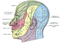
The neck is the part of the body on many vertebrates that connects the head with the torso. The neck supports the weight of the head and protects the nerves that carry sensory and motor information from the brain down to the rest of the body. In addition, the neck is highly flexible and allows the head to turn and flex in all directions. The structures of the human neck are anatomically grouped into four compartments: vertebral, visceral and two vascular compartments. Within these compartments, the neck houses the cervical vertebrae and cervical part of the spinal cord, upper parts of the respiratory and digestive tracts, endocrine glands, nerves, arteries and veins. Muscles of the neck are described separately from the compartments. They bound the neck triangles.

Articles related to anatomy include:

<span class="mw-page-title-main">Subclavian artery</span> Major arteries of the upper thorax, below the clavicle

In human anatomy, the subclavian arteries are paired major arteries of the upper thorax, below the clavicle. They receive blood from the aortic arch. The left subclavian artery supplies blood to the left arm and the right subclavian artery supplies blood to the right arm, with some branches supplying the head and thorax. On the left side of the body, the subclavian comes directly off the aortic arch, while on the right side it arises from the relatively short brachiocephalic artery when it bifurcates into the subclavian and the right common carotid artery.

<span class="mw-page-title-main">Internal carotid artery</span> Artery of the human brain

The internal carotid artery is an artery in the neck which supplies the anterior circulation of the brain.

<span class="mw-page-title-main">Infrahyoid muscles</span> Group of muscles

The infrahyoid muscles, or strap muscles, are a group of four pairs of muscles in the anterior (frontal) part of the neck. The four infrahyoid muscles are the sternohyoid, sternothyroid, thyrohyoid and omohyoid muscles.

<span class="mw-page-title-main">Omohyoid muscle</span> Human neck muscle

The omohyoid muscle is a muscle in the neck. It is one of the infrahyoid muscles. It consists of two bellies separated by an intermediate tendon. Its inferior belly is attached to the scapula; its superior belly is attached to the hyoid bone. Its intermediate tendon is anchored to the clavicle and first rib by a fascial sling. The omohyoid is innervated by the ansa cervicalis of the cervical plexus. It acts to depress the hyoid bone.

<span class="mw-page-title-main">Cervical plexus</span> Network of nerves in the neck

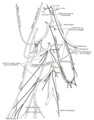
The cervical plexus is a nerve plexus of the anterior rami of the first four cervical spinal nerves C1-C4. The cervical plexus provides motor innervation to some muscles of the neck, and the diaphragm; it provides sensory innervation to parts of the head, neck, and chest.

<span class="mw-page-title-main">Common carotid artery</span> One of the two arteries that supply the head and neck with blood

In anatomy, the left and right common carotid arteries (carotids) are arteries that supply the head and neck with oxygenated blood; they divide in the neck to form the external and internal carotid arteries.

<span class="mw-page-title-main">Sternohyoid muscle</span>

The sternohyoid muscle is a bilaterally paired, long, thin, narrow strap muscle of the anterior neck. It is one of the infrahyoid muscles. It is innervated by the ansa cervicalis. It acts to depress the hyoid bone.

<span class="mw-page-title-main">Sternothyroid muscle</span>

The sternothyroid muscle is an infrahyoid muscle of the neck. It acts to depress the hyoid bone.

<span class="mw-page-title-main">Thyrohyoid muscle</span> Neck muscle that depresses the hyoid bone and elevates the larynx

The thyrohyoid muscle is a small skeletal muscle of the neck. Above, it attaches onto the greater cornu of the hyoid bone; below, it attaches onto the oblique line of the thyroid cartilage. It is innervated by fibres derived from the cervical spinal nerve 1 that run with the hypoglossal nerve to reach this muscle. The thyrohyoid muscle depresses the hyoid bone and elevates the larynx during swallowing. By controlling the position and shape of the larynx, it aids in making sound.

<span class="mw-page-title-main">Carotid sheath</span>

The carotid sheath is a condensation of the deep cervical fascia enveloping multiple vital neurovascular structures of the neck, including the common and internal carotid arteries, the internal jugular vein, the vagus nerve, and ansa cervicalis. The carotid sheath helps protects the structures contained therein.

<span class="mw-page-title-main">Superior thyroid artery</span>

The superior thyroid artery arises from the external carotid artery just below the level of the greater cornu of the hyoid bone and ends in the thyroid gland.

<span class="mw-page-title-main">Anterior triangle of the neck</span>

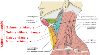
The anterior triangle is a region of the neck.

<span class="mw-page-title-main">Deep cervical fascia</span>

The deep cervical fascia lies under cover of the platysma, and invests the muscles of the neck; it also forms sheaths for the carotid vessels, and for the structures situated in front of the vertebral column. Its attachment to the hyoid bone prevents the formation of a dewlap.

<span class="mw-page-title-main">Carotid triangle</span>

The carotid triangle is a portion of the anterior triangle of the neck.

<span class="mw-page-title-main">Muscular triangle</span>

The inferior carotid triangle, is bounded, in front, by the median line of the neck from the hyoid bone to the sternum; behind, by the anterior margin of the sternocleidomastoid; above, by the superior belly of the omohyoid.

<span class="mw-page-title-main">Outline of human anatomy</span> Overview of and topical guide to human anatomy

The following outline is provided as an overview of and topical guide to human anatomy:

<span class="mw-page-title-main">Cervical spinal nerve 1</span> Spinal nerve of the cervical segment

The cervical spinal nerve 1 (C1) is a spinal nerve of the cervical segment. C1 carries predominantly motor fibres, but also a small meningeal branch that supplies sensation to parts of the dura around the foramen magnum.

<span class="mw-page-title-main">Cervical spinal nerve 2</span> Spinal nerve of the cervical segment

The cervical spinal nerve 2 (C2) is a spinal nerve of the cervical segment. It is a part of the ansa cervicalis along with the C1 and C3 nerves sometimes forming part of superior root of the ansa cervicalis. it also connects into the inferior root of the ansa cervicalis with the C3.

References

  1. 1 2 3 4 5 6 7 8 9 10 11 12 13 14 15 16 17 Sinnatamby, Chummy S. (2011). Last's Anatomy (12th ed.). Elsevier Australia. ISBN   978-0-7295-3752-0.
  2. Standring, Susan (2020). Gray's Anatomy: The Anatomical Basis of Clinical Practice (42nd ed.). New York. ISBN   978-0-7020-7707-4. OCLC   1201341621.{{cite book}}: CS1 maint: location missing publisher (link)
  3. 1 2 Moore, Keith L.; Dalley, Arthur F.; Agur, Anne M. R. (2018). Clinically Oriented Anatomy (8th ed.). Wolters Kluwer. p. 1005. ISBN   978-1-4963-4721-3.