The axillary nerve or the circumflex nerve is a nerve of the human body, that originates from the brachial plexus at the level of the axilla (armpit) and carries nerve fibers from C5 and C6. The axillary nerve travels through the quadrangular space with the posterior circumflex humeral artery and vein to innervate the deltoid and teres minor.

The lesser occipital nerve is a cutaneous spinal nerve of the cervical plexus. It arises from second cervical (spinal) nerve (C2). It innervates the skin of the back of the upper neck and of the scalp posterior to the ear.

The great auricular nerve is a cutaneous (sensory) nerve of the head. It originates from the second and third cervical (spinal) nerves (C2-C3) of the cervical plexus. It provides sensory innervation to the skin over the parotid gland and the mastoid process, parts of the outer ear, and to the parotid gland and its fascia.

The dorsal scapular nerve is a branch of the brachial plexus, usually derived from the ventral ramus of cervical nerve C5. It provides motor innervation to the rhomboid major muscle, rhomboid minor muscle, and levator scapulae muscle.
The long thoracic nerve is a branch of the brachial plexus derived from cervical nerves C5-C7 that innervates the serratus anterior muscle.
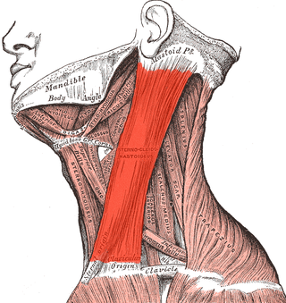
The sternocleidomastoid muscle is one of the largest and most superficial cervical muscles. The primary actions of the muscle are rotation of the head to the opposite side and flexion of the neck. The sternocleidomastoid is innervated by the accessory nerve.

The internal carotid artery is an artery in the neck which supplies the anterior circulation of the brain.

The infrahyoid muscles, or strap muscles, are a group of four pairs of muscles in the anterior (frontal) part of the neck. The four infrahyoid muscles are the sternohyoid, sternothyroid, thyrohyoid and omohyoid muscles.

The levator scapulae is a slender skeletal muscle situated at the back and side of the neck. It originates from the transverse processes of the four uppermost cervical vertebrae; it inserts onto the upper portion of the medial border of the scapula. It is innervated by the cervical nerves C3-C4, and frequently also by the dorsal scapular nerve. As the Latin name suggests, its main function is to lift the scapula.

The omohyoid muscle is a muscle in the neck. It is one of the infrahyoid muscles. It consists of two bellies separated by an intermediate tendon. Its inferior belly is attached to the scapula; its superior belly is attached to the hyoid bone. Its intermediate tendon is anchored to the clavicle and first rib by a fascial sling. The omohyoid is innervated by the ansa cervicalis of the cervical plexus. It acts to depress the hyoid bone.

The scalene muscles are a group of three muscles on each side of the neck, identified as the anterior, the middle, and the posterior. They are innervated by the third to the eighth cervical spinal nerves (C3-C8).

The ansa cervicalis is a loop formed by muscular branches of the cervical plexus formed by branches of cervical spinal nerves C1-C3. The ansa cervicalis has two roots - a superior root and an inferior root - that unite distally, forming a loop. It is situated within the carotid sheath.

The thyrohyoid muscle is a small skeletal muscle of the neck. Above, it attaches onto the greater cornu of the hyoid bone; below, it attaches onto the oblique line of the thyroid cartilage. It is innervated by fibres derived from the cervical spinal nerve 1 that run with the hypoglossal nerve to reach this muscle. The thyrohyoid muscle depresses the hyoid bone and elevates the larynx during swallowing. By controlling the position and shape of the larynx, it aids in making sound.

The carotid sheath is a condensation of the deep cervical fascia enveloping multiple vital neurovascular structures of the neck, including the common and internal carotid arteries, the internal jugular vein, the vagus nerve, and ansa cervicalis. The carotid sheath helps protects the structures contained therein.

The posterior triangle is a region of the neck.
The supraclavicular nerve is a cutaneous (sensory) nerve of the cervical plexus that arises from the third and fourth cervical (spinal) nerves. It emerges from beneath the posterior border of the sternocleidomastoid muscle, then split into multiple branches. Together, these innervate the skin over the shoulder.

The piriformis nerve, also known as the nerve to piriformis, is the peripheral nerve that provides motor innervation to the piriformis muscle.

The cervical spinal nerve 1 (C1) is a spinal nerve of the cervical segment. C1 carries predominantly motor fibres, but also a small meningeal branch that supplies sensation to parts of the dura around the foramen magnum.
The cervical spinal nerve 2 (C2) is a spinal nerve of the cervical segment. It is a part of the ansa cervicalis along with the C1 and C3 nerves sometimes forming part of superior root of the ansa cervicalis. it also connects into the inferior root of the ansa cervicalis with the C3.
The thyrohyoid branch (also: thyrohyoid branch of ansa cervicalis, or nerve to thyrohyoid (muscle)) is a motor branch derived from the cervical plexus formed by fibres of (the anterior ramus of) the cervical spinal nerve 1 (C1) (and - according to some sources - cervical spinal nerve 2 (C2) as well) that join and travel with the hypoglossal nerve (cranial nerve XII) to reach the suprahyoid region, branching away from CN XII distal to the superior root of ansa cervicalis (which is a branching other fibres of C1-C2 that had traveled with the CN XII), near the posterior border of the hyoglossus muscle. The thyrohyoid branch of ansa cervicalis innervates the thyrohyoid muscle.