Scalene muscles

Last updated

Scalene muscles
Scalenus.png
The anterior vertebral muscles.
Details
Origin Cervical vertebrae (CII-CVII)
Insertion First and second ribs
Artery Ascending cervical artery
(branch of Inferior thyroid artery)
Nerve Cervical nerves (C3-C8)
Actions Elevation of first and second ribs
Identifiers
Latin mm. scalenii
Anatomical terms of muscle

The scalene muscles are a group of three muscles on each side of the neck, identified as the anterior, the middle, and the posterior. They are innervated by the third to the eighth cervical spinal nerves (C3-C8).

Contents

The anterior and middle scalene muscles lift the first rib and bend the neck to the side they are on. The posterior scalene lifts the second rib and tilts the neck to the same side.

The muscles are named from Ancient Greek σκαληνός (skalēnós) 'uneven'.

Structure

The scalene muscles are attached at one end to bony protrusions on vertebrae C2 to C7 and at the other end to the first and second ribs. [1]

Anterior scalene Scalenus anterior01.png
Anterior scalene

Anterior scalene

The anterior scalene muscle (Latin : scalenus anterior), lies deeply at the side of the neck, behind the sternocleidomastoid muscle. It arises from the anterior tubercles of the transverse processes of the third, fourth, fifth, and sixth cervical vertebrae, and descending, almost vertically, is inserted by a narrow, flat tendon into the scalene tubercle on the inner border of the first rib, and into the ridge on the upper surface of the second rib in front of the subclavian groove. It is supplied by the anterior ramus of cervical nerve 5 and 6.

Middle scalene Gray385 - Scalenus medius muscle.png
Middle scalene

Middle scalene

The middle scalene, (Latin : scalenus medius), is the largest and longest of the three scalene muscles. The middle scalene arises from the posterior tubercles of the transverse processes of the lower six cervical vertebrae. It descends along the side of the vertebral column to insert by a broad attachment into the upper surface of the first rib, posterior to the subclavian groove. The brachial plexus and the subclavian artery pass anterior to it.

Posterior scalene Scalenus posterior04.png
Posterior scalene

Posterior scalene

The posterior scalene, (Latin : scalenus posterior) is the smallest and most deeply seated of the scalene muscles. It arises, by two or three separate tendons, from the posterior tubercles of the transverse processes of the lower two or three cervical vertebrae, and is inserted by a thin tendon into the outer surface of the second rib, behind the attachment of the anterior scalene. It is supplied by cervical nerves C5, C6 and C7. It is occasionally blended with the middle scalene.

Variation

A fourth muscle, the scalenus minimus (Sibson's muscle), is sometimes present behind the lower portion of the anterior scalene. [2]

Function

The anterior and middle scalene muscles lift the first rib and bend the neck to the same side as the acting muscle; [3] the posterior scalene lifts the second rib and tilts the neck to the same side.

Because they elevate the upper ribs, they also act as accessory muscles of respiration, along with the sternocleidomastoids.

Relations

The scalene muscles have an important relationship to other structures in the neck. The brachial plexus and subclavian artery pass between the anterior and middle scalenes. [4] The subclavian vein and phrenic nerve pass anteriorly to the anterior scalene as the muscle crosses over the first rib. The phrenic nerve is oriented vertically as it passes in front of the anterior scalene, while the subclavian vein is oriented horizontally as it passes in front of the anterior scalene muscle. [4]

The passing of the brachial plexus and the subclavian artery through the space of the anterior and middle scalene muscles constitute the scalene hiatus (the term "scalene fissure" is also used). The region in which this lies is referred to as the scaleotracheal fossa. It is bounded by the clavicle inferior anteriorly, the trachea medially, posteriorly by the trapezius, and anteriorly by the platysma muscle.

Clinical significance

The anterior and middle scalene muscles can be involved in certain forms of thoracic outlet syndrome as well as myofascial pain syndrome, the symptoms of which may mimic a spinal disc herniation of the cervical vertebrae. [5]

Since the nerves of the brachial plexus pass through the space between the anterior and middle scalene muscles, that area is sometimes targeted with the administration of regional anesthesia by an anesthesia provider. The nerve block, called an interscalene block, may be performed prior to arm or shoulder surgery. [6]

According to the medical codes in the 2016 Procedural Coding Expert, published by the American Academy of Professional Coders, for Current Procedural Terminology (CPT) and other medical codes, the scalenus anticus muscle can be divided by reparative or reconstructive surgery, with (# 21705) or without (# 21700) resection of the cervical rib.

History

The scalenes used to be known as the lateral vertebral muscles. [7]

Etymology

The muscles are named from Greek σκαληνός, or skalēnós, meaning uneven [8] as the pairs are all of differing length [2]

Additional images

Related Research Articles

<span class="mw-page-title-main">Rib cage</span> Bone structure of the thorax

The rib cage or thoracic cage, is an endoskeletal enclosure in the thorax of most vertebrates that comprises the ribs, vertebral column and sternum, which protect the vital organs of the thoracic cavity, such as the heart, lungs and great vessels and support the shoulder girdle to form the core part of the axial skeleton.

Articles related to anatomy include:

<span class="mw-page-title-main">Phrenic nerve</span> Nerve controlling the diaphragm

The phrenic nerve is a mixed motor/sensory nerve that originates from the C3-C5 spinal nerves in the neck. The nerve is important for breathing because it provides exclusive motor control of the diaphragm, the primary muscle of respiration. In humans, the right and left phrenic nerves are primarily supplied by the C4 spinal nerve, but there is also a contribution from the C3 and C5 spinal nerves. From its origin in the neck, the nerve travels downward into the chest to pass between the heart and lungs towards the diaphragm.

<span class="mw-page-title-main">Subclavian artery</span> Major arteries of the upper thorax, below the clavicle

In human anatomy, the subclavian arteries are paired major arteries of the upper thorax, below the clavicle. They receive blood from the aortic arch. The left subclavian artery supplies blood to the left arm and the right subclavian artery supplies blood to the right arm, with some branches supplying the head and thorax. On the left side of the body, the subclavian comes directly off the aortic arch, while on the right side it arises from the relatively short brachiocephalic artery when it bifurcates into the subclavian and the right common carotid artery.

<span class="mw-page-title-main">Sternocleidomastoid muscle</span> Cervical muscle

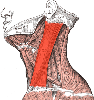
The sternocleidomastoid muscle is one of the largest and most superficial cervical muscles. The primary actions of the muscle are rotation of the head to the opposite side and flexion of the neck. The sternocleidomastoid is innervated by the accessory nerve.

<span class="mw-page-title-main">Levator scapulae muscle</span> Slender skeletal muscle at the back and side of the neck

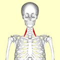
The levator scapulae is a slender skeletal muscle situated at the back and side of the neck. It originates from the transverse processes of the four uppermost cervical vertebrae; it inserts onto the upper portion of the medial border of the scapula. It is innervated by the cervical nerves C3-C4, and frequently also by the dorsal scapular nerve. As the Latin name suggests, its main function is to lift the scapula.

<span class="mw-page-title-main">Thoracic outlet syndrome</span> Medical condition

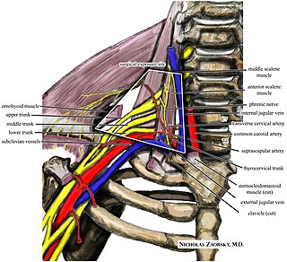
Thoracic outlet syndrome (TOS) is a condition in which there is compression of the nerves, arteries, or veins in the superior thoracic aperture, the passageway from the lower neck to the armpit, also known as the thoracic outlet. There are three main types: neurogenic, venous, and arterial. The neurogenic type is the most common and presents with pain, weakness, paraesthesia, and occasionally loss of muscle at the base of the thumb. The venous type results in swelling, pain, and possibly a bluish coloration of the arm. The arterial type results in pain, coldness, and pallor of the arm.

<span class="mw-page-title-main">Cervical plexus</span> Network of nerves in the neck

The cervical plexus is a nerve plexus of the anterior rami of the first four cervical spinal nerves C1-C4. The cervical plexus provides motor innervation to some muscles of the neck, and the diaphragm; it provides sensory innervation to parts of the head, neck, and chest.

<span class="mw-page-title-main">Cervical vertebrae</span> Vertebrae of the neck

In tetrapods, cervical vertebrae are the vertebrae of the neck, immediately below the skull. Truncal vertebrae lie caudal of cervical vertebrae. In sauropsid species, the cervical vertebrae bear cervical ribs. In lizards and saurischian dinosaurs, the cervical ribs are large; in birds, they are small and completely fused to the vertebrae. The vertebral transverse processes of mammals are homologous to the cervical ribs of other amniotes. Most mammals have seven cervical vertebrae, with the only three known exceptions being the manatee with six, the two-toed sloth with five or six, and the three-toed sloth with nine.

<span class="mw-page-title-main">Vertebral artery</span> Major arteries of the neck

The vertebral arteries are major arteries of the neck. Typically, the vertebral arteries originate from the subclavian arteries. Each vessel courses superiorly along each side of the neck, merging within the skull to form the single, midline basilar artery. As the supplying component of the vertebrobasilar vascular system, the vertebral arteries supply blood to the upper spinal cord, brainstem, cerebellum, and posterior part of brain.

<span class="mw-page-title-main">Common carotid artery</span> One of the two arteries that supply the head and neck with blood

In anatomy, the left and right common carotid arteries (carotids) are arteries that supply the head and neck with oxygenated blood; they divide in the neck to form the external and internal carotid arteries.

<span class="mw-page-title-main">Superior thoracic aperture</span>

The superior thoracic aperture, also known as the thoracic outlet, or thoracic inlet refers to the opening at the top of the thoracic cavity. It is also clinically referred to as the thoracic outlet, in the case of thoracic outlet syndrome. A lower thoracic opening is the inferior thoracic aperture.

<span class="mw-page-title-main">Posterior triangle of the neck</span> Region of the neck

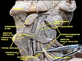
The posterior triangle is a region of the neck.

<span class="mw-page-title-main">Transverse cervical artery</span> Artery of the neck

The transverse cervical artery is an artery in the neck and a branch of the thyrocervical trunk, running at a higher level than the suprascapular artery.

<span class="mw-page-title-main">Suprascapular artery</span> Artery of the neck

The suprascapular artery is a branch of the thyrocervical trunk on the neck.

<span class="mw-page-title-main">Subclavian nerve</span>

The subclavian nerve, also known as the nerve to the subclavius, is a small branch of the upper trunk of the brachial plexus. It contains axons from C5 and C6. It innervates the subclavius muscle.

<span class="mw-page-title-main">Subclavian triangle</span>

The subclavian triangle, the smaller division of the posterior triangle, is bounded, above, by the inferior belly of the omohyoideus; below, by the clavicle; its base is formed by the posterior border of the sternocleidomastoideus.

<span class="mw-page-title-main">Prevertebral fascia</span> Layer of deep cervical fascia that surrounds the vertebral column

The prevertebral fascia is the layer of deep cervical fascia that surrounds the vertebral column. It is the deepest layer of deep cervical fascia.

<span class="mw-page-title-main">Outline of human anatomy</span> Overview of and topical guide to human anatomy

The following outline is provided as an overview of and topical guide to human anatomy:

References

  1. Henry Gray (1913). Anatomy: Descriptive and Applied. Lea & Febiger.
  2. 1 2 Davies, Clair; Davies, Amber (2013). The Trigger Point Therapy Workbook (Third ed.). New Harbinger Publications. ISBN   9781608824960.
  3. Buford JA; Yoder SM; Heiss DG; Chidley JV (October 2002). "Actions of the scalene muscles for rotation of the cervical spine in macaque and human". J Orthop Sports Phys Ther. 32 (10): 488–96. doi: 10.2519/jospt.2002.32.10.488 . PMID   12403200.
  4. 1 2 Albertine, David A. Morton, K. Bo Foreman, Kurt H. (2011). "Chapter 25: Overview of the Neck, Muscles of the Neck". Gross anatomy: the big picture. New York: McGraw-Hill. ISBN   978-0071476720.{{cite book}}: CS1 maint: multiple names: authors list (link)
  5. Abd Jalil, N; Awang, MS; Omar, M (2010). "Scalene myofascial pain syndrome mimicking cervical disc prolapse: a report of two cases". Malays J Med Sci. 17 (1): 60–6. PMC   3216145 . PMID   22135529.
  6. Graber, Raymound. "Interscalene Nerve Block". WebMD, LLC. Medscape. Retrieved December 10, 2012.
  7. Henry Gray (1913). Anatomy: Descriptive and Applied.
  8. Mosby's Medical, Nursing & Allied Health Dictionary, Fourth Edition, Mosby-Year Book Inc., 1994, p. 1395