
The larynx, commonly called the voice box, is an organ in the top of the neck involved in breathing, producing sound and protecting the trachea against food aspiration. The opening of larynx into pharynx known as the laryngeal inlet is about 4–5 centimeters in diameter. The larynx houses the vocal cords, and manipulates pitch and volume, which is essential for phonation. It is situated just below where the tract of the pharynx splits into the trachea and the esophagus. The word 'larynx' comes from the Ancient Greek word lárunx ʻlarynx, gullet, throat.ʼ

The omohyoid muscle is a muscle in the neck. It is one of the infrahyoid muscles. It consists of two bellies separated by an intermediate tendon. Its inferior belly is attached to the scapula; its superior belly is attached to the hyoid bone. Its intermediate tendon is anchored to the clavicle and first rib by a fascial sling. The omohyoid is innervated by the ansa cervicalis of the cervical plexus. It acts to depress the hyoid bone.

The rima glottidis is the opening between the two true vocal cords anteriorly, and the two arytenoid cartilages posteriorly. It is part of the larynx.

The posterior cricoarytenoid muscle is a intrinsic muscle of the larynx. It arises from the cricoid cartilage; it inserts onto the arytenoid cartilage of the same side. It is innervated by the recurrent laryngeal nerve. Each acts to open the vocal folds by pulling the vocal fold of the same side laterally. It participates in the production of sounds.

The cricoid cartilage, or simply cricoid or cricoid ring, is the only complete ring of cartilage around the trachea. It forms the back part of the voice box and functions as an attachment site for muscles, cartilages, and ligaments involved in opening and closing the airway and in producing speech.

The arytenoid cartilages are a pair of small three-sided pyramids which form part of the larynx. They are the site of attachment of the vocal cords. Each is pyramidal or ladle-shaped and has three surfaces, a base, and an apex. The arytenoid cartilages allow for movement of the vocal cords by articulating with the cricoid cartilage. They may be affected by arthritis, dislocations, or sclerosis.

The sternothyroid muscle is an infrahyoid muscle of the neck. It acts to depress the hyoid bone.

The thyrohyoid muscle is a small skeletal muscle of the neck. Above, it attaches onto the greater cornu of the hyoid bone; below, it attaches onto the oblique line of the thyroid cartilage. It is innervated by fibres derived from the cervical spinal nerve 1 that run with the hypoglossal nerve to reach this muscle. The thyrohyoid muscle depresses the hyoid bone and elevates the larynx during swallowing. By controlling the position and shape of the larynx, it aids in making sound.

The stylopharyngeus muscle is a muscle in the head. It originates from the temporal styloid process. Some of its fibres insert onto the thyroid cartilage, while others end by intermingling with proximal structures. It is innervated by the glossopharyngeal nerve. It acts to elevate the larynx and pharynx, and dilate the pharynx, thus facilitating swallowing.

The arytenoid muscle or interarytenoid muscle is a composite intrinsic muscle of the larynx, consisting of a transverse part and an oblique part - the two parts may be considered as separate muscles: an unpaired transverse arytenoid muscle, and a bilaterally paired oblique arytenoid muscle.

The aryepiglottic muscle or aryepiglotticus muscle is an intrinsic muscle of the larynx.
The oblique arytenoid is bilaterally paired intrinsic muscle of the larynx. It is superficial to the transverse arytenoid; the oblique and transverse arytenoids are often considered two parts of a single muscle - the interarytenoid muscle.

The inferior thyroid artery is an artery in the neck. It arises from the thyrocervical trunk and passes upward, in front of the vertebral artery and longus colli muscle. It then turns medially behind the carotid sheath and its contents, and also behind the sympathetic trunk, the middle cervical ganglion resting upon the vessel.

The posterior auricular artery is a small artery that arises from the external carotid artery. It ascends along the side of the head. It supplies several muscles of the neck and several structures of the head.
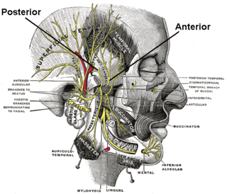
The deep temporal nerves are typically two nerves (one anterior and one posterior) which arise from the mandibular nerve (CN V3) and provide motor innervation to the temporalis muscle.
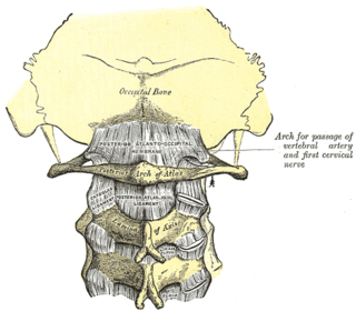
The posterior atlantooccipital membrane is a broad but thin membrane extending between the to the posterior margin of the foramen magnum above, and posterior arch of atlas below. It forms the floor of the suboccipital triangle.
The hyoepiglottic ligament is an extrinsic ligament of the larynx connecting the epiglottis and the thyroid cartilage.

The transverse arytenoid is an unpaired intrinsic muscle of the larynx. It is situated deep to the two oblique arytenoids; the oblique and transverse arytenoids are often considered two parts of a single muscle - the interarytenoid (arytenoid) muscle.

The quadrangular membrane is a layer of submucosa. It extends between the lateral margin of the epiglottis, and the apex and fovea triangularis of the ipsilateral arytenoid cartilage. It has free superior and inferior borders.
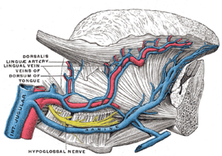
The deep lingual vein is one of the lingual veins. It commences near the apex of the tongue. It passes posterior-ward close to the inferior surface of the tongue. It terminates near the anterior border of the hyoglossus muscle by uniting with the sublingual vein to form the vena comitans of the hypoglossal nerve ; this vein then passes posterior-ward alongside the nerve to empty into either a lingual vein, the (common) facial vein, or the internal jugular vein.
