
The larynx, commonly called the voice box, is an organ in the top of the neck involved in breathing, producing sound and protecting the trachea against food aspiration. The opening of larynx into pharynx known as the laryngeal inlet is about 4–5 centimeters in diameter. The larynx houses the vocal cords, and manipulates pitch and volume, which is essential for phonation. It is situated just below where the tract of the pharynx splits into the trachea and the esophagus. The word ʻlarynxʼ comes from the Ancient Greek word lárunx ʻlarynx, gullet, throat.ʼ
Articles related to anatomy include:

The rima glottidis is the opening between the two true vocal cords anteriorly, and the two arytenoid cartilages posteriorly. It is part of the larynx.
The lateral cricoarytenoid muscles extend from the lateral cricoid cartilage to the muscular process of the arytenoid cartilage. By rotating the arytenoid cartilages medially, these muscles adduct the vocal cords and thereby close the rima glottidis, protecting the airway. The lateral cricoarytenoid muscles receive innervation from the recurrent laryngeal branch of the vagus nerve.

The posterior cricoarytenoid muscles are small, paired intrinsic muscles of the larynx that extend between cricoid cartilage to the arytenoid cartilages in the larynx.
The cricothyroid ligament is a ligament in the neck. It connects the cricoid cartilage to the thyroid cartilage. It prevents these cartilages from moving too far apart. It is cut during an emergency cricothyrotomy to treat upper airway obstruction.

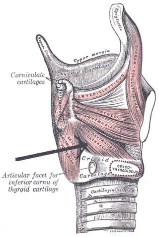
The arytenoid cartilages are a pair of small three-sided pyramids which form part of the larynx. They are the site of attachment of the vocal cords. Each is pyramidal or ladle-shaped and has three surfaces, a base, and an apex. The arytenoid cartilages allow for movement of the vocal cords by articulating with the cricoid cartilage. They may be affected by arthritis, dislocations, or sclerosis.

The stylopharyngeus is a muscle in the head. It originates from the temporal styloid process. It some of its fibres insert onto the thyroid cartilage, while others end by intermingling with proximal structures. It is innervated by the glossopharyngeal nerve. It acts to elevate the larynx and pharynx, and dilate the pharynx, thus facilitating swallowing.

The arytenoid muscle is a single muscle of the larynx. It passes from one arytenoid cartilage to the opposite arytenoid cartilage. It has oblique and transverse fibres. It is supplied by the recurrent laryngeal nerve. It approximates the arytenoid cartilages. Continuous electromyography may be used during neck surgeries such as thyroidectomy.

The thyroarytenoid muscle is a broad, thin muscle that forms the body of the vocal fold and that supports the wall of the ventricle and its appendix. It functions to shorten the vocal folds.
Cricoarytenoid muscles are muscles that connect the cricoid cartilage and arytenoid cartilage.

In the human larynx, the cuneiform cartilages are two small, elongated pieces of yellow elastic cartilage, placed one on either side, in the aryepiglottic fold.

The aryepiglottic muscle, or aryepiglotticus muscle is an intrinsic muscle of the larynx.
The oblique arytenoid, the more superficial arytenoid muscle, forms two fasciculi, which pass from the base of one cartilage to the apex of the opposite one, and therefore cross each other like the limbs of the letter X; a few fibers are continued around the lateral margin of the cartilage, and are prolonged into the aryepiglottic fold; they are sometimes described as a separate muscle, the Aryepiglotticus.

The corniculate cartilages are two small conical nodules consisting of elastic cartilage, which articulate with the summits of the arytenoid cartilages and serve to prolong them posteriorly and medially.
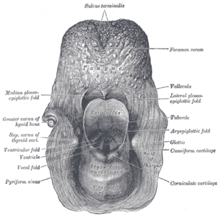
The aryepiglottic folds are triangular folds of mucous membrane of the larynx. They enclose ligamentous and muscular fibres. They extend from the lateral borders of the epiglottis to the arytenoid cartilages, hence the name 'aryepiglottic'. They contain the aryepiglottic muscles and form the upper borders of the quadrangular membrane. They have a role in growling as a form of phonation. They may be narrowed and cause stridor, or be shortened and cause laryngomalacia.
The cricoarytenoid ligament extends from the lamina of the cricoid cartilage to the medial surface of the base and muscular process of the arytenoid cartilage.
The cricoarytenoid joint is a joint connecting the cricoid cartilage and the arytenoid cartilage. It is a very shallow ball-and-socket joint. It allows for rotation and gliding motion. This controls the abduction and adduction of the vocal cords.
In the human larynx, the vocal process is the anterior angle of the base of the arytenoid cartilage, as it projects horizontally forward and gives attachment to the vocal ligament.
Arytenoid adduction is a surgical procedure used to treat vocal cord paralysis. A suture is used to emulate the action of the lateral cricoarytenoid muscle and position the paralyzed vocal cord closer to the midline. This allows the two vocal cords to meet and can improve speaking and swallowing ability for affected patients. Arytenoid adduction is often performed in conjunction with medialization thyroplasty.
