Anti-Hu associated encephalitis

Last updated
Anti-Hu associated encephalitis
Other namesAnti-ANNA1 associated encephalitis
Specialty Psychiatry, neurology
Symptoms Depression, anxiety, hallucinations, confusion, memory loss, weakness, numbness, ataxia, seizures, pain
Causes Paraneoplastic syndrome
Risk factors Smoking, male gender
Diagnostic method Western blot, EEG, MRI
Differential diagnosis Autoimmune encephalitis, metastatic cancer, viral encephalitis, Creutzfeldt-Jakob disease, cerebrovascular disease, Whipple disease, schizophrenia, toxic-metabolic encephalopathy, Wernicke encephalopathy, dementia, multiple sclerosis, Behçet's disease
Treatment Immunotherapy, chemotherapy
Prognosis Average survival less than 12 months

Anti-Hu associated encephalitis, also known as Anti-ANNA1 associated encephalitis, is an uncommon form of brain inflammation that is associated with an underlying cancer. It can cause psychiatric symptoms such as depression, anxiety, and hallucinations. [1] It can also produce neurological symptoms such as confusion, memory loss, weakness, sensory loss, pain, seizures, and problems coordinating the movement of the body. [2]

Contents

While its cause is unknown, the most common hypothesis is that it is caused by an immune system attack on the nervous system. This immune system attack is linked to cancer in most cases, usually small cell lung carcinoma. The condition's namesake, the anti-Hu antibody, is a protein made by the host's immune system, and it is present in virtually all cases. Treatment is focused on removing the underlying cancer and suppressing the immune system. Its prognosis remains quite poor, with most patients dying less than a year after diagnosis. [3]

Signs and symptoms

The signs and symptoms can vary in onset, quality, duration, severity, and response to treatment. Symptoms tend to present acutely over days to weeks. Its symptoms depend on which areas of the brain the disease affects, because specific parts of the brain have particular functions. [4]

Many cases involve an attack on the limbic system, which includes structures like the amygdala, hippocampus, and thalamus. Respectively these brain regions regulate anger, fear, memory formation, and motor and sensory signaling. Affected persons may develop memory loss and may have sudden changes in personality. This is often accompanied by headaches, delusions, or hallucinations. [5]

In some cases, the antibodies created by this illness attack another structure of the brain called the brainstem. The brainstem is responsible for basic bodily functions like breathing, but not more complex actions and emotions, which is why the presentation is different when the disease affects the limbic system than when it affects the brainstem. Symptoms may include dizziness, nausea, vomiting, and decreased breathing which may progress to respiratory failure. [6] [7]

Cause

The condition can involve the limbic system of the brain, which mediates key functions within motivation, emotion, learning, and memory. The Limbic System and Nearby Structures - John Taylor.jpg
The condition can involve the limbic system of the brain, which mediates key functions within motivation, emotion, learning, and memory.

Anti-Hu associated encephalitis is a syndrome associated with cancer. However, occasionally it occurs without cancer being present. Proteins react within the brain and change behavior and basic biological functions. Primarily adults contract this illness, and typically they have an underlying cancer that is either undiagnosed, diagnosed, in remission, or cured. [3]

The condition can occur at any point during cancer. Small cell lung cancer is a particularly aggressive cancer more common in smokers and is associated with anti-Hu encephalitis. Neuroblastoma is a cancer more frequently affecting children, and despite the relatively low rates of anti-Hu among children with neuroblastoma, these are the most likely children to have anti-Hu associated encephalitis. [8] [9]

Pathophysiology

Nearly all people with the condition have anti-Hu antibodies in their serum. The antibody is produced by the body as an immune system response to Hu proteins, which are naturally clustered within the nuclei of neurons in the central and peripheral nervous system. The condition can involve a number of neural structures including the brainstem, cerebellum, spinal cord, dorsal roots, peripheral nerves, and the limbic system of the brain. [1]

There is a debate about whether the antibody is a cause of, rather than an effect of, the disease process. Older studies suggested the antibodies caused the disease, pointing to the discovery of antibody deposition in the brain tissue of patients at autopsy. However, the injection of the antibodies into mice did not produce any disease, and the deposition of antibody was often not at the places where brain damage was greatest. [3] Newer studies suggest the antibodies are an effect, not a cause, of the condition, with a consensus that a patient's own T cells are playing a major role in the disease process. These T cells may be activated by the Hu proteins. [10] [11]

In people with cancer, the cancer has a likely role in the cause of the encephalitis. In a paraneoplastic syndrome, a cancer cell can create proteins that are normally only found as naturally-occurring proteins in other cell types in other parts of the body. In patients with small cell carcinoma of the lung, cancer cells in the lung can produce Hu proteins that are usually only found inside of the body's own neurons. It is hypothesized that through these cancer-produced Hu proteins, the body creates an immune system response. This reaction includes T cells, which then attack nervous tissue. [12] The cancer-produced Hu proteins are found in nearly all small-cell lung carcinomas, 70 percent of neuroblastomas, and a small percentage of other tumors. [13]

Diagnosis

Anti-Hu encephalitis is a disease characterized by production of anti-Hu antibodies and rapid development of particular signs and symptoms. Therefore, the diagnosis usually involves detecting its associated psychiatric and neurologic deficits and then performing diagnostic testing. If these signs and symptoms occur in a person who is suspected of having cancer, then anti-Hu associated encephalitis is also suspected. Because small cell lung cancer commonly occurs together with anti-Hu encephalitis, a diagnosis of small cell lung cancer confers a greater suspicion. [1]

In suspected cases, physicians perform diagnostic testing using a protein-detecting test that identify anti-Hu antibodies, if present. Another test involves examining the fluid that bathes the brain and spine, although this test is less specific for the disease. Physicians may also use a special imaging device, known as magnetic resonance imaging (MRI), which can take pictures of the patient's brain and detect signs of inflammation that suggest ongoing disease. An electroencephalogram (EEG) is another tool that can be done to clarify whether anti-Hu encephalitis is the underlying cause of a patient's symptoms. This is a test that involves placing probes on a person's head to detect electrical brain activity. Certain patterns of activity can be indicative of brain disease. In the case of anti-Hu encephalitis, temporal lobe electrical activity changes and the length of certain electrical waves known as delta and theta waves become slowed. [1]

Before the diagnosis can be made, other causes of disease need to be ruled out. They could be the sole cause or a co-contributor to a patient's new symptoms, in addition to anti-Hu encephalitis. Examples include—but are not limited to—problems with metabolism, a brain tumor, or inflammation of tissue coating around the brain. [14]

Treatment

The mainstay of treatment involves two broad strategies: treat the cancer that usually occurs with the disease, and give medications that suppress the body's immune system attack on the nervous system. Because current treatments are not successful at eliminating the disease, the goal of treatment is often to reduce symptoms rather than attempt to cure it. To date, treatments have been unsuccessful in achieving a sustained reduction of symptoms or survival in the vast majority of patients. [2]

Some treatments may directly combat the mechanisms by which the disease may be caused. To suppress the immune system, steroids, antibodies, or even human cells may be injected into a patient. Certain types of antibodies called intravenous immunoglobulins (IVIG) also have shown to lead to reduced symptoms due to their ability to reduce and eliminate anti-Hu antibodies. [15] A drug called rituximab, a molecule that targets B cells, helps reduce the symptoms of anti-Hu encephalitis and decreases the number of anti-Hu antibodies. [16] Cancer treatment may involve surgical removal of the tumor, or medications that may shrink or eliminate the tumor. [17] [2] Treatment with cyclophosphamide, a chemotherapy drug, has shown promise, in addition to adrenocorticotropic hormone (ACTH). [18] [19] [2] This hormone is involved in regulating many body functions including stress level and blood pressure. Steroids such as dexamethasone may help reduce disease burden by reducing the antibody-building activity of the disease. Despite the fact that steroids can be used to reduce the immunological antibody-building activity of the disease in all people, many other anti-Hu encephalitis treatments are most effective in children. [15]

Treatments may also be focused purely on symptoms rather than targeting the potential causes of the disease. For seizures, anticonvulsant medications may be used, such as valproic acid, levetiracetam, or lamotrigine. For hallucinations, delusions, and mood disturbances, second generation antipsychotic agents (e.g., olanzapine, clozapine) are also used for symptom control. [2]

Prognosis

Although many patients have an underlying cancer, the prognosis is determined by the severity of the neurological symptoms produced by the encephalitis. Compared to other paraneoplastic encephalitides, anti-Hu associated encephalitis has an especially poor prognosis. Several studies reporting an average survival time of less than a year, from the time of diagnosis. Much of the prognosis depends on the efficacy of treatment, which is directed at the underlying cancer, if present. [20] [21] [22] Patients with lower titers of the anti-Hu antibody tend to have a better prognosis. [13]

Epidemiology

Histopathology of small cell carcinoma of the lung, which is strongly associated with anti-Hu antibodies. Small cell carcinoma (3923396504).jpg
Histopathology of small cell carcinoma of the lung, which is strongly associated with anti-Hu antibodies.

The typical age at diagnosis is 63 years old. It is three times more common in men than women. Of those diagnosed with the condition, about 85 percent also had a cancer diagnosis, with 86 percent being lung cancers (mostly small-cell carcinoma) and 14 percent being outside the lung (most commonly prostate, gastrointestinal, breast, and bladder cancer). [13] However, other cancers have been known to co-occur with the disease, including spindle cell carcinoma of the sinus and a seminoma of the mediastinum. [23] People with small cell carcinoma often have other diseases caused by an immune response to the cancer, including Cushing syndrome, SIADH, and Lambert-Eaton myasthenic syndrome. [24]

History

The condition was first identified in 1985 at the Memorial Sloan Kettering Cancer Center at Cornell University, by three physicians, Francesc Graus, Carlos Cordon-Cardo, and Jerome Posner. They identified the anti-Hu antibody in two patients who had sensory neuronopathy and small cell carcinoma of the lung. [25]

Special populations

Children, in addition to adults, also can develop anti-Hu encephalitis; however, the disease manifests differently in children. As with adults, anti-Hu encephalitis is associated with malignancy. The cancers most associated with anti-Hu encephalitis are neuroblastoma and ganglioneuroblastoma. Opsoclonus-myoclonus syndrome (OMS) is a condition that develops in children as a result of anti-Hu antibodies. The illness afflicts younger children, with one study showing an age range of about 2 months to 10 years, with the majority of cases falling between 6 months to 3 years. The first symptoms are nonspecific. For instance, it can present like an upper airway infection, with cough and fever, or like an intestinal infection, with vomiting, diarrhea, and fever. Crying, particularly in younger children, can be an early sign. [26] Other symptoms include problems with eye movement, irritability, and insomnia. [8] [9]

See also

Related Research Articles

<span class="mw-page-title-main">Lambert–Eaton myasthenic syndrome</span> Medical condition

Lambert–Eaton myasthenic syndrome (LEMS) is a rare autoimmune disorder characterized by muscle weakness of the limbs.

Neuromyotonia (NMT) is a form of peripheral nerve hyperexcitability that causes spontaneous muscular activity resulting from repetitive motor unit action potentials of peripheral origin. NMT along with Morvan's syndrome are the most severe types in the Peripheral Nerve Hyperexciteability spectrum. Example of two more common and less severe syndromes in the spectrum are Cramp Fasciculation Syndrome and Benign Fasciculation Syndrome. NMT can have both hereditary and acquired forms. The prevalence of NMT is unknown.

Morvan's syndrome is a rare, life-threatening autoimmune disease named after the nineteenth century French physician Augustin Marie Morvan. "La chorée fibrillaire" was first coined by Morvan in 1890 when describing patients with multiple, irregular contractions of the long muscles, cramping, weakness, pruritus, hyperhidrosis, insomnia and delirium. It normally presents with a slow insidious onset over months to years. Approximately 90% of cases spontaneously go into remission, while the other 10% of cases lead to death.

Opsoclonus myoclonus syndrome (OMS), also known as opsoclonus-myoclonus-ataxia (OMA), is a rare neurological disorder of unknown cause which appears to be the result of an autoimmune process involving the nervous system. It is an extremely rare condition, affecting as few as 1 in 10,000,000 people per year. It affects 2 to 3% of children with neuroblastoma and has been reported to occur with celiac disease and diseases of neurologic and autonomic dysfunction.

<span class="mw-page-title-main">Rituximab</span> Biopharmaceutical drug

Rituximab, sold under the brand name Rituxan among others, is a monoclonal antibody medication used to treat certain autoimmune diseases and types of cancer. It is used for non-Hodgkin lymphoma, chronic lymphocytic leukemia, rheumatoid arthritis, granulomatosis with polyangiitis, idiopathic thrombocytopenic purpura, pemphigus vulgaris, myasthenia gravis and Epstein–Barr virus-positive mucocutaneous ulcers. It is given by slow intravenous infusion. Biosimilars of Rituxan include Blitzima, Riabni, Ritemvia, Rituenza, Rixathon, Ruxience, and Truxima.

<span class="mw-page-title-main">Stiff-person syndrome</span> Human medical condition

Stiff-person syndrome (SPS), also known as stiff-man syndrome, is a rare neurologic disorder of unclear cause characterized by progressive muscular rigidity and stiffness. The stiffness primarily affects the truncal muscles and is superimposed by spasms, resulting in postural deformities. Chronic pain, impaired mobility, and lumbar hyperlordosis are common symptoms.

Paraneoplastic cerebellar degeneration (PCD) is a paraneoplastic syndrome associated with a broad variety of tumors including lung cancer, ovarian cancer, breast cancer, Hodgkin’s lymphoma and others. PCD is a rare condition that occurs in less than 1% of cancer patients.

<span class="mw-page-title-main">Non-small-cell lung cancer</span> Any type of epithelial lung cancer other than small-cell lung carcinoma

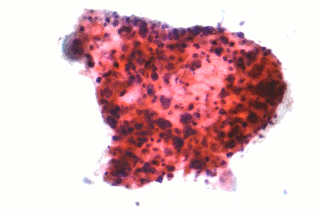
Non-small-cell lung cancer (NSCLC), or non-small-cell lung carcinoma, is any type of epithelial lung cancer other than small-cell lung cancer (SCLC). NSCLC accounts for about 85% of all lung cancers. As a class, NSCLCs are relatively insensitive to chemotherapy, compared to small-cell carcinoma. When possible, they are primarily treated by surgical resection with curative intent, although chemotherapy has been used increasingly both preoperatively and postoperatively.

<span class="mw-page-title-main">Hashimoto's encephalopathy</span> Human disease (neurological condition)

Hashimoto's encephalopathy, also known as steroid-responsive encephalopathy associated with autoimmune thyroiditis (SREAT), is a neurological condition characterized by encephalopathy, thyroid autoimmunity, and good clinical response to corticosteroids. It is associated with Hashimoto's thyroiditis, and was first described in 1966. It is sometimes referred to as a neuroendocrine disorder, although the condition's relationship to the endocrine system is widely disputed. It is recognized as a rare disease by the NIH Genetic and Rare Diseases Information Center.

<span class="mw-page-title-main">Limbic encephalitis</span> Inflammation involving the limbic system in the brain

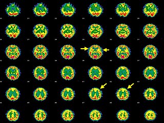
Limbic encephalitis is a form of encephalitis, a disease characterized by inflammation of the brain. Limbic encephalitis is caused by autoimmunity: an abnormal state where the body produces antibodies against itself. Some cases are associated with cancer and some are not. Although the disease is known as "limbic" encephalitis, it is seldom limited to the limbic system and post-mortem studies usually show involvement of other parts of the brain. The disease was first described by Brierley and others in 1960 as a series of three cases. The link to cancer was first noted in 1968 and confirmed by later investigators.

A paraneoplastic syndrome is a syndrome that is the consequence of a tumor in the body. It is specifically due to the production of chemical signaling molecules by tumor cells or by an immune response against the tumor. Unlike a mass effect, it is not due to the local presence of cancer cells.

Guillain–Barré syndrome (GBS) is a rapid-onset muscle weakness caused by the immune system damaging the peripheral nervous system. Typically, both sides of the body are involved, and the initial symptoms are changes in sensation or pain often in the back along with muscle weakness, beginning in the feet and hands, often spreading to the arms and upper body. The symptoms may develop over hours to a few weeks. During the acute phase, the disorder can be life-threatening, with about 15% of people developing weakness of the breathing muscles and, therefore, requiring mechanical ventilation. Some are affected by changes in the function of the autonomic nervous system, which can lead to dangerous abnormalities in heart rate and blood pressure.

<span class="mw-page-title-main">ELAV-like protein 3</span> Protein-coding gene in the species Homo sapiens

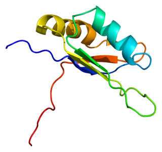
ELAV-like protein 3 is a protein that in humans is encoded by the ELAVL3 gene.

<span class="mw-page-title-main">Anti-NMDA receptor encephalitis</span> Rare disease which results in brain inflammation

Anti-NMDA receptor encephalitis is a type of brain inflammation caused by antibodies. Early symptoms may include fever, headache, and feeling tired. This is then typically followed by psychosis which presents with false beliefs (delusions) and seeing or hearing things that others do not see or hear (hallucinations). People are also often agitated or confused. Over time, seizures, decreased breathing, and blood pressure and heart rate variability typically occur. In some cases, patients may develop catatonia.

Bickerstaff brainstem encephalitis is a rare inflammatory disorder of the central nervous system, first described by Edwin Bickerstaff in 1951. It may also affect the peripheral nervous system, and has features in common with both Miller Fisher syndrome and Guillain–Barré syndrome.

Targeted molecular therapy for neuroblastoma involves treatment aimed at molecular targets that have a unique expression in this form of cancer. Neuroblastoma, the second most common pediatric malignant tumor, often involves treatment through intensive chemotherapy. A number of molecular targets have been identified for the treatment of high-risk forms of this disease. Aiming treatment in this way provides a more selective way to treat the disease, decreasing the risk for toxicities that are associated with the typical treatment regimen. Treatment using these targets can supplement or replace some of the intensive chemotherapy that is used for neuroblastoma. These molecular targets of this disease include GD2, ALK, and CD133. GD2 is a target of immunotherapy, and is the most fully developed of these treatment methods, but is also associated with toxicities. ALK has more recently been discovered, and drugs in development for this target are proving to be successful in neuroblastoma treatment. The role of CD133 in neuroblastoma has also been more recently discovered and is an effective target for treatment of this disease.

<span class="mw-page-title-main">Autoimmune encephalitis</span> Type of encephalitis

Autoimmune encephalitis (AIE) is a type of encephalitis, and one of the most common causes of noninfectious encephalitis. It can be triggered by tumors, infections, or it may be cryptogenic. The neurological manifestations can be either acute or subacute and usually develop within six weeks. The clinical manifestations include behavioral and psychiatric symptoms, autonomic disturbances, movement disorders, and seizures.

<span class="mw-page-title-main">Autoimmune autonomic ganglionopathy</span> Medical condition

Autoimmune autonomic ganglionopathy is a type of immune-mediated autonomic failure that is associated with antibodies against the ganglionic nicotinic acetylcholine receptor present in sympathetic, parasympathetic, and enteric ganglia. Typical symptoms include gastrointestinal dysmotility, orthostatic hypotension, and tonic pupils. Many cases have a sudden onset, but others worsen over time, resembling degenerative forms of autonomic dysfunction. For milder cases, supportive treatment is used to manage symptoms. Plasma exchange, intravenous immunoglobulin, corticosteroids, or immunosuppression have been used successfully to treat more severe cases.

Autoimmune retinopathy (AIR) is a rare disease in which the patient's immune system attacks proteins in the retina, leading to loss of eyesight. The disease is poorly understood, but may be the result of cancer or cancer chemotherapy. The disease is an autoimmune condition characterized by vision loss, blind spots, and visual field abnormalities. It can be divided into cancer-associated retinopathy (CAR) and melanoma-associated retinopathy (MAR). The condition is associated with retinal degeneration caused by autoimmune antibodies recognizing retinal proteins as antigens and targeting them. AIR's prevalence is extremely rare, with CAR being more common than MAR. It is more commonly diagnosed in females in the age range of 50–60.

Anti-VGKC-complex encephalitis are caused by antibodies against the voltage gated potassium channel-complex (VGKC-complex) and are implicated in several autoimmune conditions including limbic encephalitis, epilepsy and neuromyotonia.

References

  1. 1 2 3 4 Kaplan PW, Sutter R (2013). "Electroencephalography of autoimmune limbic encephalopathy". J Clin Neurophysiol. 30 (5): 490–504. doi:10.1097/WNP.0b013e3182a73d47. PMID   24084182. S2CID   24130688.
  2. 1 2 3 4 5 Dubey D, Blackburn K, Greenberg B, Stuve O, Vernino S (2016). "Diagnostic and therapeutic strategies for management of autoimmune encephalopathies". Expert Rev Neurother. 16 (8): 937–49. doi:10.1080/14737175.2016.1189328. PMID   27171736. S2CID   13759612.
  3. 1 2 3 Didelot A, Honnorat J (2014). "Paraneoplastic disorders of the central and peripheral nervous systems". Neurologic Aspects of Systemic Disease Part III. Handbook of Clinical Neurology. Vol. 121. pp. 1159–79. doi:10.1016/B978-0-7020-4088-7.00078-X. ISBN   9780702040887. PMID   24365410.{{cite book}}: |journal= ignored (help)
  4. Helpman L, Zhu X, Suarez-Jimenez B, Lazarov A, Monk C, Neria Y (2017). "Sex Differences in Trauma-Related Psychopathology: a Critical Review of Neuroimaging Literature (2014-2017)". Curr Psychiatry Rep. 19 (12): 104. doi:10.1007/s11920-017-0854-y. PMC   5737777 . PMID   29116470.
  5. Langer JE, Lopes MB, Fountain NB, Pranzatelli MR, Thiele EA, Rust RS, Goodkin HP (2012). "An unusual presentation of anti-Hu-associated paraneoplastic limbic encephalitis". Dev Med Child Neurol. 54 (9): 863–6. doi: 10.1111/j.1469-8749.2012.04221.x . PMID   22320677. S2CID   22388381.
  6. Vigliani MC, Novero D, Cerrato P, Daniele D, Crasto S, Berardino M, Mutani R (2009). "Double step paraneoplastic brainstem encephalitis: a clinicopathological study". J Neurol Neurosurg Psychiatry. 80 (6): 693–5. doi:10.1136/jnnp.2008.145961. PMID   19448098. S2CID   24747904.
  7. Najjar M, Taylor A, Agrawal S, Fojo T, Merkler AE, Rosenblum MK, Lennihan L, Kluger MD (2017). "Anti-Hu paraneoplastic brainstem encephalitis caused by a pancreatic neuroendocrine tumor presenting with central hypoventilation". J Clin Neurosci. 40: 72–73. doi:10.1016/j.jocn.2017.02.015. PMID   28256369. S2CID   26309502.
  8. 1 2 Blaes F, Fühlhuber V, Preissner KT (2007). "Identification of autoantigens in pediatric opsoclonus-myoclonus syndrome". Expert Rev Clin Immunol. 3 (6): 975–82. doi:10.1586/1744666X.3.6.975. PMID   20477144. S2CID   43047140.
  9. 1 2 Meena JP, Seth R, Chakrabarty B, Gulati S, Agrawala S, Naranje P (2016). "Neuroblastoma presenting as opsoclonus-myoclonus: A series of six cases and review of literature". J Pediatr Neurosci. 11 (4): 373–377. doi: 10.4103/1817-1745.199462 . PMC   5314861 . PMID   28217170.
  10. Dalmau J, Rosenfeld MR (2014). "Autoimmune encephalitis update". Neuro Oncol. 16 (6): 771–8. doi:10.1093/neuonc/nou030. PMC   4022229 . PMID   24637228.
  11. Bernal F, Graus F, Pifarré A, Saiz A, Benyahia B, Ribalta T (2002). "Immunohistochemical analysis of anti-Hu-associated paraneoplastic encephalomyelitis". Acta Neuropathol. 103 (5): 509–15. doi:10.1007/s00401-001-0498-0. PMID   11935268. S2CID   25157807.
  12. de Jongste AH, de Graaf MT, van den Broek PD, Kraan J, Smitt PA, Gratama JW (2013). "Elevated numbers of regulatory T cells, central memory T cells and class-switched B cells in cerebrospinal fluid of patients with anti-Hu antibody associated paraneoplastic neurological syndromes". J Neuroimmunol. 258 (1–2): 85–90. doi:10.1016/j.jneuroim.2013.02.006. PMID   23566401. S2CID   38435280.
  13. 1 2 3 Graus F, Keime-Guibert F, Reñe R, Benyahia B, Ribalta T, Ascaso C, Escaramis G, Delattre JY (2001). "Anti-Hu-associated paraneoplastic encephalomyelitis: analysis of 200 patients". Brain. 124 (Pt 6): 1138–48. doi: 10.1093/brain/124.6.1138 . PMID   11353730.
  14. Ochenduszko S, Wilk B, Dabrowska J, Herman-Sucharska I, Dubis A, Puskulluoglu M (2017). "Paraneoplastic limbic encephalitis in a patient with extensive disease small-cell lung cancer". Mol Clin Oncol. 6 (4): 575–578. doi:10.3892/mco.2017.1162. PMC   5374934 . PMID   28413671.
  15. 1 2 Pranzatelli MR (1996). "The immunopharmacology of the opsoclonus-myoclonus syndrome". Clin Neuropharmacol. 19 (1): 1–47. doi:10.1097/00002826-199619010-00001. PMID   8867515.
  16. Ketterl TG, Messinger YH, Niess DR, Gilles E, Engel WK, Perkins JL (2013). "Ofatumumab for refractory opsoclonus-myoclonus syndrome following treatment of neuroblastoma". Pediatr Blood Cancer. 60 (12): E163-5. doi: 10.1002/pbc.24646 . PMID   23813921. S2CID   12279928.
  17. Lancaster E (2017). "Paraneoplastic Disorders". Continuum (Minneap Minn). 23 (6, Neuro–oncology): 1653–1679. doi:10.1212/CON.0000000000000542. PMID   29200116. S2CID   46599983.
  18. Armstrong MB, Robertson PL, Castle VP (2005). "Delayed, recurrent opsoclonus-myoclonus syndrome responding to plasmapheresis". Pediatr Neurol. 33 (5): 365–7. doi:10.1016/j.pediatrneurol.2005.05.018. PMID   16243225.
  19. Blaes F (2013). "Paraneoplastic brain stem encephalitis". Curr Treat Options Neurol. 15 (2): 201–9. doi:10.1007/s11940-013-0221-1. PMID   23378230. S2CID   21581917.
  20. Huemer F, Melchardt T, Tränkenschuh W, Neureiter D, Moser G, Magnes T, Weiss L, Schlattau A, Hufnagl C, Ricken G, Höftberger R, Greil R, Egle A (2015). "Anti-Hu Antibody Associated Paraneoplastic Cerebellar Degeneration in Head and Neck Cancer". BMC Cancer. 15: 996. doi: 10.1186/s12885-015-2020-4 . PMC   4687318 . PMID   26694863.
  21. Shams'ili S, Grefkens J, de Leeuw B, van den Bent M, Hooijkaas H, van der Holt B, Vecht C, Sillevis Smitt P (2003). "Paraneoplastic cerebellar degeneration associated with antineuronal antibodies: analysis of 50 patients". Brain. 126 (Pt 6): 1409–18. doi: 10.1093/brain/awg133 . hdl: 1765/10135 . PMID   12764061.
  22. Graus F, Dalmou J, Reñé R, Tora M, Malats N, Verschuuren JJ, Cardenal F, Viñolas N, Garcia del Muro J, Vadell C, Mason WP, Rosell R, Posner JB, Real FX (1997). "Anti-Hu antibodies in patients with small-cell lung cancer: association with complete response to therapy and improved survival". J Clin Oncol. 15 (8): 2866–72. doi:10.1200/JCO.1997.15.8.2866. PMID   9256130.
  23. Pohley I, Roesler K, Wittstock M, Bitsch A, Benecke R, Wolters A (2015). "NMDA-receptor antibody and anti-Hu antibody positive paraneoplastic syndrome associated with a primary mediastinal seminoma". Acta Neurol Belg. 115 (1): 81–3. doi:10.1007/s13760-014-0296-9. PMID   24696410. S2CID   11672970.
  24. Kanaji N, Watanabe N, Kita N, Bandoh S, Tadokoro A, Ishii T, Dobashi H, Matsunaga T (2014). "Paraneoplastic syndromes associated with lung cancer". World J Clin Oncol. 5 (3): 197–223. doi: 10.5306/wjco.v5.i3.197 . PMC   4127595 . PMID   25114839.
  25. Graus F, Cordon-Cardo C, Posner JB (1985). "Neuronal antinuclear antibody in sensory neuronopathy from lung cancer". Neurology. 35 (4): 538–43. doi: 10.1212/wnl.35.4.538 . PMID   2984600.
  26. Pranzatelli MR, Tate ED, McGee NR (2017). "Demographic, Clinical, and Immunologic Features of 389 Children with Opsoclonus-Myoclonus Syndrome: A Cross-sectional Study". Front Neurol. 8: 468. doi: 10.3389/fneur.2017.00468 . PMC   5604058 . PMID   28959231.