Aspartoacylase

Last updated
ASPA
Available structures
PDB Ortholog search: PDBe RCSB
Identifiers
Aliases ASPA , ACY2, ASP, aspartoacylase
External IDs OMIM: 608034 MGI: 87914 HomoloGene: 33 GeneCards: ASPA
Orthologs
SpeciesHumanMouse
Entrez
Ensembl
UniProt
RefSeq (mRNA)

NM_000049
NM_001128085

NM_023113

RefSeq (protein)

NP_000040
NP_001121557

NP_075602

Location (UCSC) Chr 17: 3.47 – 3.5 Mb Chr 11: 73.2 – 73.22 Mb
PubMed search [3] [4]
Wikidata
View/Edit Human View/Edit Mouse
Aspartoacylase
ASPA dimer.png
Structure of aspartoacylase dimer. Generated from 2I3C. [5]
Identifiers
EC no. 3.5.1.15
Databases
IntEnz IntEnz view
BRENDA BRENDA entry
ExPASy NiceZyme view
KEGG KEGG entry
MetaCyc metabolic pathway
PRIAM profile
PDB structures RCSB PDB PDBe PDBsum
Search
PMC articles
PubMed articles
NCBI proteins

Aspartoacylase is a hydrolytic enzyme (EC 3.5.1.15, also called aminoacylase II, ASPA and other names [lower-alpha 1] ) that in humans is encoded by the ASPA gene. ASPA catalyzes the deacylation of N-acetyl-l-aspartate (N-acetylaspartate) into aspartate and acetate. [7] [8] It is a zinc-dependent hydrolase that promotes the deprotonation of water to use as a nucleophile in a mechanism analogous to many other zinc-dependent hydrolases. [9] It is most commonly found in the brain, where it controls the levels of N-acetyl-l-aspartate. Mutations that result in loss of aspartoacylase activity are associated with Canavan disease, a rare autosomal recessive neurodegenerative disease. [10]

Contents

Structure

Aspartoacylase is a dimer of two identical monomers of 313 amino acids and uses a zinc cofactor in each. [5] [11] There are two distinct domains in each monomer: the N-terminal domain from residues 1-212 and the C-terminal domain from residues 213–313. [12] The N-terminal domain of aspartoacylase is similar to that of zinc-dependent hydrolases such as carboxypeptidaseA. However, carboxypeptidases do not have something similar to the C-domain. In carboxypeptidase A, the active site is accessible to large substrates like the bulky C-terminal residue of polypeptides, whereas the C-domain sterically hinders access to the active site in aspartoacylase. Instead, the N-domain and C-domain of aspartoacylase form a deep narrow channel that leads to the active site. [5]

The zinc cofactor is found at the active site and is held by Glu-24, His-21, and His 116. [13] The substrate is held in place by Arg-63, Asn-70, Arg-71, Tyr-164, Arg-168, and Tyr-288. [5] The zinc cofactor is used to lower the pKa of a ligated water molecule so that an attack on N-acetyl-L-aspartate may occur and to stabilize the resulting tetrahedral intermediate along with Arg-63, and Glu-178. [13]

A monomer of aspartoacylase with the N-domain in green, C-domain in yellow, and zinc cofactor in red. Generated from 2I3C. ASPA domains.png
A monomer of aspartoacylase with the N-domain in green, C-domain in yellow, and zinc cofactor in red. Generated from 2I3C.
Active site of aspartoacylase with a bound N-phosphonamidate-L-aspartate. This is a tetrahedral intermediate analogue with phosphorus replacing the attacked carbon. In the structure, zinc, Arg-63, and Glu-178 are stabilizing the tetrahedral intermediate. Generated from 2O4H. ASPA bound to an intermediate analogue.png
Active site of aspartoacylase with a bound N-phosphonamidate-L-aspartate. This is a tetrahedral intermediate analogue with phosphorus replacing the attacked carbon. In the structure, zinc, Arg-63, and Glu-178 are stabilizing the tetrahedral intermediate. Generated from 2O4H.

Mechanism

There are two types of possible mechanisms for zinc-dependent hydrolases depending on what is the nucleophile. The first uses deprotonated water and the second attacks with an aspartate or glutamate first forming an anhydride. [14] Aspartoacylase follows the deprotonated water mechanism. [13] Zinc lowers the pKa of a ligated water molecule and the reaction proceeds via an attack on N-acetyl-l-aspartate when the water molecule is deprotonated by Glu-178. [5] This leads to a tetrahedral intermediate that is stabilized by the zinc, Arg-63, and Glu-178. [13] Finally, the carbonyl is then reformed, the bond with nitrogen is broken, and the nitrogen is protonated by the proton taken by Glu-178 all in one concerted step. [14]

Aspartoacylase mechanism. All the coordinating residues are not shown for clarity. ASPA mechanism.jpg
Aspartoacylase mechanism. All the coordinating residues are not shown for clarity.

Biological function

Aspartoacylase is used to metabolize N-acetyl-L-aspartate by catalyzing its deacylation. Aspartoacylase prevents the buildup of N-acetyl-L-aspartate in the brain. It is believed that controlling N-acetyl-L-aspartate levels is essential for developing and maintaining white matter. [5] It is not known why so much N-acetyl-L-aspartate is produced in the brain nor what its primary function is. [15] However, one hypothesis is that it is potentially used as a chemical reservoir that can be tapped into for acetate for acetyl-CoA synthesis or aspartate for glutamate synthesis. [15] [16] This way, N-acetyl-L-aspartate can be used to transport these precursor molecules and aspartoacylase is used to release them. For example, N-acetyl-L-aspartate produced in neurons can be transported into oligodendrocytes and the acetate released can be used for myelin synthesis. [12] [17] Another hypothesis is that N-acetyl-L-aspartate is essential osmolyte that acts as a molecular water pump that helps maintain a proper fluid balance in the brain. [18]

Disease relevance

Mutations that lead to loss of aspartoacylase activity have been identified as the cause of Canavan disease. [19] Canavan disease is a rare autosomal recessive disorder that causes spongy degeneration of the white matter in the brain and severe psychomotor retardation, usually leading to death at a young age. [12] [20] The loss of aspartoacylase activity leads to the buildup of N-acetyl-L-aspartate in the brain and an increase in urine concentration by up to 60 times normal levels. [19] Though the exact mechanism of how loss of aspartoacylase activity leads to Canavan disease is not fully understood, there are two primary competing explanations. The first is that it leads to defective myelin synthesis due to a deficiency of acetyl-CoA derived from the acetate product. [20] Another explanation is that the elevated levels of N-acetyl-l-aspartate interfere with its normal brain osmoregulatory mechanism leading to osmotic disequilibrium. [21]

There are over 70 reported mutations of this enzyme, but the most common ones are the amino acid substitutions E285A and A305E. [12] E285A reduces activity of aspartoacylase down to as low as 0.3% of its normal function and is found in 98% of cases with Ashkenazi Jewish ancestry. [22] The mutation A305E is found in about 40% of non-Jewish patients and reduces activity to about 10%. [22] Of these two mutations, a crystal structure of the E285A mutant has been taken, showing that the loss of the hydrogen bonding from glutamate leads to a conformational change that distorts the active site and alters the substrate binding, leading to the much lower catalytic activity. [12]

Distortion of the active site caused by the E285A mutation. Wild type ASPA is on the left (2O4H ) and E285A on the right (4NFR ). E282a mutation.JPG
Distortion of the active site caused by the E285A mutation. Wild type ASPA is on the left (2O4H ) and E285A on the right (4NFR ).

See also

Notes

  1. The enzyme is also known as N-acetylaspartate amidohydrolase, acetyl-aspartic deaminase or acylase II [6]

Related Research Articles

The urea cycle (also known as the ornithine cycle) is a cycle of biochemical reactions that produces urea (NH2)2CO from ammonia (NH3). Animals that use this cycle, mainly amphibians and mammals, are called ureotelic.

Canavan disease, or Canavan–Van Bogaert–Bertrand disease, is a rare and fatal autosomal recessive degenerative disease that causes progressive damage to nerve cells and loss of white matter in the brain. It is one of the most common degenerative cerebral diseases of infancy. It is caused by a deficiency of the enzyme aminoacylase 2, and is one of a group of genetic diseases referred to as leukodystrophies. It is characterized by degeneration of myelin in the phospholipid layer insulating the axon of a neuron and is associated with a gene located on human chromosome 17.

<span class="mw-page-title-main">Angiotensin-converting enzyme</span> Mammalian protein found in humans

Angiotensin-converting enzyme, or ACE, is a central component of the renin–angiotensin system (RAS), which controls blood pressure by regulating the volume of fluids in the body. It converts the hormone angiotensin I to the active vasoconstrictor angiotensin II. Therefore, ACE indirectly increases blood pressure by causing blood vessels to constrict. ACE inhibitors are widely used as pharmaceutical drugs for treatment of cardiovascular diseases.

In molecular biology, biosynthesis is a multi-step, enzyme-catalyzed process where substrates are converted into more complex products in living organisms. In biosynthesis, simple compounds are modified, converted into other compounds, or joined to form macromolecules. This process often consists of metabolic pathways. Some of these biosynthetic pathways are located within a single cellular organelle, while others involve enzymes that are located within multiple cellular organelles. Examples of these biosynthetic pathways include the production of lipid membrane components and nucleotides. Biosynthesis is usually synonymous with anabolism.

<span class="mw-page-title-main">Leukodystrophy</span> Group of disorders characterised by degeneration of white matter in the brain

Leukodystrophies are a group of, usually, inherited disorders, characterized by degeneration of the white matter in the brain. The word leukodystrophy comes from the Greek roots leuko, "white", dys, "abnormal" and troph, "growth". The leukodystrophies are caused by imperfect growth or development of the glial cells which produce the myelin sheath, the fatty insulating covering around nerve fibers. Leukodystrophies may be classified as hypomyelinating or demyelinating diseases, respectively, depending on whether the damage is present before birth or occurs after. Other demyelinating diseases are usually not congenital and have a toxic or autoimmune cause.

<span class="mw-page-title-main">Catalytic triad</span> Set of three coordinated amino acids

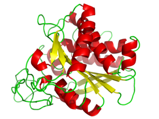
A catalytic triad is a set of three coordinated amino acids that can be found in the active site of some enzymes. Catalytic triads are most commonly found in hydrolase and transferase enzymes. An acid-base-nucleophile triad is a common motif for generating a nucleophilic residue for covalent catalysis. The residues form a charge-relay network to polarise and activate the nucleophile, which attacks the substrate, forming a covalent intermediate which is then hydrolysed to release the product and regenerate free enzyme. The nucleophile is most commonly a serine or cysteine amino acid, but occasionally threonine or even selenocysteine. The 3D structure of the enzyme brings together the triad residues in a precise orientation, even though they may be far apart in the sequence.

<span class="mw-page-title-main">Glucocerebrosidase</span> Mammalian protein found in humans

β-Glucocerebrosidase is an enzyme with glucosylceramidase activity that cleaves by hydrolysis the β-glycosidic linkage of the chemical glucocerebroside, an intermediate in glycolipid metabolism that is abundant in cell membranes. It is localized in the lysosome, where it remains associated with the lysosomal membrane. β-Glucocerebrosidase is 497 amino acids in length and has a molecular mass of 59,700 Da.

<span class="mw-page-title-main">Carboxypeptidase</span>

A carboxypeptidase is a protease enzyme that hydrolyzes (cleaves) a peptide bond at the carboxy-terminal (C-terminal) end of a protein or peptide. This is in contrast to an aminopeptidases, which cleave peptide bonds at the N-terminus of proteins. Humans, animals, bacteria and plants contain several types of carboxypeptidases that have diverse functions ranging from catabolism to protein maturation. At least two mechanisms have been discussed.

<span class="mw-page-title-main">Hexosaminidase</span> Class of enzymes

Hexosaminidase is an enzyme involved in the hydrolysis of terminal N-acetyl-D-hexosamine residues in N-acetyl-β-D-hexosaminides.

<i>N</i>-Acetylaspartic acid Derivative of aspartic acid found in the brain

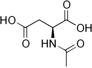
N-Acetylaspartic acid, or N-acetylaspartate (NAA), is a derivative of aspartic acid with a formula of C6H9NO5 and a molecular weight of 175.139.

<span class="mw-page-title-main">CAD protein</span> Protein-coding gene in the species Homo sapiens

CAD protein is a trifunctional multi-domain enzyme involved in the first three steps of pyrimidine biosynthesis. De-novo synthesis starts with cytosolic carbamoylphosphate synthetase II which uses glutamine, carbon dioxide and ATP. This enzyme is inhibited by uridine triphosphate.

<span class="mw-page-title-main">Myelin protein zero</span> Protein-coding gene in the species Homo sapiens

Myelin protein zero is a single membrane glycoprotein which in humans is encoded by the MPZ gene. P0 is a major structural component of the myelin sheath in the peripheral nervous system (PNS). Myelin protein zero is expressed by Schwann cells and accounts for over 50% of all proteins in the peripheral nervous system, making it the most common protein expressed in the PNS. Mutations in myelin protein zero can cause myelin deficiency and are associated with neuropathies like Charcot–Marie–Tooth disease and Dejerine–Sottas disease.

<i>N</i>-Acetylaspartylglutamic acid Peptide neurotransmitter

N-Acetylaspartylglutamic acid is a peptide neurotransmitter and the third-most-prevalent neurotransmitter in the mammalian nervous system. NAAG consists of N-acetylaspartic acid (NAA) and glutamic acid coupled via a peptide bond.

Pantothenate kinase (EC 2.7.1.33, PanK; CoaA) is the first enzyme in the Coenzyme A (CoA) biosynthetic pathway. It phosphorylates pantothenate (vitamin B5) to form 4'-phosphopantothenate at the expense of a molecule of adenosine triphosphate (ATP). It is the rate-limiting step in the biosynthesis of CoA.

<span class="mw-page-title-main">Carboxypeptidase A</span>

Carboxypeptidase A usually refers to the pancreatic exopeptidase that hydrolyzes peptide bonds of C-terminal residues with aromatic or aliphatic side-chains. Most scientists in the field now refer to this enzyme as CPA1, and to a related pancreatic carboxypeptidase as CPA2.

<span class="mw-page-title-main">Palmitoyl(protein) hydrolase</span> Class of enzymes

Palmitoyl protein hydrolase/thioesterases is an enzyme (EC 3.1.2.22) that removes thioester-linked fatty acyl groups such as palmitate from modified cysteine residues in proteins or peptides during lysosomal degradation. It catalyzes the reaction

In enzymology, an aminoacylase (EC 3.5.1.14) is an enzyme that catalyzes the chemical reaction

Protein <i>O</i>-GlcNAcase Protein-coding gene in the species Homo sapiens

Protein O-GlcNAcase (EC 3.2.1.169, OGA, glycoside hydrolase O-GlcNAcase, O-GlcNAcase, BtGH84, O-GlcNAc hydrolase) is an enzyme with systematic name (protein)-3-O-(N-acetyl-D-glucosaminyl)-L-serine/threonine N-acetylglucosaminyl hydrolase. OGA is encoded by the OGA gene. This enzyme catalyses the removal of the O-GlcNAc post-translational modification in the following chemical reaction:

  1. [protein]-3-O-(N-acetyl-β-D-glucosaminyl)-L-serine + H2O ⇌ [protein]-L-serine + N-acetyl-D-glucosamine
  2. [protein]-3-O-(N-acetyl-β-D-glucosaminyl)-L-threonine + H2O ⇌ [protein]-L-threonine + N-acetyl-D-glucosamine
<span class="mw-page-title-main">ABHD12</span> Protein-coding gene in the species Homo sapiens

alpha/beta-Hydrolase domain containing 12 (ABHD12) is a serine hydrolase encoded by the ABHD12 gene that participates in the breakdown of the endocannabinoid neurotransmitter 2-arachidonylglycerol (2-AG) in the central nervous system. It is responsible for about 9% of brain 2-AG hydrolysis. Together, ABHD12 along with two other enzymes, monoacylglycerol lipase (MAGL) and ABHD6, control 99% of 2-AG hydrolysis in the brain. ABHD12 also serves as a lysophospholipase and metabolizes lysophosphatidylserine (LPS).

<span class="mw-page-title-main">Spongy degeneration of the central nervous system</span> Neurodegenerative disorder

Spongy degeneration of the central nervous system, also known as Canavan's disease, Van Bogaert-Bertrand type or Aspartoacylase (AspA) deficiency, is a rare autosomal recessive neurodegenerative disorder. It belongs to a group of genetic disorders known as leukodystrophies, where the growth and maintenance of myelin sheath in the central nervous system (CNS) are impaired. There are three types of spongy degeneration: infantile, congenital and juvenile, with juvenile being the most severe type. Common symptoms in infants include lack of motor skills, weak muscle tone, and macrocephaly. It may also be accompanied by difficulties in feeding and swallowing, seizures and sleep disturbances. Affected children typically die before the age of 10, but life expectancy can vary.

References

  1. 1 2 3 GRCh38: Ensembl release 89: ENSG00000108381 - Ensembl, May 2017
  2. 1 2 3 GRCm38: Ensembl release 89: ENSMUSG00000020774 - Ensembl, May 2017
  3. "Human PubMed Reference:". National Center for Biotechnology Information, U.S. National Library of Medicine.
  4. "Mouse PubMed Reference:". National Center for Biotechnology Information, U.S. National Library of Medicine.
  5. 1 2 3 4 5 6 7 8 Bitto E, Bingman CA, Wesenberg GE, McCoy JG, Phillips GN (January 2007). "Structure of aspartoacylase, the brain enzyme impaired in Canavan disease". Proceedings of the National Academy of Sciences of the United States of America. 104 (2): 456–61. doi: 10.1073/pnas.0607817104 . PMC   1766406 . PMID   17194761.
  6. "Aspartoacylase (EC 3.5.1.15)". biocyc.org. Archived from the original on 22 July 2022. Retrieved 2022-07-22.
  7. Birnbaum SM (1955). [12] Aminoacylase. Methods in Enzymology. Vol. 2. pp. 115–119. doi:10.1016/S0076-6879(55)02176-9. ISBN   9780121818029.
  8. Birnbaum SM, Levintow L, Kingsley RB, Greenstein JP (January 1952). "Specificity of amino acid acylases". The Journal of Biological Chemistry. 194 (1): 455–70. doi: 10.1016/S0021-9258(18)55898-1 . PMID   14927637.
  9. Le Coq J, An HJ, Lebrilla C, Viola RE (May 2006). "Characterization of human aspartoacylase: the brain enzyme responsible for Canavan disease". Biochemistry. 45 (18): 5878–84. doi:10.1021/bi052608w. PMC   2566822 . PMID   16669630.
  10. Hershfield JR, Pattabiraman N, Madhavarao CN, Namboodiri MA (May 2007). "Mutational analysis of aspartoacylase: implications for Canavan disease". Brain Research. 1148: 1–14. doi:10.1016/j.brainres.2007.02.069. PMC   1933483 . PMID   17391648.
  11. Herga S, Berrin JG, Perrier J, Puigserver A, Giardina T (October 2006). "Identification of the zinc binding ligands and the catalytic residue in human aspartoacylase, an enzyme involved in Canavan disease". FEBS Letters. 580 (25): 5899–904. doi: 10.1016/j.febslet.2006.09.056 . PMID   17027983. S2CID   41351955.
  12. 1 2 3 4 5 6 Wijayasinghe YS, Pavlovsky AG, Viola RE (August 2014). "Aspartoacylase catalytic deficiency as the cause of Canavan disease: a structural perspective". Biochemistry. 53 (30): 4970–8. doi: 10.1021/bi500719k . PMID   25003821.
  13. 1 2 3 4 5 6 Le Coq J, Pavlovsky A, Malik R, Sanishvili R, Xu C, Viola RE (March 2008). "Examination of the mechanism of human brain aspartoacylase through the binding of an intermediate analogue". Biochemistry. 47 (11): 3484–92. doi:10.1021/bi702400x. PMC   2666850 . PMID   18293939.
  14. 1 2 Zhang C, Liu X, Xue Y (January 2012). "A general acid–general base reaction mechanism for human brain aspartoacylase: A QM/MM study". Computational and Theoretical Chemistry. 980: 85–91. doi:10.1016/j.comptc.2011.11.023.
  15. 1 2 Moffett JR, Arun P, Ariyannur PS, Namboodiri AM (December 2013). "N-Acetylaspartate reductions in brain injury: impact on post-injury neuroenergetics, lipid synthesis, and protein acetylation". Frontiers in Neuroenergetics. 5: 11. doi: 10.3389/fnene.2013.00011 . PMC   3872778 . PMID   24421768.
  16. Hershfield JR, Madhavarao CN, Moffett JR, Benjamins JA, Garbern JY, Namboodiri A (October 2006). "Aspartoacylase is a regulated nuclear-cytoplasmic enzyme". FASEB Journal. 20 (12): 2139–41. doi: 10.1096/fj.05-5358fje . PMID   16935940. S2CID   36296718.
  17. Chakraborty G, Mekala P, Yahya D, Wu G, Ledeen RW (August 2001). "Intraneuronal N-acetylaspartate supplies acetyl groups for myelin lipid synthesis: evidence for myelin-associated aspartoacylase". Journal of Neurochemistry. 78 (4): 736–45. doi: 10.1046/j.1471-4159.2001.00456.x . PMID   11520894. S2CID   23338456.
  18. Baslow MH (April 2002). "Evidence supporting a role for N-acetyl-L-aspartate as a molecular water pump in myelinated neurons in the central nervous system. An analytical review". Neurochemistry International. 40 (4): 295–300. doi:10.1016/s0197-0186(01)00095-x. PMID   11792458. S2CID   34902757.
  19. 1 2 Moore RA, Le Coq J, Faehnle CR, Viola RE (May 2003). "Purification and preliminary characterization of brain aspartoacylase". Archives of Biochemistry and Biophysics. 413 (1): 1–8. CiteSeerX   10.1.1.600.6321 . doi:10.1016/s0003-9861(03)00055-9. PMID   12706335.
  20. 1 2 Namboodiri AM, Peethambaran A, Mathew R, Sambhu PA, Hershfield J, Moffett JR, Madhavarao CN (June 2006). "Canavan disease and the role of N-acetylaspartate in myelin synthesis". Molecular and Cellular Endocrinology. 252 (1–2): 216–23. doi:10.1016/j.mce.2006.03.016. PMID   16647192. S2CID   12255670.
  21. Baslow MH, Guilfoyle DN (April 2013). "Canavan disease, a rare early-onset human spongiform leukodystrophy: insights into its genesis and possible clinical interventions". Biochimie. 95 (4): 946–56. doi:10.1016/j.biochi.2012.10.023. PMID   23151389.
  22. 1 2 Zano S, Wijayasinghe YS, Malik R, Smith J, Viola RE (January 2013). "Relationship between enzyme properties and disease progression in Canavan disease". Journal of Inherited Metabolic Disease. 36 (1): 1–6. doi:10.1007/s10545-012-9520-z. PMID   22850825. S2CID   34211521.