aminoacylase | |||||||||
---|---|---|---|---|---|---|---|---|---|
Identifiers | |||||||||
EC no. | 3.5.1.14 | ||||||||
CAS no. | 9012-37-7 | ||||||||
Databases | |||||||||
IntEnz | IntEnz view | ||||||||
BRENDA | BRENDA entry | ||||||||
ExPASy | NiceZyme view | ||||||||
KEGG | KEGG entry | ||||||||
MetaCyc | metabolic pathway | ||||||||
PRIAM | profile | ||||||||
PDB structures | RCSB PDB PDBe PDBsum | ||||||||
Gene Ontology | AmiGO / QuickGO | ||||||||
|
In enzymology, an aminoacylase (EC 3.5.1.14) is an enzyme that catalyzes the chemical reaction
Thus, the two substrates of this enzyme are N-acyl-L-amino acid and H2O, whereas its two products are carboxylate and L-amino acid.
This enzyme belongs to the family of hydrolases, those acting on carbon-nitrogen bonds other than peptide bonds, specifically in linear amides. The systematic name of this enzyme class is N-acyl-L-amino acid amidohydrolase. Other names in common use include dehydropeptidase II, histozyme, hippuricase, benzamidase, acylase I, hippurase, amido acid deacylase, L-aminoacylase, acylase, aminoacylase I, L-amino-acid acylase, alpha-N-acylaminoacid hydrolase , long acyl amidoacylase, and short acyl amidoacylase. This enzyme participates in urea cycle and metabolism of amino groups.
As of late 2007, two structures have been solved for this class of enzymes, with PDB accession codes 1Q7L and 1YSJ. These structures also correspond to two known primary amino acid sequences for aminoacylases. The associated papers identify two types of domains comprising aminoacylases: Zinc binding domains - which bind Zn2+ ions - and domains that facilitate dimerization of Zinc binding domains. [1] [2] It is this dimerization that allows catalysis to occur, since aminoacylase's active site lies between its two Zinc binding domains. [1]
Bound Zinc facilitates the binding of the N-acyl-L-amino acid substrate, causing a conformational shift that brings the protein's subunits together around the substrate and allowing catalysis to occur. [3] Aminoacylase 1 exists in a heterotetrameric structure, meaning 2 Zinc binding domains and 2 dimerization domains come together to make aminoacylase 1's quaternary structure.
Aminoacylase is a metallo-enzyme that needs Zinc (Zn2+) as a cofactor to function. [3] [4] The Zinc ions inside of aminoacylase are each coordinated to histidine, glutamate, aspartate, and water. [1] [3] [5] The Zinc ion polarizes the water, facilitating its deprotonation by a nearby basic residue. [3] [5] The negatively charged hydroxide ion is nucleophilic and attacks the electrophilic carbonyl carbon of the substrate's acyl group. [5] The exact mechanism after this point is unknown, with one possibility being that the carbonyl then reforms, breaks the amide bond, and forms the two products. At some point in the mechanism, another water molecule enters and coordinates with Zinc, returning the enzyme to its original state. [5]
The nucleophilic attack by water is the rate-limiting step of aminoacylase's catalytic mechanism. [6] This nucleophilic attack is reversible while the subsequent steps are fast and irreversible. [6] This reaction sequence is an example of Michaelis–Menten kinetics, allowing one to determine KM, Kcat, Vmax, turnover number, and substrate specificity through classic Michaelis-Menten enzyme experiments. [6] The second and third forward steps cause the formation and release of the reaction's products. [6]
Aminoacylases are expressed in the kidney, where they recycle N-acyl-L-amino acids as L-amino acids and aid in urea cycle regulation.
N-acyl-L-amino acids are formed when L-amino acids have their N-terminus covalently bonded to an acyl group. The acyl group provides stability for the amino acid, making it more resistant to degradation. Additionally, N-acyl-L-amino acids cannot be used directly as building blocks for proteins and must first be converted to L-amino acids by aminoacylase. Again, the L-amino acid products can be used for biosynthesis or catabolized energy.
Aminoacylase is involved in the regulation of the urea cycle. N-acetyl-L-glutamate is an allosteric activator of carbamoyl phosphate synthetase, a crucial enzyme that commits NH4+ molecules to the urea cycle. [7] The urea cycle gets rid of excess ammonia (NH4+) in the body, a process that must be up-regulated during times of increased protein catabolism, as amino acid breakdown produces large amounts of NH4+. [7] When amino acid catabolism increases, N-Acetylglutamate synthase is up-regulated, producing more N-acetyl-L-glutamate, which up-regulates carbamoyl phosphate synthetase and allows it to dispose of the excess NH4+ from catabolism. [7]
Aminoacylase is up-regulated during times of nutrient deficit or starvation, causing N-acetyl-L-glutamate breakdown, which down-regulates carbamoyl phosphate synthetase and the rest of the urea cycle. This response is evolutionarily advantageous, since a nutrient deficit means there isn't as much NH4+ that needs to be disposed of and since the body wants to salvage as many amino acids as it can. [7]
Aminoacylase 1 deficiency (A1D) is a rare disease caused by an autosomal recessive mutation in the aminoacylase 1 gene ( ACY1 ) on chromosome 3p21. [8] [9] [10] [11] [12] The lack of functional aminoacylase 1 caused by A1D results in a dysfunctional urea cycle, causing an array of neurological disorders including seizures, muscular hypotonia, mental retardation, and impaired psychomotor development. [8] [13] [14] [15] A1D has also been associated with autism . [16] Patients with A1D often start expressing symptoms shortly after birth but seem to recover fully in the next few years. [13] [14] [15]
Aminoacylase 2 deficiency - also known as Canavan's disease - is another rare disease caused by a mutation in the ASPA gene (on chromosome 17) that leads to a deficiency in the enzyme aminoacylase 2. Aminoacylase 2 is known for the fact that it can hydrolyze N-acetylaspartate while aminoacylase 1 cannot. [17]
Aminoacylases have been used for the production of L-amino acids in industrial settings since the late 1950s. [18] Since aminoacylases are substrate specific for N-acyl-L-amino acids and not N-acyl-D-amino acids, aminoacylases can be used to reliably take a mixture of these two reactants and only convert the L enantiomers into products - which can then be isolated by solubility from the unreacted N-acyl-D-amino acids. [18] [19] While this process was done in a batch reactor for many years, a faster and less wasteful process was developed in the late 1970s that placed aminoacylases in a column that N-acyl-amino acids were then continuously washed through. [18] [20] This process is still used in industrial settings today to convert N-acyl-amino acids to amino acids in an enantiomerically specific way.
Many scientific studies throughout the past half century have used porcine aminoacylase as their model aminoacylase enzyme. [21] The amino acid sequence and primary structure of porcine aminoacylase have been determined. [4] Porcine aminoacylase 1 is composed of two identical heterodimeric subunits each consisting of 406 amino acids, with acetylalanine at the N-terminus of each. [4] Porcine aminoacylase differs from human aminoacylase in structure but replicates its function. [1] [4] [22] It can be inferred from this data that these two enzymes evolved from a common ancestral protein, retaining function but diverging in structure over time. [1] [4]
The urea cycle (also known as the ornithine cycle) is a cycle of biochemical reactions that produces urea (NH2)2CO from ammonia (NH3). Animals that use this cycle, mainly amphibians and mammals, are called ureotelic.
Aromatic L-amino acid decarboxylase, also known as DOPA decarboxylase (DDC), tryptophan decarboxylase, and 5-hydroxytryptophan decarboxylase, is a lyase enzyme, located in region 7p12.2-p12.1.
A metalloproteinase, or metalloprotease, is any protease enzyme whose catalytic mechanism involves a metal. An example is ADAM12 which plays a significant role in the fusion of muscle cells during embryo development, in a process known as myogenesis.
Malate dehydrogenase (EC 1.1.1.37) (MDH) is an enzyme that reversibly catalyzes the oxidation of malate to oxaloacetate using the reduction of NAD+ to NADH. This reaction is part of many metabolic pathways, including the citric acid cycle. Other malate dehydrogenases, which have other EC numbers and catalyze other reactions oxidizing malate, have qualified names like malate dehydrogenase (NADP+).
In molecular biology, biosynthesis is a multi-step, enzyme-catalyzed process where substrates are converted into more complex products in living organisms. In biosynthesis, simple compounds are modified, converted into other compounds, or joined to form macromolecules. This process often consists of metabolic pathways. Some of these biosynthetic pathways are located within a single cellular organelle, while others involve enzymes that are located within multiple cellular organelles. Examples of these biosynthetic pathways include the production of lipid membrane components and nucleotides. Biosynthesis is usually synonymous with anabolism.
A catalytic triad is a set of three coordinated amino acids that can be found in the active site of some enzymes. Catalytic triads are most commonly found in hydrolase and transferase enzymes. An acid-base-nucleophile triad is a common motif for generating a nucleophilic residue for covalent catalysis. The residues form a charge-relay network to polarise and activate the nucleophile, which attacks the substrate, forming a covalent intermediate which is then hydrolysed to release the product and regenerate free enzyme. The nucleophile is most commonly a serine or cysteine amino acid, but occasionally threonine or even selenocysteine. The 3D structure of the enzyme brings together the triad residues in a precise orientation, even though they may be far apart in the sequence.
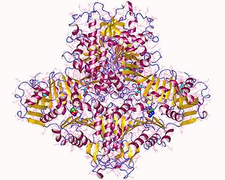
Glutamine synthetase (GS) is an enzyme that plays an essential role in the metabolism of nitrogen by catalyzing the condensation of glutamate and ammonia to form glutamine:
Methionine synthase also known as MS, MeSe, MTR is responsible for the regeneration of methionine from homocysteine. In humans it is encoded by the MTR gene (5-methyltetrahydrofolate-homocysteine methyltransferase). Methionine synthase forms part of the S-adenosylmethionine (SAMe) biosynthesis and regeneration cycle, and is the enzyme responsible for linking the cycle to one-carbon metabolism via the folate cycle. There are two primary forms of this enzyme, the Vitamin B12 (cobalamin)-dependent (MetH) and independent (MetE) forms, although minimal core methionine synthases that do not fit cleanly into either category have also been described in some anaerobic bacteria. The two dominant forms of the enzymes appear to be evolutionary independent and rely on considerably different chemical mechanisms. Mammals and other higher eukaryotes express only the cobalamin-dependent form. In contrast, the distribution of the two forms in Archaeplastida (plants and algae) is more complex. Plants exclusively possess the cobalamin-independent form, while algae have either one of the two, depending on species. Many different microorganisms express both the cobalamin-dependent and cobalamin-independent forms.
Glucose-6-phosphate dehydrogenase (G6PD or G6PDH) (EC 1.1.1.49) is a cytosolic enzyme that catalyzes the chemical reaction
N-Acetylglutamate synthase (NAGS) is an enzyme that catalyses the production of N-acetylglutamate (NAG) from glutamate and acetyl-CoA.
Carbamoyl phosphate synthetase I is a ligase enzyme located in the mitochondria involved in the production of urea. Carbamoyl phosphate synthetase I transfers an ammonia molecule to a molecule of bicarbonate that has been phosphorylated by a molecule of ATP. The resulting carbamate is then phosphorylated with another molecule of ATP. The resulting molecule of carbamoyl phosphate leaves the enzyme.
Acyl-CoA dehydrogenases (ACADs) are a class of enzymes that function to catalyze the initial step in each cycle of fatty acid β-oxidation in the mitochondria of cells. Their action results in the introduction of a trans double-bond between C2 (α) and C3 (β) of the acyl-CoA thioester substrate. Flavin adenine dinucleotide (FAD) is a required co-factor in addition to the presence of an active site glutamate in order for the enzyme to function.
Aspartoacylase is a hydrolytic enzyme that in humans is encoded by the ASPA gene. ASPA catalyzes the deacylation of N-acetyl-l-aspartate (N-acetylaspartate) into aspartate and acetate. It is a zinc-dependent hydrolase that promotes the deprotonation of water to use as a nucleophile in a mechanism analogous to many other zinc-dependent hydrolases. It is most commonly found in the brain, where it controls the levels of N-acetyl-l-aspartate. Mutations that result in loss of aspartoacylase activity are associated with Canavan disease, a rare autosomal recessive neurodegenerative disease.
Dihydrolipoamide dehydrogenase (DLD), also known as dihydrolipoyl dehydrogenase, mitochondrial, is an enzyme that in humans is encoded by the DLD gene. DLD is a flavoprotein enzyme that oxidizes dihydrolipoamide to lipoamide.
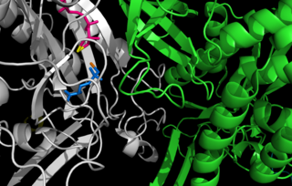
Carboxypeptidase A usually refers to the pancreatic exopeptidase that hydrolyzes peptide bonds of C-terminal residues with aromatic or aliphatic side-chains. Most scientists in the field now refer to this enzyme as CPA1, and to a related pancreatic carboxypeptidase as CPA2.
Carbamoyl phosphate synthetase catalyzes the ATP-dependent synthesis of carbamoyl phosphate from glutamine or ammonia and bicarbonate. This enzyme catalyzes the reaction of ATP and bicarbonate to produce carboxy phosphate and ADP. Carboxy phosphate reacts with ammonia to give carbamic acid. In turn, carbamic acid reacts with a second ATP to give carbamoyl phosphate plus ADP.
Citrin, also known as solute carrier family 25, member 13 (citrin) or SLC25A13, is a protein which in humans is encoded by the SLC25A13 gene.
In enzymology, a N-acyl-D-amino-acid deacylase (EC 3.5.1.81) is an enzyme that catalyzes the chemical reaction
The human ETFB gene encodes the Electron-transfer-flavoprotein, beta subunit, also known as ETF-β. Together with Electron-transfer-flavoprotein, alpha subunit, encoded by the 'ETFA' gene, it forms the heterodimeric Electron transfer flavoprotein (ETF). The native ETF protein contains one molecule of FAD and one molecule of AMP, respectively.
Aminoacylase-1 is an enzyme that in humans is encoded by the ACY1 gene.