Aurora kinase B

Last updated
AURKB
Available structures
PDB Ortholog search: PDBe RCSB
Identifiers
Aliases AURKB , AIK2, AIM-1, AIM1, ARK2, AurB, IPL1, PPP1R48, STK12, STK5, aurkb-sv1, aurkb-sv2, aurora kinase B, STK-1, ARK-2
External IDs OMIM: 604970 MGI: 107168 HomoloGene: 55807 GeneCards: AURKB
Orthologs
SpeciesHumanMouse
Entrez
Ensembl
UniProt
RefSeq (mRNA)

NM_011496

RefSeq (protein)

NP_035626

Location (UCSC) Chr 17: 8.2 – 8.21 Mb Chr 11: 68.94 – 68.94 Mb
PubMed search [3] [4]
Wikidata
View/Edit Human View/Edit Mouse

Aurora kinase B is a protein that functions in the attachment of the mitotic spindle to the centromere.

Contents

Function

Chromosomal segregation during mitosis as well as meiosis is regulated by kinases and phosphatases. The Aurora kinases associate with microtubules during chromosome movement and segregation. Aurora kinase B localizes to microtubules near kinetochores, specifically to the specialized microtubules called K-fibers, and Aurora kinase A (MIM 603072) localizes to centrosomes (Lampson et al., 2004).[supplied by OMIM] [5]

In cancerous cells, over-expression of these enzymes causes unequal distribution of genetic information, creating aneuploid cells, a hallmark of cancer.

Discovery

In 1998, Aurora kinase B was identified in humans by a polymerase chain reaction screen for kinases that are overexpressed in cancers. [6] In the same year, rat Aurora kinase B was identified in a screen designed to find kinases that altered S. cerevisiae proliferation when overexpressed. [7]

Expression and subcellular localization

Aurora kinase B (in green) localizes in a cell cycle dependent manner. (DNA is in blue) Aurora B localization.jpg
Aurora kinase B (in green) localizes in a cell cycle dependent manner. (DNA is in blue)

The expression and activity of Aurora B are regulated according to the cell cycle. Expression of Aurora B reaches a maximum at the G2-M transition, whereas Aurora B protein is most active during mitosis. [6]

Aurora B is a chromosomal passenger protein. Specifically, Aurora B localizes to the chromosomes in prophase, the centromere in prometaphase and metaphase, and the central mitotic spindle in anaphase. [8] This localization has been determined by indirect immunofluorescence in mammalian, C. elegans, and Drosophila cells. A more detailed analysis of Aurora B localization has been carried out in mammalian cells by tagging Aurora B with green fluorescent protein. [9] This analysis showed that the association of Aurora B with centromeres is dynamic (Aurora B at the centromere is constantly exchanging with a pool of cytoplasmic Aurora B). The analysis of tagged Aurora B also suggested that it associates with spindle microtubules during anaphase of mitosis and this association significantly limits its mobility. Finally, a portion of the tagged Aurora B localized to the equatorial cell cortex, having been transported to this location by astral microtubules.

Regulation of Aurora B

Aurora B complexes with three other proteins, Survivin, Borealin and INCENP. Each of the four components of the complex is required for the proper localization and function of the other three. [10] INCENP stimulates Aurora B kinase activity. Survivin might do the same. [11]

Localization of Aurora B to the centromere during prometaphase and metaphase requires phosphorylation of the mammalian kinetochore-specific histone-H3 variant centromere protein A (CENP-A). [12] CENP-A associates with the centromere and is necessary for assembly of the kinetochore. Phosphorylation of CENP-A at serine 7 by Aurora A kinase recruits Aurora B to the centromere. [13] Aurora B, itself, can also phosphorylate CENP-A at the same residue once it is recruited (see below).

Additionally, topoisomerase II has been implicated in the regulation of Aurora B localization and enzymatic activity. [14] This regulatory role may be directly associated with the role of topoisomerase II in disjoining sister chromatids prior to anaphase. In topoisomerase II-depleted cells, Aurora B and INCENP do not transfer to the central spindle in late mitosis. Instead, they remain tightly associated with the centromeres of non-disjoined sister chromatids. Also, cells deficient in topoisomerase II show significantly reduced Aurora B kinase activity. Inhibition of Aurora B due to loss of topoisomerase II seems to depend on BubR1 activity (see below).

Aurora B has been shown to bind to end-binding protein 1 (EB1), a protein that regulates microtubule dynamics. [15] Indirect immunofluorescence showed that Aurora B and EB1 colocalize during anaphase on the central spindle and in the midbody during cytokinesis. Intriguingly, EB1 overexpression enhances Aurora B kinase activity, at least in part because EB1 blocks the dephosphorylation/inactivation of Aurora B by protein phosphatase 2A.

Role in chromosome biorientation

Studies in several organisms indicate that Aurora B oversees chromosome biorientation by ensuring that appropriate connections are made between spindle microtubules and kinetochores.

Inhibition of Aurora B function by RNA interference [16] or microinjection of blocking antibodies [17] impairs the alignment of chromosomes at the equator of the mitotic spindle. This process of alignment is referred to as chromosome congression. The reason for this defect is a subject of ongoing study. Aurora B inhibition may lead to an increase in the number of syntelic attachments (sister chromatid pairs in which both sister kinetochores are attached to microtubules radiating from the same spindle pole). [18] Intriguingly, expression of a dominant-negative and catalytically inactive form of Aurora B disrupted microtubule attachment to the kinetochore and prevented the association of dynein and centromere protein E (CENP-E) with kinetochores.

Numerous kinetochore targets of Aurora kinases have been determined in organisms ranging from yeast to man. Most notably, CENP-A is a target of Aurora B. [12] The phosphorylation of CENP-A by Aurora B reaches a maximum in prometaphase. In fact, Aurora A targets the same CENP-A phosphorylation site as Aurora B, and CENP-A phosphorylation by Aurora A is thought to precede that by Aurora B. Thus, a model has been proposed in which CENP-A phosphorylation by Aurora A recruits Aurora B to the centromere, the latter maintaining the phosphorylation state of CENP-A in a positive feedback loop. Oddly, mutation of this phosphorylation site in CENP-A leads to defects in cytokinesis.

Aurora B also interacts with mitotic centromere-associated kinesin (MCAK). Both Aurora B and MCAK localize to the inner centromere during prometaphase. [19] Aurora B has been shown to recruit MCAK to the centromere and directly phosphorylate MCAK on various residues. [20] Phosphorylation of MCAK by Aurora B limits the ability of MCAK to depolymerize microtubules. Importantly, inhibition of MCAK by a number of approaches leads to improper attachment of kinetochores to spindle microtubules. [21]

It has been hypothesized that tension generated by amphitelic attachment (biorientation; the attachment of sister kinetochores to opposite spindle poles) pulls sister kinetochores apart, thus disrupting the interaction of Aurora B at the innermost portion of the centromere with microtubule binding sites on the fibrous corona of the outermost centromere. Specifically, the tension generated by biorientation pulls MCAK outside of the area of Aurora B localization. [20] Thus, mitosis proceeds upon biorientation and dissociation of Aurora B from its substrates.

Role in chromosome condensation and chromosome cohesion

Aurora B is responsible for phosphorylation of histone-H3 on serine 10 during mitosis. [22] This modification is conserved from yeast (where the kinase is known as Ipl1) to man. Notably, histone-H3 phosphorylation by Aurora B seems not to be responsible for chromatin condensation. Though Aurora B is enriched at centromeres, it localizes diffusely to all chromatin.

In Drosophila cells, Aurora B depletion disrupts chromosome structure and compaction. [23] In these cells, the condensin complex does not localize appropriately to the chromosomes. Similarly, in C. elegans, condensin activity is dependent on Aurora B in metaphase. [24] However, in Xenopus egg cell-free extracts, condensin binding and chromosome condensation occur normally even in the absence of Aurora B. [25] Likewise, after treating cells with an Aurora B enzyme inhibitor (Aurora B localization is not affected), the condensin complex localizes normally.

Aurora B localizes to the paired arms of homologous chromosomes in metaphase I of C. elegans meiosis, and perturbs microtubule dynamics in mitosis. [26] Release of this cohesion, which is dependent on Aurora B, is required for progression to anaphase I and segregation of homologous chromosomes. [27] In mitotic vertebrate B lymphocytes, the proper centromeric localization of a number of Aurora B binding partners requires cohesin. [28]

Role in cytokinesis

The Aurora B complex is necessary for cytokinesis in vertebrates, C. elegans, Drosophila, and fission yeast.

In various cell types, overexpression of a catalytically inactive Aurora B prevents cytokinesis. [7] Disruption of cytokinesis can also arise from Aurora B mislocalization due to mutation of Aurora B binding partners. [29]

Aurora B targets a number of proteins that localize to the cleavage furrow, including the type-III intermediate filament proteins vimentin, [30] desmin, and glial fibrillary acidic protein (GFAP). [31] In general, phosphorylation destabilizes intermediate filaments. Therefore, it has been proposed that phosphorylation of intermediate filaments at the cleavage furrow destabilizes the filaments in preparation for cytokinesis. [31] In agreement with this hypothesis, mutation of Aurora B target sites in intermediate filament proteins leads to defects in filament deformation and prevents the final stage of cytokinesis.

Aurora B also phosphorylates myosin II regulatory light chain at the cleavage furrow. Inhibition of Aurora B activity prevents proper myosin II localization to the cleavage furrow and disrupts spindle midzone organization. [32]

Role in the spindle assembly checkpoint

The spindle assembly checkpoint inhibits progression of mitosis from metaphase to anaphase until all sister chromatid pairs are bioriented.

Presence of Aurora B halts metaphase-to-anaphase transition until proper biorientation is achieved. This is demonstrated in Aurora B deficit cells, where inhibition of Aurora B by hesperadin causes the cell to progress from metaphase to anaphase even when there are misaligned chromosomes in the cell. [33] [34]

Aurora B may be involved in the localization of MAD2 and BubR1, proteins that recognize correct chromosome attachment to spindle microtubules. Loss of Aurora B lowers the concentration of Mad2 and BubR1 at the kinetochores. In particular, Aurora B seems to be responsible for maintaining the localization of Mad2 and BubR1 to the kinetochore following their initial recruitment, which occurs independent of Aurora B. [35] Aurora B may be directly or indirectly involved in the hyper-phosphorylation of BubR1 seen in mitosis in wild-type cells. [36]

Interactions

Aurora B kinase has been shown to interact with:

Role in cancer

Abnormally elevated levels of Aurora B kinase cause unequal chromosomal separation during cell division, resulting in the formation of cells with abnormal numbers of chromosomes, which are both a cause and driver of cancer.

Inhibition of Aurora B kinase by BI811283 in cancer cells leads to the formation of cells with severely abnormal numbers of chromosomes (polyploid). Counterintuitively, inhibition of Aurora B kinase actually causes the polyploid cells formed to continue dividing however, because these cells have severe chromosomal abnormalities, they eventually stop dividing or undergo cell death. [44] [45]

Role in axonal outgrowth and axon regeneration

A novel function for Aurora B kinase has recently been reported in neurons. Following axotomy of cultured neurons, significant upregulation in Aurora B kinase gene expression was observed coinciding with regenerative axonal sprouting. [46] Furthermore, overexpression of Aurora B kinase results in accelerated axonal outgrowth of spinal motor neurons in developing zebrafish. [47]

See also

Related Research Articles

<span class="mw-page-title-main">Mitosis</span> Process in which chromosomes are replicated and separated into two new identical nuclei

Mitosis is a part of the cell cycle in which replicated chromosomes are separated into two new nuclei. Cell division by mitosis is an equational division which gives rise to genetically identical cells in which the total number of chromosomes is maintained. Mitosis is preceded by the S phase of interphase and is followed by telophase and cytokinesis, which divide the cytoplasm, organelles, and cell membrane of one cell into two new cells containing roughly equal shares of these cellular components. The different stages of mitosis altogether define the mitotic phase of a cell cycle—the division of the mother cell into two daughter cells genetically identical to each other.

<span class="mw-page-title-main">Cell division</span> Process by which living cells divide

Cell division is the process by which a parent cell divides into two daughter cells. Cell division usually occurs as part of a larger cell cycle in which the cell grows and replicates its chromosome(s) before dividing. In eukaryotes, there are two distinct types of cell division: a vegetative division (mitosis), producing daughter cells genetically identical to the parent cell, and a cell division that produces haploid gametes for sexual reproduction (meiosis), reducing the number of chromosomes from two of each type in the diploid parent cell to one of each type in the daughter cells. Mitosis is a part of the cell cycle, in which, replicated chromosomes are separated into two new nuclei. Cell division gives rise to genetically identical cells in which the total number of chromosomes is maintained. In general, mitosis is preceded by the S stage of interphase and is followed by telophase and cytokinesis; which divides the cytoplasm, organelles, and cell membrane of one cell into two new cells containing roughly equal shares of these cellular components. The different stages of mitosis all together define the M phase of an animal cell cycle—the division of the mother cell into two genetically identical daughter cells. To ensure proper progression through the cell cycle, DNA damage is detected and repaired at various checkpoints throughout the cycle. These checkpoints can halt progression through the cell cycle by inhibiting certain cyclin-CDK complexes. Meiosis undergoes two divisions resulting in four haploid daughter cells. Homologous chromosomes are separated in the first division of meiosis, such that each daughter cell has one copy of each chromosome. These chromosomes have already been replicated and have two sister chromatids which are then separated during the second division of meiosis. Both of these cell division cycles are used in the process of sexual reproduction at some point in their life cycle. Both are believed to be present in the last eukaryotic common ancestor.

<span class="mw-page-title-main">Anaphase</span> Stage of a cell division

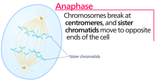
Anaphase is the stage of mitosis after the process of metaphase, when replicated chromosomes are split and the newly-copied chromosomes are moved to opposite poles of the cell. Chromosomes also reach their overall maximum condensation in late anaphase, to help chromosome segregation and the re-formation of the nucleus.

<span class="mw-page-title-main">Spindle apparatus</span> Feature of biological cell structure

In cell biology, the spindle apparatus is the cytoskeletal structure of eukaryotic cells that forms during cell division to separate sister chromatids between daughter cells. It is referred to as the mitotic spindle during mitosis, a process that produces genetically identical daughter cells, or the meiotic spindle during meiosis, a process that produces gametes with half the number of chromosomes of the parent cell.

<span class="mw-page-title-main">Telophase</span> Final stage of a cell division for eukaryotic cells both in mitosis and meiosis

Telophase is the final stage in both meiosis and mitosis in a eukaryotic cell. During telophase, the effects of prophase and prometaphase are reversed. As chromosomes reach the cell poles, a nuclear envelope is re-assembled around each set of chromatids, the nucleoli reappear, and chromosomes begin to decondense back into the expanded chromatin that is present during interphase. The mitotic spindle is disassembled and remaining spindle microtubules are depolymerized. Telophase accounts for approximately 2% of the cell cycle's duration.

<span class="mw-page-title-main">Spindle checkpoint</span> Cell cycle checkpoint

The spindle checkpoint, also known as the metaphase-to-anaphase transition, the spindle assembly checkpoint (SAC), the metaphase checkpoint, or the mitotic checkpoint, is a cell cycle checkpoint during metaphase of mitosis or meiosis that prevents the separation of the duplicated chromosomes (anaphase) until each chromosome is properly attached to the spindle. To achieve proper segregation, the two kinetochores on the sister chromatids must be attached to opposite spindle poles. Only this pattern of attachment will ensure that each daughter cell receives one copy of the chromosome. The defining biochemical feature of this checkpoint is the stimulation of the anaphase-promoting complex by M-phase cyclin-CDK complexes, which in turn causes the proteolytic destruction of cyclins and proteins that hold the sister chromatids together.

<span class="mw-page-title-main">Kinetochore</span> Protein complex that allows microtubules to attach to chromosomes during cell division

A kinetochore is a disc-shaped protein structure associated with duplicated chromatids in eukaryotic cells where the spindle fibers attach during cell division to pull sister chromatids apart. The kinetochore assembles on the centromere and links the chromosome to microtubule polymers from the mitotic spindle during mitosis and meiosis. The term kinetochore was first used in a footnote in a 1934 Cytology book by Lester W. Sharp and commonly accepted in 1936. Sharp's footnote reads: "The convenient term kinetochore has been suggested to the author by J. A. Moore", likely referring to John Alexander Moore who had joined Columbia University as a freshman in 1932.

<span class="mw-page-title-main">Aurora kinase A</span> Protein-coding gene in the species Homo sapiens

Aurora kinase A also known as serine/threonine-protein kinase 6 is an enzyme that in humans is encoded by the AURKA gene.

Polo-like kinases (Plks) are regulatory serine/threonine kinases of the cell cycle involved in mitotic entry, mitotic exit, spindle formation, cytokinesis, and meiosis. Only one Plk is found in the genomes of the fly Drosophila melanogaster (Polo), budding yeast (Cdc5) and fission yeast (Plo1). Vertebrates and other animals, however, have many Plk family members including Plk1, Plk2/Snk, Plk3/Prk/FnK, Plk4/Sak and Plk5. Of the vertebrate Plk family members, the mammalian Plk1 has been most extensively studied. During mitosis and cytokinesis, Plks associate with several structures including the centrosome, kinetochores, and the central spindle.

<span class="mw-page-title-main">PLK1</span> Mammalian protein found in Homo sapiens

Serine/threonine-protein kinase PLK1, also known as polo-like kinase 1 (PLK-1) or serine/threonine-protein kinase 13 (STPK13), is an enzyme that in humans is encoded by the PLK1 gene.

<span class="mw-page-title-main">BUB1</span> Protein-coding gene in the species Homo sapiens

Mitotic checkpoint serine/threonine-protein kinase BUB1 also known as BUB1 is an enzyme that in humans is encoded by the BUB1 gene.

<span class="mw-page-title-main">BUB1B</span> Protein-coding gene in the species Homo sapiens

Mitotic checkpoint serine/threonine-protein kinase BUB1 beta is an enzyme that in humans is encoded by the BUB1B gene. Also known as BubR1, this protein is recognized for its mitotic roles in the spindle assembly checkpoint (SAC) and kinetochore-microtubule interactions that facilitate chromosome migration and alignment. BubR1 promotes mitotic fidelity and protects against aneuploidy by ensuring proper chromosome segregation between daughter cells. BubR1 is proposed to prevent tumorigenesis.

<span class="mw-page-title-main">CENPA</span> Protein-coding gene in the species Homo sapiens

Centromere protein A, also known as CENPA, is a protein which in humans is encoded by the CENPA gene. CENPA is a histone H3 variant which is the critical factor determining the kinetochore position(s) on each chromosome in most eukaryotes including humans.

<span class="mw-page-title-main">CENPF</span> Centromere- and microtubule-associated protein

Centromere protein F is a protein that in humans is encoded by the CENPF gene. It is involved in chromosome segregation during cell division. It also has a role in the orientation of microtubules to form cellular cilia.

<span class="mw-page-title-main">INCENP</span> Protein-coding gene in the species Homo sapiens

Inner centromere protein is a protein that in humans is encoded by the INCENP gene. It is a regulatory protein in the chromosome passenger complex (CPC). It is involved in regulation of the catalytic proteins Aurora B and Aurora C. It acts in association with two other proteins - Survivin and Borealin. These proteins form a tight three-helical bundle. The N-terminal domain of INCENP is the domain involved in formation of this three-helical bundle while its C-terminal domain is responsible for the interaction with Aurora B.

<span class="mw-page-title-main">Centromere protein E</span> Centromere- and microtubule-associated protein

Centromere-associated protein E is a protein that in humans is encoded by the CENPE gene.

<span class="mw-page-title-main">KIF2C</span> Protein-coding gene in the species Homo sapiens

Kinesin-like protein KIF2C is a protein that in humans is encoded by the KIF2C gene.

<span class="mw-page-title-main">Aurora kinase C</span> Protein-coding gene in the species Homo sapiens

Aurora kinase C, also Serine/threonine-protein kinase 13 is an enzyme that in humans is encoded by the AURKC gene.

Syntelic attachment occurs when both sister chromosomes are attached to a single spindle pole.

<span class="mw-page-title-main">Tim J. Yen</span> American molecular biologist

Tim J. Yen is an American molecular biologist and cancer biologist. Yen held the rank of Professor and in 2023, became Emeritus at Fox Chase Cancer Center in Philadelphia, Pennsylvania. Yen is known for pioneering work in the field of mitosis.

References

  1. 1 2 3 GRCh38: Ensembl release 89: ENSG00000178999 - Ensembl, May 2017
  2. 1 2 3 GRCm38: Ensembl release 89: ENSMUSG00000020897 - Ensembl, May 2017
  3. "Human PubMed Reference:". National Center for Biotechnology Information, U.S. National Library of Medicine.
  4. "Mouse PubMed Reference:". National Center for Biotechnology Information, U.S. National Library of Medicine.
  5. "Entrez Gene: AURKB aurora kinase B".
  6. 1 2 Bischoff JR, Anderson L, Zhu Y, Mossie K, Ng L, Souza B, et al. (1998). "A homologue of Drosophila aurora kinase is oncogenic and amplified in human colorectal cancers". The EMBO Journal. 17 (11): 3052–65. doi:10.1093/emboj/17.11.3052. PMC   1170645 . PMID   9606188.
  7. 1 2 Terada Y, Tatsuka M, Suzuki F, Yasuda Y, Fujita S, Otsu M (1998). "AIM-1: A mammalian midbody-associated protein required for cytokinesis". The EMBO Journal. 17 (3): 667–76. doi:10.1093/emboj/17.3.667. PMC   1170416 . PMID   9450992.
  8. Adams RR, Carmena M, Earnshaw WC (2001). "Chromosomal passengers and the (aurora) ABCs of mitosis". Trends in Cell Biology. 11 (2): 49–54. doi:10.1016/s0962-8924(00)01880-8. PMID   11166196.
  9. Murata-Hori M, Tatsuka M, Wang YL (2002). "Probing the dynamics and functions of aurora B kinase in living cells during mitosis and cytokinesis". Molecular Biology of the Cell. 13 (4): 1099–108. doi:10.1091/mbc.01-09-0467. PMC   102254 . PMID   11950924.
  10. Honda R, Körner R, Nigg EA (2003). "Exploring the functional interactions between Aurora B, INCENP, and survivin in mitosis". Molecular Biology of the Cell. 14 (8): 3325–41. doi:10.1091/mbc.E02-11-0769. PMC   181570 . PMID   12925766.
  11. Chen J, Jin S, Tahir SK, Zhang H, Liu X, Sarthy AV, et al. (2003). "Survivin enhances Aurora-B kinase activity and localizes Aurora-B in human cells". Journal of Biological Chemistry. 278 (1): 486–90. doi: 10.1074/jbc.M211119200 . PMID   12419797.
  12. 1 2 Zeitlin SG, Shelby RD, Sullivan KF (2001). "CENP-A is phosphorylated by Aurora B kinase and plays an unexpected role in completion of cytokinesis". The Journal of Cell Biology. 155 (7): 1147–57. doi:10.1083/jcb.200108125. PMC   2199334 . PMID   11756469.
  13. Kunitoku N, Sasayama T, Marumoto T, Zhang D, Honda S, Kobayashi O, et al. (2003). "CENP-A phosphorylation by Aurora-A in prophase is required for enrichment of Aurora-B at inner centromeres and for kinetochore function". Developmental Cell. 5 (6): 853–64. doi: 10.1016/s1534-5807(03)00364-2 . PMID   14667408.
  14. Coelho PA, Queiroz-Machado J, Carmo AM, Moutinho-Pereira S, Maiato H, Sunkel CE (2008). "Dual role of topoisomerase II in centromere resolution and aurora B activity". PLOS Biology. 6 (8): e207. doi: 10.1371/journal.pbio.0060207 . PMC   2525683 . PMID   18752348.
  15. Sun L, Gao J, Dong X, Liu M, Li D, Shi X, et al. (2008). "EB1 promotes Aurora-B kinase activity through blocking its inactivation by protein phosphatase 2A". Proceedings of the National Academy of Sciences. 105 (20): 7153–8. Bibcode:2008PNAS..105.7153S. doi: 10.1073/pnas.0710018105 . PMC   2438220 . PMID   18477699.
  16. Adams RR, Maiato H, Earnshaw WC, Carmena M (2001). "Essential roles of Drosophila inner centromere protein (INCENP) and aurora B in histone H3 phosphorylation, metaphase chromosome alignment, kinetochore disjunction, and chromosome segregation". The Journal of Cell Biology. 153 (4): 865–80. doi:10.1083/jcb.153.4.865. PMC   2192373 . PMID   11352945.
  17. Kallio MJ, McCleland ML, Stukenberg PT, Gorbsky GJ (2002). "Inhibition of aurora B kinase blocks chromosome segregation, overrides the spindle checkpoint, and perturbs microtubule dynamics in mitosis". Current Biology. 12 (11): 900–5. Bibcode:2002CBio...12..900K. doi: 10.1016/s0960-9822(02)00887-4 . PMID   12062053. S2CID   13939857.
  18. Hauf S, Cole RW, Laterra S, Zimmer C, Schnapp G, Walter R, et al. (2003). "The small molecule Hesperadin reveals a role for Aurora B in correcting kinetochore-microtubule attachment and in maintaining the spindle assembly checkpoint". The Journal of Cell Biology. 161 (2): 281–94. doi:10.1083/jcb.200208092. PMC   2172906 . PMID   12707311.
  19. Wordeman L, Wagenbach M, Maney T (1999). "Mutations in the ATP-binding domain affect the subcellular distribution of mitotic centromere-associated kinesin (MCAK)". Cell Biology International. 23 (4): 275–86. doi:10.1006/cbir.1999.0359. PMID   10600236. S2CID   29403722.
  20. 1 2 Andrews PD, Ovechkina Y, Morrice N, Wagenbach M, Duncan K, Wordeman L, et al. (2004). "Aurora B regulates MCAK at the mitotic centromere". Developmental Cell. 6 (2): 253–68. doi: 10.1016/s1534-5807(04)00025-5 . PMID   14960279.
  21. Kline-Smith SL, Khodjakov A, Hergert P, Walczak CE (2004). "Depletion of centromeric MCAK leads to chromosome congression and segregation defects due to improper kinetochore attachments". Molecular Biology of the Cell. 15 (3): 1146–59. doi:10.1091/mbc.E03-08-0581. PMC   363095 . PMID   14699064.
  22. Hsu JY, Sun ZW, Li X, Reuben M, Tatchell K, Bishop DK, et al. (2000). "Mitotic phosphorylation of histone H3 is governed by Ipl1/aurora kinase and Glc7/PP1 phosphatase in budding yeast and nematodes". Cell. 102 (3): 279–91. doi: 10.1016/s0092-8674(00)00034-9 . PMID   10975519. S2CID   16057773.
  23. Giet R, Glover DM (2001). "Drosophila aurora B kinase is required for histone H3 phosphorylation and condensin recruitment during chromosome condensation and to organize the central spindle during cytokinesis". The Journal of Cell Biology. 152 (4): 669–82. doi:10.1083/jcb.152.4.669. PMC   2195771 . PMID   11266459.
  24. Crosio C, Fimia GM, Loury R, Kimura M, Okano Y, Zhou H, et al. (2002). "Mitotic phosphorylation of histone H3: Spatio-temporal regulation by mammalian Aurora kinases". Molecular and Cellular Biology. 22 (3): 874–85. doi:10.1128/mcb.22.3.874-885.2002. PMC   133550 . PMID   11784863.
  25. MacCallum DE, Losada A, Kobayashi R, Hirano T (2002). "ISWI remodeling complexes in Xenopus egg extracts: Identification as major chromosomal components that are regulated by INCENP-aurora B". Molecular Biology of the Cell. 13 (1): 25–39. doi:10.1091/mbc.01-09-0441. PMC   65070 . PMID   11809820.
  26. Kaitna S, Pasierbek P, Jantsch M, Loidl J, Glotzer M (2002). "The aurora B kinase AIR-2 regulates kinetochores during mitosis and is required for separation of homologous Chromosomes during meiosis". Current Biology. 12 (10): 798–812. Bibcode:2002CBio...12..798K. doi: 10.1016/s0960-9822(02)00820-5 . PMID   12015116. S2CID   2215023.
  27. Rogers E, Bishop JD, Waddle JA, Schumacher JM, Lin R (2002). "The aurora kinase AIR-2 functions in the release of chromosome cohesion in Caenorhabditis elegans meiosis". The Journal of Cell Biology. 157 (2): 219–29. doi:10.1083/jcb.200110045. PMC   1855215 . PMID   11940606.
  28. Sonoda E, Matsusaka T, Morrison C, Vagnarelli P, Hoshi O, Ushiki T, et al. (2001). "Scc1/Rad21/Mcd1 is required for sister chromatid cohesion and kinetochore function in vertebrate cells". Developmental Cell. 1 (6): 759–70. doi: 10.1016/s1534-5807(01)00088-0 . PMID   11740938.
  29. MacKay AM, Ainsztein AM, Eckley DM, Earnshaw WC (1998). "A dominant mutant of inner centromere protein (INCENP), a chromosomal protein, disrupts prometaphase congression and cytokinesis". The Journal of Cell Biology. 140 (5): 991–1002. doi:10.1083/jcb.140.5.991. PMC   2132686 . PMID   9490714.
  30. Goto H, Yasui Y, Kawajiri A, Nigg EA, Terada Y, Tatsuka M, et al. (2003). "Aurora-B regulates the cleavage furrow-specific vimentin phosphorylation in the cytokinetic process". Journal of Biological Chemistry. 278 (10): 8526–30. doi: 10.1074/jbc.M210892200 . PMID   12458200.
  31. 1 2 Kawajiri A, Yasui Y, Goto H, Tatsuka M, Takahashi M, Nagata K, et al. (2003). "Functional significance of the specific sites phosphorylated in desmin at cleavage furrow: Aurora-B may phosphorylate and regulate type III intermediate filaments during cytokinesis coordinatedly with Rho-kinase". Molecular Biology of the Cell. 14 (4): 1489–500. doi:10.1091/mbc.E02-09-0612. PMC   153117 . PMID   12686604.
  32. Murata-Hori M, Fumoto K, Fukuta Y, Iwasaki T, Kikuchi A, Tatsuka M, et al. (2000). "Myosin II regulatory light chain as a novel substrate for AIM-1, an aurora/Ipl1p-related kinase from rat". Journal of Biochemistry. 128 (6): 903–7. doi:10.1093/oxfordjournals.jbchem.a022840. PMID   11098131.
  33. Hauf S, Cole RW, Laterra S, Zimmer C, Schnapp G, Walter R, et al. (2003). "The small molecule Hesperadin reveals a role for Aurora B in correcting kinetochore-microtubule attachment and in maintaining the spindle assembly checkpoint". The Journal of Cell Biology. 161 (2): 281–94. doi:10.1083/jcb.200208092. PMC   2172906 . PMID   12707311.
  34. Santaguida S, Vernieri C, Villa F, Ciliberto A, Musacchio A (2011-04-20). "Evidence that Aurora B is implicated in spindle checkpoint signalling independently of error correction". The EMBO Journal. 30 (8): 1508–1519. doi:10.1038/emboj.2011.70. ISSN   1460-2075. PMC   3102279 . PMID   21407176.
  35. Lens SM, Wolthuis RM, Klompmaker R, Kauw J, Agami R, Brummelkamp T, et al. (2003). "Survivin is required for a sustained spindle checkpoint arrest in response to lack of tension". The EMBO Journal. 22 (12): 2934–47. doi:10.1093/emboj/cdg307. PMC   162159 . PMID   12805209.
  36. Ditchfield C, Johnson VL, Tighe A, Ellston R, Haworth C, Johnson T, et al. (2003). "Aurora B couples chromosome alignment with anaphase by targeting BubR1, Mad2, and Cenp-E to kinetochores". The Journal of Cell Biology. 161 (2): 267–80. doi:10.1083/jcb.200208091. PMC   2172902 . PMID   12719470.
  37. Ryser S, Dizin E, Jefford CE, Delaval B, Gagos S, Christodoulidou A, et al. (February 2009). "Distinct roles of BARD1 isoforms in mitosis: full-length BARD1 mediates Aurora B degradation, cancer-associated BARD1beta scaffolds Aurora B and BRCA2". Cancer Research. 69 (3): 1125–34. doi: 10.1158/0008-5472.CAN-08-2134 . PMID   19176389.
  38. Wheatley SP, Carvalho A, Vagnarelli P, Earnshaw WC (June 2001). "INCENP is required for proper targeting of Survivin to the centromeres and the anaphase spindle during mitosis". Current Biology. 11 (11): 886–90. Bibcode:2001CBio...11..886W. doi: 10.1016/S0960-9822(01)00238-X . PMID   11516652. S2CID   381637.
  39. Chen J, Jin S, Tahir SK, Zhang H, Liu X, Sarthy AV, et al. (January 2003). "Survivin enhances Aurora-B kinase activity and localizes Aurora-B in human cells". J. Biol. Chem. 278 (1): 486–90. doi: 10.1074/jbc.M211119200 . PMID   12419797.
  40. Sampath SC, Ohi R, Leismann O, Salic A, Pozniakovski A, Funabiki H (July 2004). "The chromosomal passenger complex is required for chromatin-induced microtubule stabilization and spindle assembly". Cell. 118 (2): 187–202. doi: 10.1016/j.cell.2004.06.026 . PMID   15260989. S2CID   17795816.
  41. Gassmann R, Carvalho A, Henzing AJ, Ruchaud S, Hudson DF, Honda R, et al. (July 2004). "Borealin: a novel chromosomal passenger required for stability of the bipolar mitotic spindle". J. Cell Biol. 166 (2): 179–91. doi:10.1083/jcb.200404001. PMC   2172304 . PMID   15249581.
  42. Delaval B, Ferrand A, Conte N, Larroque C, Hernandez-Verdun D, Prigent C, et al. (June 2004). "Aurora B -TACC1 protein complex in cytokinesis". Oncogene. 23 (26): 4516–22. doi: 10.1038/sj.onc.1207593 . PMID   15064709.
  43. Chen BB, Glasser JR, Coon TA, Mallampalli RK (August 2013). "Skp-cullin-F box E3 ligase component FBXL2 ubiquitinates Aurora B to inhibit tumorigenesis". Cell Death & Disease. 4 (8): e759. doi:10.1038/cddis.2013.271. ISSN   2041-4889. PMC   3763433 . PMID   23928698.
  44. Gürtler U, Tontsch-Grunt U, Jarvis M, Zahn SK, Boehmelt G, Quant J, et al. (2010). "Effect of BI 811283, a novel inhibitor of Aurora B kinase, on tumor senescence and apoptosis". J. Clin. Oncol. 28 (15 Suppl e13632): e13632. doi:10.1200/jco.2010.28.15_suppl.e13632.
  45. Sorrentino R, Libertini S, Pallante PL, Troncone G, Palombini L, Bavetsias V, et al. (2005). "Aurora B overexpression associates with the thyroid carcinoma undifferentiated phenotype and is required for thyroid carcinoma cell proliferation". J. Clin. Endocrinol. Metab. 90 (2): 928–35. doi: 10.1210/jc.2004-1518 . PMID   15562011.
  46. Ng JM, Chen MJ, Leung JY, Peng ZF, Manikandan J, Qi RZ, et al. (Apr 2012). "Transcriptional insights on the regenerative mechanics of axotomized neurons in vitro". Journal of Cellular and Molecular Medicine. 16 (4): 789–811. doi:10.1111/j.1582-4934.2011.01361.x. PMC   3822849 . PMID   21711447.
  47. Gwee SS, Radford RA, Chow S, Syal MD, Morsch M, Formella I, et al. (Dec 2017). "Aurora kinase B regulates axonal outgrowth and regeneration in the spinal motor neurons of developing zebrafish". Cellular and Molecular Life Sciences. 75 (23): 4269–4285. doi:10.1007/s00018-018-2780-5. PMID   29468257. S2CID   3408586.

Further reading

This article incorporates text from the United States National Library of Medicine, which is in the public domain.