Automated analyser

Last updated

An automated analyser is a medical laboratory instrument designed to measure various substances and other characteristics in a number of biological samples quickly, with minimal human assistance. These measured properties of blood and other fluids may be useful in the diagnosis of disease.

Contents

Roche Cobas 6000
Roche Cobas u 411 Cobas u 411.JPG
Roche Cobas u 411
Beckman Chemistry analysers: Access (left); Synchron (right). LabMachines.jpg
Beckman Chemistry analysers: Access (left); Synchron (right).
Racks: for putting samples, quality controls or calibrations. Cobas 6000 RACKS.jpg
Racks: for putting samples, quality controls or calibrations. Cobas 6000
These tubes are put in the racks for testing Tube vacuette.jpg
These tubes are put in the racks for testing

Photometry is the most common method for testing the amount of a specific analyte in a sample. In this technique, the sample undergoes a reaction to produce a color change. Then, a photometer measures the absorbance of the sample to indirectly measure the concentration of analyte present in the sample. The use of an ion-selective electrode (ISE) is another common analytical method that specifically measures ion concentrations. This typically measures the concentrations of sodium, calcium or potassium present in the sample. [1]

There are various methods of introducing samples into the analyser. Test tubes of samples are often loaded into racks. These racks can be inserted directly into some analysers or, in larger labs, moved along an automated track. More manual methods include inserting tubes directly into circular carousels that rotate to make the sample available. Some analysers require samples to be transferred to sample cups. However, the need to protect the health and safety of laboratory staff has prompted many manufacturers to develop analysers that feature closed tube sampling, preventing workers from direct exposure to samples. [2] [3] Samples can be processed singly, in batches, or continuously.

The automation of laboratory testing does not remove the need for human expertise (results must still be evaluated by medical technologists and other qualified clinical laboratory professionals), but it does ease concerns about error reduction, staffing concerns, and safety.

Routine biochemistry analysers

These are machines that process a large portion of the samples going into a hospital or private medical laboratory. Automation of the testing process has reduced testing time for many analytes from days to minutes. The history of discrete sample analysis for the clinical laboratory began with the introduction of the "Robot Chemist" invented by Hans Baruch and introduced commercially in 1959[1].

The AutoAnalyzer is an early example of an automated chemistry analyzer using a special flow technique named "continuous flow analysis (CFA)", invented in 1957 by Leonard Skeggs, PhD and first made by the Technicon Corporation. The first applications were for clinical (medical) analysis. The AutoAnalyzer profoundly changed the character of the chemical testing laboratory by allowing significant increases in the numbers of samples that could be processed. Samples used in the analyzers include, but are not limited to, blood, serum, plasma, urine, cerebrospinal fluid, and other fluids from within the body. [4] The design based on separating a continuously flowing stream with air bubbles largely reduced slow, clumsy, and error-prone manual methods of analysis. The types of tests include enzyme levels (such as many of the liver function tests), ion levels (e.g. sodium and potassium, and other tell-tale chemicals (such as glucose, serum albumin, or creatinine).

Simple ions are often measured with ion selective electrodes, which let one type of ion through, and measure voltage differences. [5] Enzymes may be measured by the rate they change one coloured substance to another; in these tests, the results for enzymes are given as an activity, not as a concentration of the enzyme. Other tests use colorimetric changes to determine the concentration of the chemical in question. Turbidity may also be measured.

Immuno-based analysers

Antibodies are used by some analysers to detect many substances by immunoassay and other reactions that employ the use of antibody-antigen reactions.

When concentration of these compounds is too low to cause a measurable increase in turbidity when bound to antibody, more specialised methods must be used.

Recent developments include automation for the immunohaematology lab, also known as transfusion medicine.

Hematology analysers

These are used to perform complete blood counts, erythrocyte sedimentation rates (ESRs), or coagulation tests.

Cell counters

Automated cell counters sample the blood, and quantify, classify, and describe cell populations using both electrical and optical techniques. Electrical analysis involves passing a dilute solution of the blood through an aperture across which an electrical current is flowing. The passage of cells through the current changes the impedance between the terminals (the Coulter principle). [6] A lytic reagent is added to the blood solution to selectively lyse the red cells (RBCs), leaving only white cells (WBCs), and platelets intact. Then the solution is passed through a second detector. This allows the counts of RBCs, WBCs, and platelets to be obtained. The platelet count is easily separated from the WBC count by the smaller impedance spikes they produce in the detector due to their lower cell volumes.

Optical detection may be utilised to gain a differential count of the populations of white cell types. A dilute suspension of cells is passed through a flow cell, which passes cells one at a time through a capillary tube past a laser beam. The reflectance, transmission and scattering of light from each cell is analysed by sophisticated software giving a numerical representation of the likely overall distribution of cell populations.

Some of the latest hematology instruments may report Cell Population Data that consist in Leukocyte morphological information that may be used for flagging Cell abnormalities that trigger the suspect of some diseases.

Reticulocyte counts can now be performed by many analysers, giving an alternative to time-consuming manual counts. Many automated reticulocyte counts, like their manual counterparts, employ the use of a supravital dye such as new methylene blue to stain the red cells containing reticulin prior to counting. [7] Some analysers have a modular slide maker which is able to both produce a blood film of consistent quality and stain the film, which is then reviewed by a medical laboratory professional.

Coagulometers

Automated coagulation machines or Coagulometers measure the ability of blood to clot by performing any of several types of tests including Partial thromboplastin times, Prothrombin times (and the calculated INRs commonly used for therapeutic evaluation), Lupus anticoagulant screens, D dimer assays, and factor assays.

Coagulometers require blood samples that have been drawn in tubes containing sodium citrate as an anticoagulant. These are used because the mechanism behind the anticoagulant effect of sodium citrate is reversible. Depending on the test, different substances can be added to the blood plasma to trigger a clotting reaction. The progress of clotting may be monitored optically by measuring the absorbance of a particular wavelength of light by the sample and how it changes over time.

StaRRsed Inversa, automated Westergren-based ESR 'analyzer' StaRRsed Inversa, automated Westergren based ESR 'analyzer'.jpg
StaRRsed Inversa, automated Westergren-based ESR 'analyzer'

..

Other hematology apparatus

Automatic erythrocyte sedimentation rate (ESR) readers, while not strictly analysers, do preferably have to comply to the 2011-published CLSI (Clinical and Laboratory Standards Institute) "Procedures for the Erythrocyte Sedimentation Rate Test: H02-A5 and to the ICSH (International Council for Standardization in Haematology) published "ICSH review of the measurement of the erythrocyte sedimentation rate", both indicating the only reference method, being Westergren, explicitly indicating the use of diluted blood (with sodium citrate), in 200 mm pipettes, bore 2.55 mm. After 30 or 60 minutes being in a vertical position, with no draughts and vibration or direct sunlight allowed, an optical reader determines how far the red cells have fallen by detecting the level.

Miscellaneous analysers

Some tests and test categories are unique in their mechanism or scope, and require a separate analyser for only a few tests, or even for only one test. Other tests are esoteric in nature—they are performed less frequently than other tests, and are generally more expensive and time-consuming to perform. Even so, the current[ when? ] shortage of qualified clinical laboratory professionals [8] has spurred manufacturers to develop automated systems for even these rarely performed tests.

Analysers that fall into this category include instruments that perform:

See also

Notes

1. Rosenfeld, Louis. Four Centuries of Clinical Chemistry. Gordon and Breach Science Publishers, 1999. ISBN   90-5699-645-2. Pp. 490–492

Related Research Articles

<span class="mw-page-title-main">Analytical chemistry</span> Study of the separation, identification, and quantification of matter

Analytical chemistry studies and uses instruments and methods to separate, identify, and quantify matter. In practice, separation, identification or quantification may constitute the entire analysis or be combined with another method. Separation isolates analytes. Qualitative analysis identifies analytes, while quantitative analysis determines the numerical amount or concentration.

<span class="mw-page-title-main">Clinical chemistry</span> Area of clinical pathology that is generally concerned with analysis of bodily fluids

Clinical chemistry is a division in medical laboratory sciences focusing on qualitative tests of important compounds, referred to as analytes or markers, in bodily fluids and tissues using analytical techniques and specialized instruments. This interdisciplinary field includes knowledge from medicine, biology, chemistry, biomedical engineering, informatics, and an applied form of biochemistry.

<span class="mw-page-title-main">Blood cell</span> Cell produced by hematopoiesis

A blood cell, also called a hematopoietic cell, hemocyte, or hematocyte, is a cell produced through hematopoiesis and found mainly in the blood. Major types of blood cells include red blood cells (erythrocytes), white blood cells (leukocytes), and platelets (thrombocytes). Together, these three kinds of blood cells add up to a total 45% of the blood tissue by volume, with the remaining 55% of the volume composed of plasma, the liquid component of blood.

<span class="mw-page-title-main">Erythrocyte sedimentation rate</span> Physiological quantity

The erythrocyte sedimentation rate is the rate at which red blood cells in anticoagulated whole blood descend in a standardized tube over a period of one hour. It is a common hematology test, and is a non-specific measure of inflammation. To perform the test, anticoagulated blood is traditionally placed in an upright tube, known as a Westergren tube, and the distance which the red blood cells fall is measured and reported in millimetres at the end of one hour.

<span class="mw-page-title-main">Complete blood count</span> Routine laboratory test of blood cells

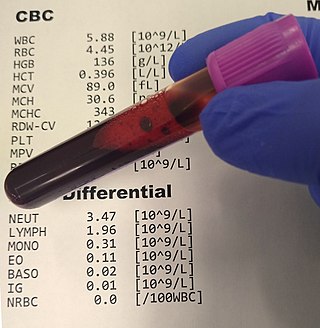
A complete blood count (CBC), also known as a full blood count (FBC), is a set of medical laboratory tests that provide information about the cells in a person's blood. The CBC indicates the counts of white blood cells, red blood cells and platelets, the concentration of hemoglobin, and the hematocrit. The red blood cell indices, which indicate the average size and hemoglobin content of red blood cells, are also reported, and a white blood cell differential, which counts the different types of white blood cells, may be included.

<span class="mw-page-title-main">Hitachi 917</span>

The Hitachi 917 is an automated biochemistry analyser utilise medical laboratories to process biological fluid specimens, such as urine, cerebrospinal fluid, and most commonly, blood.

<span class="mw-page-title-main">Serum (blood)</span> Fluid and solute component of blood

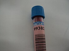
Serum is the fluid and solvent component of blood which does not play a role in clotting. It may be defined as blood plasma without the clotting factors, or as blood with all cells and clotting factors removed. Serum contains all proteins except clotting factors, including all electrolytes, antibodies, antigens, hormones; and any exogenous substances. Serum also does not contain all the formed elements of blood, which include blood cells and platelets.

A blood bank is a center where blood gathered as a result of blood donation is stored and preserved for later use in blood transfusion. The term "blood bank" typically refers to a department of a hospital usually within a clinical pathology laboratory where the storage of blood product occurs and where pre-transfusion and blood compatibility testing is performed. However, it sometimes refers to a collection center, and some hospitals also perform collection. Blood banking includes tasks related to blood collection, processing, testing, separation, and storage.

<span class="mw-page-title-main">Blood smear</span> Stained blood on microscope slide

A blood smear, peripheral blood smear or blood film is a thin layer of blood smeared on a glass microscope slide and then stained in such a way as to allow the various blood cells to be examined microscopically. Blood smears are examined in the investigation of hematological (blood) disorders and are routinely employed to look for blood parasites, such as those of malaria and filariasis.

An assay is an investigative (analytic) procedure in laboratory medicine, mining, pharmacology, environmental biology and molecular biology for qualitatively assessing or quantitatively measuring the presence, amount, or functional activity of a target entity. The measured entity is often called the analyte, the measurand, or the target of the assay. The analyte can be a drug, biochemical substance, chemical element or compound, or cell in an organism or organic sample. An assay usually aims to measure an analyte's intensive property and express it in the relevant measurement unit.

<span class="mw-page-title-main">Prothrombin time</span> Assay for evaluating the extrinsic pathway & common pathway of coagulation

The prothrombin time (PT) – along with its derived measures of prothrombin ratio (PR) and international normalized ratio (INR) – is an assay for evaluating the extrinsic pathway and common pathway of coagulation. This blood test is also called protime INR and PT/INR. They are used to determine the clotting tendency of blood, in such things as the measure of warfarin dosage, liver damage, and vitamin K status. PT measures the following coagulation factors: I (fibrinogen), II (prothrombin), V (proaccelerin), VII (proconvertin), and X.

<span class="mw-page-title-main">Apheresis</span> Medical techniques to separate one or more components of blood

Apheresis is a medical technology in which the blood of a person is passed through an apparatus that separates out one particular constituent and returns the remainder to the circulation. It is thus an extracorporeal therapy.

<span class="mw-page-title-main">Immunoassay</span> Biochemical test for a protein or other molecule using an antibody

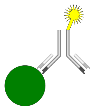
An immunoassay (IA) is a biochemical test that measures the presence or concentration of a macromolecule or a small molecule in a solution through the use of an antibody (usually) or an antigen (sometimes). The molecule detected by the immunoassay is often referred to as an "analyte" and is in many cases a protein, although it may be other kinds of molecules, of different sizes and types, as long as the proper antibodies that have the required properties for the assay are developed. Analytes in biological liquids such as serum or urine are frequently measured using immunoassays for medical and research purposes.

<span class="mw-page-title-main">AutoAnalyzer</span> Automated analyzer using a flow technique

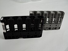
The AutoAnalyzer is an automated analyzer using a flow technique called continuous flow analysis (CFA), or more correctly segmented flow analysis (SFA) first made by the Technicon Corporation. The instrument was invented in 1957 by Leonard Skeggs, PhD and commercialized by Jack Whitehead's Technicon Corporation. The first applications were for clinical analysis, but methods for industrial and environmental analysis soon followed. The design is based on segmenting a continuously flowing stream with air bubbles.

Thromboplastin (TPL) is derived from cell membranes and is a mixture of both phospholipids and tissue factor, neither of which are enzymes. Thromboplastin acts on and accelerates the activity of Factor Xa, also known as thrombokinase, aiding blood coagulation through catalyzing the conversion of prothrombin to thrombin. Thromboplastin is found in brain, lung, and other tissues and especially in blood platelets.

Thromboelastometry (TEM), previously named rotational thromboelastography (ROTEG) or rotational thromboelastometry (ROTEM), is an established viscoelastic method for hemostasis testing in whole blood. It is a modification of traditional thromboelastography (TEG).

Pseudothrombocytopenia (PTCP) or spurious thrombocytopenia is an in-vitro sampling problem which may mislead the diagnosis towards the more critical condition of thrombocytopenia. The phenomenon occurs when the anticoagulant used while testing the blood sample causes clumping of platelets which mimics a low platelet count. The phenomenon has first been reported in 1969.

Hematology analyzers are used to count and identify blood cells at high speed with accuracy. During the 1950s, laboratory technicians counted each individual blood cell underneath a microscope. Tedious and inconsistent, this was replaced with the first, very basic hematology analyzer, engineered by Wallace H. Coulter. The early hematology analyzers relied on Coulter's principle. However, they have evolved to encompass numerous techniques.

<span class="mw-page-title-main">White blood cell differential</span> Blood test

A white blood cell differential is a medical laboratory test that provides information about the types and amounts of white blood cells in a person's blood. The test, which is usually ordered as part of a complete blood count (CBC), measures the amounts of the five normal white blood cell types – neutrophils, lymphocytes, monocytes, eosinophils and basophils – as well as abnormal cell types if they are present. These results are reported as percentages and absolute values, and compared against reference ranges to determine whether the values are normal, low, or high. Changes in the amounts of white blood cells can aid in the diagnosis of many health conditions, including viral, bacterial, and parasitic infections and blood disorders such as leukemia.

<span class="mw-page-title-main">Celloscope automated cell counter</span> Cell counting and analysis device

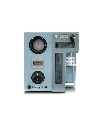
Celloscope automated cell counter was developed in the 50s for enumeration of erythrocytes, leukocytes, and thrombocytes in blood samples. Together with the Coulter counter, the Celloscope analyzer can be considered one of the predecessors of today’s automated hematology analyzers, as the principle of the electrical impedance method is still utilized in cell counters installed in clinical laboratories around the world.

References