Biliverdin

Last updated
Biliverdin
Biliverdin3.svg
Biliverdin-based-on-xtal-3D-bs-17.png
Names
IUPAC name
3,3′-(2,17-Diethenyl-3,7,13,18-tetramethyl-1,19-dioxo-19,21,22,24-tetrahydro-1H-biline-8,12-diyl)dipropanoic acid
Systematic IUPAC name
3,3′-([12(2)Z,4(52)Z,6(72)Z]-13,74-Diethenyl-14,33,54,73-tetramethyl-15,75-dioxo-11,15,71,75-tetrahydro-31H-1,7(2),3,5(2,5)-tetrapyrrolaheptaphane-12(2),4(52),6(72)-triene-34,53-diyl)dipropanoic acid
Identifiers
3D model (JSmol)
ChEBI
ChEMBL
ChemSpider
ECHA InfoCard 100.003.675 OOjs UI icon edit-ltr-progressive.svg
MeSH Biliverdin
PubChem CID
UNII
  • InChI=1S/C33H34N4O6/c1-7-20-19(6)32(42)37-27(20)14-25-18(5)23(10-12-31(40)41)29(35-25)15-28-22(9-11-30(38)39)17(4)24(34-28)13-26-16(3)21(8-2)33(43)36-26/h7-8,13-15,35H,1-2,9-12H2,3-6H3,(H,36,43)(H,37,42)(H,38,39)(H,40,41)/b26-13-,27-14-,28-15- Yes check.svgY
    Key: QBUVFDKTZJNUPP-BBROENKCSA-N Yes check.svgY
  • InChI=1/C33H34N4O6/c1-7-20-19(6)32(42)37-27(20)14-25-18(5)23(10-12-31(40)41)29(35-25)15-28-22(9-11-30(38)39)17(4)24(34-28)13-26-16(3)21(8-2)33(43)36-26/h7-8,13-15,35H,1-2,9-12H2,3-6H3,(H,36,43)(H,37,42)(H,38,39)(H,40,41)/b26-13-,27-14-,28-15-
    Key: QBUVFDKTZJNUPP-BBROENKCBK
  • CC\1=C(/C(=C/C2=C(C(=C(N2)/C=C\3/C(=C(C(=O)N3)C)C=C)C)CCC(=O)O)/N/C1=C\C4=NC(=O)C(=C4C)C=C)CCC(=O)O
Properties
C33H34N4O6
Molar mass 582.646
AppearanceDark green plates or prisms with violet surface color
Melting point > 300 °C
Hazards
Occupational safety and health (OHS/OSH):
Main hazards
Irritant
Safety data sheet (SDS) Sigma-Aldrich
Except where otherwise noted, data are given for materials in their standard state (at 25 °C [77 °F], 100 kPa).
Yes check.svgY  verify  (what is  Yes check.svgYX mark.svgN ?)

Biliverdin (from the Latin for green bile) is a green tetrapyrrolic bile pigment, and is a product of heme catabolism. [1] [2] It is the pigment responsible for a greenish color sometimes seen in bruises. [2]

Contents

Metabolism

Heme metabolism Heme Breakdown.png
Heme metabolism

Biliverdin results from the breakdown of the heme moiety of hemoglobin in erythrocytes. Macrophages break down senescent erythrocytes and break the heme down into biliverdin along with hemosiderin, in which biliverdin normally rapidly reduces to free bilirubin. [1] [3]

Biliverdin is seen briefly in some bruises as a green color. In bruises, its breakdown into bilirubin leads to a yellowish color. [2]

Role in disease

Biliverdin has been found in excess in the blood of humans suffering from hepatic diseases. Jaundice is caused by the accumulation of biliverdin or bilirubin (or both) in the circulatory system and tissues. [1] Jaundiced skin and sclera (whites of the eyes) are characteristic of liver failure.

Role in treatment of disease

While typically regarded as a mere waste product of heme breakdown, evidence that suggests that biliverdin – and other bile pigments – has a physiological role in humans has been mounting. [4] [5]

Bile pigments such as biliverdin possess significant anti-mutagenic and antioxidant properties and therefore, may fulfil a useful physiological function. [5] Biliverdin and bilirubin have been shown to be potent scavengers of hydroperoxyl radicals. [4] [5] They have also been shown to inhibit the effects of polycyclic aromatic hydrocarbons, heterocyclic amines, and oxidants – all of which are mutagens. Some studies have found that people with higher concentration levels of bilirubin and biliverdin in their bodies have a lower frequency of cancer and cardiovascular disease. [4] It has been suggested that biliverdin – as well as many other tetrapyrrolic pigments – may function as an HIV-1 protease inhibitor [6] as well as having beneficial effects in asthma [5] though further research is needed to confirm these results. There are currently no practical implications for using biliverdin in the treatment of any disease.

In non-human animals

Biliverdin is an important pigment component in avian egg shells, especially blue and green shells. Blue egg shells have a significantly higher concentration of biliverdin than brown egg shells. [7]

Research has shown that the biliverdin of egg shells is produced from the shell gland, rather than from the breakdown of erythrocytes in the blood stream,[ citation needed ] although there is no evidence that the sources of the material are neither tetrapyrroles nor free haem from the blood plasma.[ clarification needed ][ citation needed ]

Along with its presence in avian egg shells, other studies have also shown that biliverdin is present in the blue-green blood of many marine fish, the blood of tobacco hornworm, the wings of moth and butterfly, the serum and eggs of frogs, and the placenta of dogs. [8] With dogs this can lead, in extremely rare cases, to the birth of puppies with green fur; however, the green color fades out soon after birth. [9] In the garfish (Belone belone) and related species, the bones are bright green because of biliverdin.[ citation needed ] The green coloration of many grasshoppers and lepidopteran larvae is also due to biliverdin. [10]

Biliverdin is also present in the green blood, muscles, bones, and mucosal lining of skinks of the genus Prasinohaema , found in New Guinea. It is uncertain whether this presence of biliverdin is an ecological or physiological adaptation of any kind. It has been suggested that accumulation of biliverdin might deter harmful infection by Plasmodium malaria parasites, although no statistically significant correlation has been established. [11] The Cambodian frog, Chiromantis samkosensis , also exhibits this characteristic along with turquoise bones. [12]

In fluorescence imaging

Fluorescent proteins visualize the cell cycle progression. IFP2.0-hGem(1/110) fluorescence is shown in green and highlights the S/G2/M phases. smURFP-hCdtI(30/120) fluorescence is shown in red and highlights the G0/G1 phases. Far-Red & Near-infrared Fluorescent Ubiquitination-based Cell Cycle Indicator (FUCCI).gif
Fluorescent proteins visualize the cell cycle progression. IFP2.0-hGem(1/110) fluorescence is shown in green and highlights the S/G2/M phases. smURFP-hCdtI(30/120) fluorescence is shown in red and highlights the G0/G1 phases.

In a complex with reengineered bacterial phytochrome, biliverdin has been employed as an IR-emitting chromophore for in vivo imaging. [13] [14] In contrast to fluorescent proteins which form their chromophore through posttranslational modifications of the polypeptide chain, phytochromes bind an external ligand (in this case, biliverdin), and successful imaging of the first bacteriophytochrome-based probe required addition of the exogenous biliverdin. [13] Recent studies demonstrated that bacteriophytochrome-based fluorescent proteins with high affinity to biliverdin can be imaged in vivo utilizing endogenous ligand only and, thus, with the same ease as the conventional fluorescent proteins. [14] Advent of the second and further generations of the biliverdin-binding bacteriophytochrome-based probes should broaden the possibilities for the non-invasive in vivo imaging.

A new class of fluorescent protein was evolved from a cyanobacterial ( Trichodesmium erythraeum ) phycobiliprotein, α-allophycocyanin, and named small ultra red fluorescent protein (smURFP) in 2016. smURFP autocatalytically self-incorporates the chromophore biliverdin without the need of an external protein, known as a lyase. [15] Jellyfish- and coral-derived fluorescent proteins require oxygen and produce a stoichiometric amount of hydrogen peroxide upon chromophore formation. [16] smURFP does not require oxygen or produce hydrogen peroxide and uses the chromophore biliverdin. smURFP has a large extinction coefficient (180,000 M−1 cm−1) and has a modest quantum yield (0.20), which makes it comparable biophysical brightness to eGFP and about 2-fold brighter than most red or far-red fluorescent proteins derived from coral. smURFP spectral properties are similar to the organic dye Cy5. [15]

See also

Related Research Articles

<span class="mw-page-title-main">Jaundice</span> Abnormal pigmentation symptom for disease of the liver

Jaundice, also known as icterus, is a yellowish or greenish pigmentation of the skin and sclera due to high bilirubin levels. Jaundice in adults is typically a sign indicating the presence of underlying diseases involving abnormal heme metabolism, liver dysfunction, or biliary-tract obstruction. The prevalence of jaundice in adults is rare, while jaundice in babies is common, with an estimated 80% affected during their first week of life. The most commonly associated symptoms of jaundice are itchiness, pale feces, and dark urine.

<span class="mw-page-title-main">Bilirubin</span> Red pigment of the bile

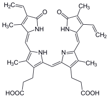
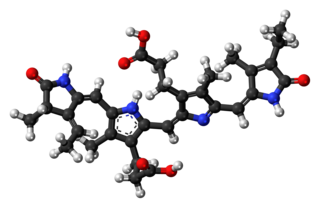
Bilirubin (BR) is a red-orange compound that occurs in the normal catabolic pathway that breaks down heme in vertebrates. This catabolism is a necessary process in the body's clearance of waste products that arise from the destruction of aged or abnormal red blood cells. In the first step of bilirubin synthesis, the heme molecule is stripped from the hemoglobin molecule. Heme then passes through various processes of porphyrin catabolism, which varies according to the region of the body in which the breakdown occurs. For example, the molecules excreted in the urine differ from those in the feces. The production of biliverdin from heme is the first major step in the catabolic pathway, after which the enzyme biliverdin reductase performs the second step, producing bilirubin from biliverdin.

<span class="mw-page-title-main">Bile</span> Dark greenish-brown fluid aiding in the digestion of fats

Bile, or gall, is a yellow-green fluid produced by the liver of most vertebrates that aids the digestion of lipids in the small intestine. In humans, bile is primarily composed of water, produced continuously by the liver, and stored and concentrated in the gallbladder. After a human eats, this stored bile is discharged into the first section of their small intestine.

<span class="mw-page-title-main">Heme</span> Chemical coordination complex of an iron ion chelated to a porphyrin

Heme, or haem, is a ring-shaped iron-containing molecular component of hemoglobin, which is necessary to bind oxygen in the bloodstream. It is composed of four pyrrole rings with 2 vinyl and 2 propionic acid side chains. Heme is biosynthesized in both the bone marrow and the liver.

<span class="mw-page-title-main">Stercobilin</span> Brown pigment of bile origin

Stercobilin is a tetrapyrrolic bile pigment and is one end-product of heme catabolism. It is the chemical responsible for the brown color of human feces and was originally isolated from feces in 1932. Stercobilin can be used as a marker for biochemical identification of fecal pollution levels in rivers.

Phycobilins are light-capturing bilins found in cyanobacteria and in the chloroplasts of red algae, glaucophytes and some cryptomonads. Most of their molecules consist of a chromophore which makes them coloured. They are unique among the photosynthetic pigments in that they are bonded to certain water-soluble proteins, known as phycobiliproteins. Phycobiliproteins then pass the light energy to chlorophylls for photosynthesis.

<span class="mw-page-title-main">Hereditary spherocytosis</span> Medical condition

Hereditary spherocytosis (HS) is a congenital hemolytic disorder wherein a genetic mutation coding for a structural membrane protein phenotype causes the red blood cells to be sphere-shaped (spherocytosis), rather than the normal biconcave disk shape. This abnormal shape interferes with the cells' ability to flex during blood circulation, and also makes them more prone to rupture under osmotic stress, mechanical stress, or both. Cells with the dysfunctional proteins are degraded in the spleen, which leads to a shortage of erythrocytes and results in hemolytic anemia.

<span class="mw-page-title-main">Phytochrome</span> Protein used by plants, bacteria and fungi to detect light

Phytochromes are a class of photoreceptor proteins found in plants, bacteria and fungi. They respond to light in the red and far-red regions of the visible spectrum and can be classed as either Type I, which are activated by far-red light, or Type II that are activated by red light. Recent advances have suggested that phytochromes also act as temperature sensors, as warmer temperatures enhance their de-activation. All of these factors contribute to the plant's ability to germinate.

<span class="mw-page-title-main">Heme oxygenase</span> Class of enzymes

Heme oxygenase, or haem oxygenase, is an enzyme that catalyzes the degradation of heme to produce biliverdin, ferrous ion, and carbon monoxide.

<span class="mw-page-title-main">Biliverdin reductase</span> Class of enzymes

Biliverdin reductase (BVR) is an enzyme found in all tissues under normal conditions, but especially in reticulo-macrophages of the liver and spleen. BVR facilitates the conversion of biliverdin to bilirubin via the reduction of a double bond between the second and third pyrrole ring into a single bond.

<span class="mw-page-title-main">Urobilin</span> Yellow pigment in urine

Urobilin or urochrome is the chemical primarily responsible for the yellow color of urine. It is a linear tetrapyrrole compound that, along with the related colorless compound urobilinogen, are degradation products of the cyclic tetrapyrrole heme.

Neonatal cholestasis refers to elevated levels of conjugated bilirubin identified in newborn infants within the first few months of life. Conjugated hyperbilirubinemia is clinically defined as >20% of total serum bilirubin or conjugated bilirubin concentration greater than 1.0 mg/dL regardless of total serum bilirubin concentration. The differential diagnosis for neonatal cholestasis can vary extensively. However, the underlying disease pathology is caused by improper transport and/or defects in excretion of bile from hepatocytes leading to an accumulation of conjugated bilirubin in the body. Generally, symptoms associated with neonatal cholestasis can vary based on the underlying cause of the disease. However, most infants affected will present with jaundice, scleral icterus, failure to thrive, acholic or pale stools, and dark urine.

<span class="mw-page-title-main">Bilin (biochemistry)</span> Class of chemical compound

Bilins, bilanes or bile pigments are biological pigments formed in many organisms as a metabolic product of certain porphyrins. Bilin was named as a bile pigment of mammals, but can also be found in lower vertebrates, invertebrates, as well as red algae, green plants and cyanobacteria. Bilins can range in color from red, orange, yellow or brown to blue or green.

<span class="mw-page-title-main">Phycocyanobilin</span> Chemical compound

Phycocyanobilin is a blue phycobilin, i.e., a tetrapyrrole chromophore found in cyanobacteria and in the chloroplasts of red algae, glaucophytes, and some cryptomonads. Phycocyanobilin is present only in the phycobiliproteins allophycocyanin and phycocyanin, of which it is the terminal acceptor of energy. It is covalently linked to these phycobiliproteins by a thioether bond.

<span class="mw-page-title-main">Roger Y. Tsien</span> American biochemist and Nobel laureate (1952–2016)

Roger Yonchien Tsien was an American biochemist. He was a professor of chemistry and biochemistry at the University of California, San Diego and was awarded the Nobel Prize in Chemistry in 2008 for his discovery and development of the green fluorescent protein, in collaboration with organic chemist Osamu Shimomura and neurobiologist Martin Chalfie. Tsien was also a pioneer of calcium imaging.

A chromoprotein is a conjugated protein that contains a pigmented prosthetic group. A common example is haemoglobin, which contains a heme cofactor, which is the iron-containing molecule that makes oxygenated blood appear red. Other examples of chromoproteins include other hemochromes, cytochromes, phytochromes and flavoproteins.

<span class="mw-page-title-main">SmURFP</span>

Small ultra red fluorescent protein (smURFP) is a class of far-red fluorescent protein evolved from a cyanobacterial phycobiliprotein, α-allophycocyanin. Native α-allophycocyanin requires an exogenous protein, known as a lyase, to attach the chromophore, phycocyanobilin. Phycocyanobilin is not present in mammalian cells. smURFP was evolved to covalently attach phycocyanobilin without a lyase and fluoresce, covalently attach biliverdin and fluoresce, blue-shift fluorescence to match the organic fluorophore, Cy5, and not inhibit E. coli growth. smURFP was found after 12 rounds of random mutagenesis and manually screening 10,000,000 bacterial colonies.

<span class="mw-page-title-main">Biliprotein</span> Class of pigment proteins in photosynthesising organisms

Biliproteins are pigment protein compounds that are located in photosynthesising organisms such as algae, and sometimes also in certain insects. They refer to any protein that contains a bilin chromophore. In plants and algae, the main function of biliproteins is to make the process of light accumulation required for photosynthesis more efficient; while in insects they play a role in growth and development. Some of their properties: including light-receptivity, light-harvesting and fluorescence have made them suitable for applications in bioimaging and as indicators; while other properties such as anti-oxidation, anti-aging and anti-inflammation in phycobiliproteins have given them potential for use in medicine, cosmetics and food technology. While research on biliproteins dates back as far as 1950, it was hindered due to issues regarding biliprotein structure, lack of methods available for isolating individual biliprotein components, as well as limited information on lyase reactions . Research on biliproteins has also been primarily focused on phycobiliproteins; but advances in technology and methodology, along with the discovery of different types of lyases, has renewed interest in biliprotein research, allowing new opportunities for investigating biliprotein processes such as assembly/disassembly and protein folding.

Hemolytic jaundice, also known as prehepatic jaundice, is a type of jaundice arising from hemolysis or excessive destruction of red blood cells, when the byproduct bilirubin is not excreted by the hepatic cells quickly enough. Unless the patient is concurrently affected by hepatic dysfunctions or is experiencing hepatocellular damage, the liver does not contribute to this type of jaundice.

Alexander Glazer was a professor of the Graduate School in the Department of Molecular and Cell Biology at the University of California, Berkeley. He had a passion for protein chemistry and structure function relationships. He also had a longstanding interest in light-harvesting complexes in cyanobacteria and red algae called phycobilisomes. He had also spent more than 10 years working on the human genome project where he has investigated methods for DNA detection and sequencing which most notably includes the development of fluorescent reagents involved in cell labeling. Most recently, he had focused his studies on issues in environmental sciences. He died on July 18, 2021, in Orinda, California

References

  1. 1 2 3 Boron W, Boulpaep E. Medical Physiology: a cellular and molecular approach, 2005. 984-986. Elsevier Saunders, United States. ISBN   1-4160-2328-3
  2. 1 2 3 Mosqueda, L; Burnight, K; Liao, S (2005). "The Life Cycle of Bruises in Older Adults". Journal of the American Geriatrics Society. 53 (8): 1339–1343. doi:10.1111/j.1532-5415.2005.53406.x. PMID   16078959. S2CID   12394659.
  3. Seyfried, H; Klicpera, M; Leithner, C; Penner, E (1976). "Bilirubin metabolism (author's transl)". Wiener Klinische Wochenschrift. 88 (15): 477–82. PMID   793184.
  4. 1 2 3 Bulmer, A. C.; Ried, K.; Blanchfield, J. T.; Wagner, K. H. (2008). "The anti-mutagenic properties of bile pigments". Mutation Research. 658 (1–2): 28–41. doi:10.1016/j.mrrev.2007.05.001. PMID   17602853.
  5. 1 2 3 4 Ohrui, T.; Yasuda, H.; Yamaya, M.; Matsui, T.; Sasaki, H. (2003). "Transient relief of asthma symptoms during jaundice: a possible beneficial role of bilirubin". The Tohoku Journal of Experimental Medicine. 199 (3): 193–196. doi: 10.1620/tjem.199.193 . PMID   12703664.
  6. McPhee, F.; Caldera, P. S.; Bemis, G. W.; McDonagh, A. F.; Kuntz, I. D.; Craik, C. S. (1996). "Bile pigments as HIV-1 protease inhibitors and their effects on HIV-1 viral maturation and infectivity in vitro". The Biochemical Journal. 320 (Pt 2): 681–686. doi:10.1042/bj3200681. PMC   1217983 . PMID   8973584.
  7. Halepas, Steven; Hamchand, Randy; Lindeyer, Samuel E. D.; Brückner, Christian (2017). "Isolation of Biliverdin IXα, as its Dimethyl Ester, from Emu Eggshells". Journal of Chemical Education. 94 (10): 1533–1537. Bibcode:2017JChEd..94.1533H. doi:10.1021/acs.jchemed.7b00449.
  8. Fang, LS; Bada, JL (1990). "The blue-green blood plasma of marine fish". Comparative Biochemistry and Physiology B. 97 (1): 37–45. doi:10.1016/0305-0491(90)90174-R. PMID   2253479.
  9. "These Puppies Were Born with Green Fur".
  10. Shamim, G; Ranjan, S; Pandey, D; Ramani, R (2014). "Biochemistry and biosynthesis of insect pigments" (PDF). European Journal of Entomology. 111(2): 155. doi:10.14411/eje.2014.021 . Retrieved 25 June 2023.
  11. Austin, C; Perkins, S (2006). "Parasites in a biodiversity hotspot: a survey of hematozoa and a molecular phyolgenetic analysis of plasmodium in New Guinea skinks". Journal of Parasitology. 92 (4): 770–777. doi:10.1645/GE-693R.1. PMID   16995395. S2CID   1937837.
  12. Lee Grismer, L.; Thy, Neang; Chav, Thou; Holden, Jeremy (2007). "A New Species of Chiromantis Peters 1854 (Anura: Rhacophoridae) from Phnom Samkos in the Northwestern Cardamom Mountains, Cambodia". Herpetologica. 63 (3): 392–400. doi:10.1655/0018-0831(2007)63[392:ANSOCP]2.0.CO;2. S2CID   84472376.
  13. 1 2 X. Shu; et al. (2009). "Mammalian expression of infrared fluorescent proteins engineered from a bacterial phytochrome". Science. 324 (5928): 804–807. Bibcode:2009Sci...324..804S. doi:10.1126/science.1168683. PMC   2763207 . PMID   19423828.
  14. 1 2 GSFilonov; Piatkevich, Kiryl D; Ting, Li-Min; Zhang, Jinghang; Kim, Kami; Verkhusha, Vladislav V; et al. (2011). "Bright and stable near infra-red fluorescent protein for in vivo imaging". Nat Biotechnol. 29 (8): 757–761. doi:10.1038/nbt.1918. PMC   3152693 . PMID   21765402.
  15. 1 2 Rodriguez, Erik A.; Tran, Geraldine N.; Gross, Larry A.; Crisp, Jessica L.; Shu, Xiaokun; Lin, John Y.; Tsien, Roger Y. (2016-08-01). "A far-red fluorescent protein evolved from a cyanobacterial phycobiliprotein". Nature Methods. 13 (9): 763–9. doi:10.1038/nmeth.3935. ISSN   1548-7105. PMC   5007177 . PMID   27479328.
  16. Tsien, Roger Y. (1998-01-01). "The Green Fluorescent Protein". Annual Review of Biochemistry. 67 (1): 509–544. doi:10.1146/annurev.biochem.67.1.509. PMID   9759496.