Canavalin

Last updated
Canavalin crystals grown on Earth (right) and in microgravity. Canavalin crystals.jpg
Canavalin crystals grown on Earth (right) and in microgravity.

Canavalin is a plant protein found in the jack bean, sword bean, and related plants. It is the major storage protein found in these plants' seeds, and is one of four proteins readily isolated from the seeds; the others are concanavalin A, concanavalin B, and urease. [2] Canavalin is a vicilin protein homologous to phaseolin. [3]

The crystallization of jack bean seed proteins has been studied extensively since the early 20th century and was of particular interest to 1946 Nobel Prize in Chemistry laureate James B. Sumner, [4] though Sumner's group never fully characterized canavalin and it remained of little interest until its crystallization properties began to be studied in the 1970s. [3] It was among the first reported examples of a protein whose tertiary structure contains two pseudo-symmetrical protein domains. [3] Canavalin has since been used as a model system for studying protein crystallization, [5] most notably in the study of protein crystal formation in space under microgravity conditions. [1]

Canavalin is found in large quantities, the protein makes up about half percent of total soluble proteins. It is part of the vicilin protein part of the seed and has similar characteristics. It is soluble in salt concentrations, and low ionic strength buffers. In saline, canavalin is insoluble and crystallizes. Crystallization occurs at 37 °C. At this temperature the crystals stay together, indicating that the molecule is highly stable. Crystallography demonstrates that the protein consists of six identical subunits organized in a hexamer. [6]

History

In 1919, J. B Sumner first recorded Canavalin to have a molecular weight to 115,000. [6] Sumner isolated the protein from a jack bean, but was not able to crystalize it. In the same research, he named two other proteins he was able to crystallize: concavalin A and concavalin B. Sumner tried multiple times to crystallize canavalin, but his attempts failed. However, in 1936 his student, S. Howell, working in his laboratory left out a sample of canavalin in a flask without regards for sterility. An extended period of time passed and they discovered crystals on the bottom of the flask. They suspected that the crystallization was caused by degradative enzymes produced from microbes. Sumner and Howell were able to reproduce the crystals using sterile solutions of adding trypsin, chymotrypsin and other proteases to the canavalin. They believed the crystallization of the protein was due to hydrolysis of surrounding contaminating proteins. It was also thought that the crystals were a proteolytic product of the native protein. [7]

More work on canavalin appeared in the literature in 1974 from researchers at the Massachusetts Institution of Technology. The researchers reproduced Sumner's work and were able to produce large crystalline structures and describe some of canavalins biochemical properties. Work in 1982 demonstrated that the native protein had a monomer that was cleaved in half by proteases and had mol weight of about 47,000. They also found that the three cleaved monomers were distributed in a perfect 3-fold axis of symmetry in a molecule with a mol weight of about 142,000 [7] and consists of 445 amino acids. [8]

Throughout studying canavalin and other plant seed proteins, it was apparent that canavalin and phaseolin had very similar properties. The differences were that phaseolin was glycosylated and had a mixture of three different subunits, while canavalin had no glycosylation and only one kind of polypeptide. The research showed that canavalin was a part of the vicilin family of seed storage proteins and that its characteristics were shared amount the family. [7]

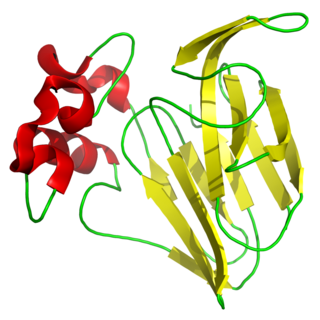
Homology of Canavalin and Phaseolin

Canavalin and phaseolin are two of the first proteins to show almost identical domain structure using X-ray diffraction analysis. The structure contains β-barrels having the Swiss roll motif. Canavalin and phaseolin share 60% of the same amino acids and have similar tertiary and quaternary structures.
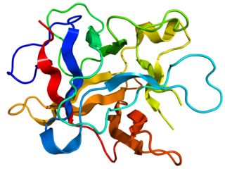
Structure

The 3-fold axis of the canavalin trimer marks a channel in the trimer. The channel is about 18 Armstrongs in diameter and runs free through the protein. The channel is lined with hydrophilic and charged amino acid residues. The trimers are stacked, resembling a preferred packing motif. [7]

Conversion of Precanavalin to Canavalin

Canavalin is derived from precanavalin by proteolytic cleavage. Precanavalin and canavalin are a large proportion of the seed’s protein and are assumed to be nutrient proteins that are a source of amino acids for any developing seedlings. Precanavalin has a monomer and exist as a continuous, single polypeptide chain. Canavalin is cleaved from precanavalin, consisting of three polypeptide chains with weights of 24,000, 13,000, and 12,000 D. Canavalin is produced when precanavalin is incubated at 37 Celsius with 2.5% for up to 24 hours. Canavalin can also be obtained by a similar process with chymotrypsin. The production of canavalin with chymotrypsin is less complex and digestion occurs slower, allowing the production to be controlled easier. The protein is first cleaved into two polypeptide chains with molecular weights of 24,000 and 25,000 D. With chymotrypsin, this cleavage is completed within 30 minutes, and trypsin occurs in about 3 minutes. When the digestion occurs at conditions allowing for crystallization, the polypeptide chain weighing 25,000 D is cleaved into two fragments of 12,000 and 13,000 D. These three subunits form the canavalin molecule, resulting in a molecular weight of 147,000 D. The fragment of 24,000 D has nearly identical amino acid composition as fragments 12,000 and 13,000D, implying that homology may exist between primary structures. [6]

Canavalins Response to Different Salt Concentrations

Canavalin extracted from sword bean in distilled water has its monomer structure; however, when placed in high concentrations of NaCl and MgCl2, a change occurs between the monomeric to the trimeric form. The solubility of the quaternary structure of canavalin differed with different concentrations of salt. To form the trimer, increasing ionic strength is an important influence. Also, in the presence of 60mM MgCl2 and 200mM CaCl2, the soluble trimer form of canavalin was aggregated. In comparison, aggregation does not occur in 200mM NaCl. These different properties seen with canavalin and different salts might indicate that salt bridges formed through divalent cations. This would prompt aggregation of the trimer form when around MgCl2.

Canavalin belongs to the vicilin fraction from the 7s globulin family. B-conglycinin is classified in the same group also has a trimer structure that can be purified and crystallized in the presence of high salt concentrations such as NaCl. This suggests that in the presence of higher salt concentrations, the trimer structure of 7s globulin family is present. It is unknown if the trimers of 7s globulin are present in the bean seeds. The extract of sword bean canavalin was found to be a monomer and not a trimer, suggesting that canavalin the seeds are in monomer form. [9]

Genetic Engineering of Canavalin

The structure of canavalin is an ideal model for protein engineering. Using recombinant DNA manipulation and x-ray crystallography, canavalin can be genetically altered. Due to site directed mutagenesis, the genetic engineering can happen very quickly. If an engineered gene for canavalin can be inserted into a plant, then nutritionally enhanced seed storage proteins may exist. This would create a possibility for improving and enhancing all of the leguminous plants, a major source of the world's dietary protein. [7]

Related Research Articles

<span class="mw-page-title-main">Chymotrypsin</span> Digestive enzyme

Chymotrypsin (EC 3.4.21.1, chymotrypsins A and B, alpha-chymar ophth, avazyme, chymar, chymotest, enzeon, quimar, quimotrase, alpha-chymar, alpha-chymotrypsin A, alpha-chymotrypsin) is a digestive enzyme component of pancreatic juice acting in the duodenum, where it performs proteolysis, the breakdown of proteins and polypeptides. Chymotrypsin preferentially cleaves peptide amide bonds where the side chain of the amino acid N-terminal to the scissile amide bond (the P1 position) is a large hydrophobic amino acid (tyrosine, tryptophan, and phenylalanine). These amino acids contain an aromatic ring in their side chain that fits into a hydrophobic pocket (the S1 position) of the enzyme. It is activated in the presence of trypsin. The hydrophobic and shape complementarity between the peptide substrate P1 side chain and the enzyme S1 binding cavity accounts for the substrate specificity of this enzyme. Chymotrypsin also hydrolyzes other amide bonds in peptides at slower rates, particularly those containing leucine at the P1 position.

<span class="mw-page-title-main">Proteolysis</span> Breakdown of proteins into smaller polypeptides or amino acids

Proteolysis is the breakdown of proteins into smaller polypeptides or amino acids. Uncatalysed, the hydrolysis of peptide bonds is extremely slow, taking hundreds of years. Proteolysis is typically catalysed by cellular enzymes called proteases, but may also occur by intra-molecular digestion.

<span class="mw-page-title-main">Protease</span> Enzyme that cleaves other proteins into smaller peptides

A protease is an enzyme that catalyzes proteolysis, breaking down proteins into smaller polypeptides or single amino acids, and spurring the formation of new protein products. They do this by cleaving the peptide bonds within proteins by hydrolysis, a reaction where water breaks bonds. Proteases are involved in numerous biological pathways, including digestion of ingested proteins, protein catabolism, and cell signaling.

<span class="mw-page-title-main">Globular protein</span> Spherical, water-soluble type of protein

In biochemistry, globular proteins or spheroproteins are spherical ("globe-like") proteins and are one of the common protein types. Globular proteins are somewhat water-soluble, unlike the fibrous or membrane proteins. There are multiple fold classes of globular proteins, since there are many different architectures that can fold into a roughly spherical shape.

<span class="mw-page-title-main">Thaumatin</span> Low-calorie sweetener and flavor modifier

Thaumatin is a low-calorie sweetener and flavor modifier. The protein is often used primarily for its flavor-modifying properties and not exclusively as a sweetener.

The globulins are a family of globular proteins that have higher molecular weights than albumins and are insoluble in pure water but dissolve in dilute salt solutions. Some globulins are produced in the liver, while others are made by the immune system. Globulins, albumins, and fibrinogen are the major blood proteins. The normal concentration of globulins in human blood is about 2.6-3.5 g/dL.

<span class="mw-page-title-main">Serine protease</span> Class of enzymes

Serine proteases are enzymes that cleave peptide bonds in proteins. Serine serves as the nucleophilic amino acid at the (enzyme's) active site. They are found ubiquitously in both eukaryotes and prokaryotes. Serine proteases fall into two broad categories based on their structure: chymotrypsin-like (trypsin-like) or subtilisin-like.

In chemistry, water(s) of crystallization or water(s) of hydration are water molecules that are present inside crystals. Water is often incorporated in the formation of crystals from aqueous solutions. In some contexts, water of crystallization is the total mass of water in a substance at a given temperature and is mostly present in a definite (stoichiometric) ratio. Classically, "water of crystallization" refers to water that is found in the crystalline framework of a metal complex or a salt, which is not directly bonded to the metal cation.

<span class="mw-page-title-main">Concanavalin A</span> Lectin (carbohydrate-binding protein) originally extracted from the jack-bean

Concanavalin A (ConA) is a lectin originally extracted from the jack-bean. It is a member of the legume lectin family. It binds specifically to certain structures found in various sugars, glycoproteins, and glycolipids, mainly internal and nonreducing terminal α-D-mannosyl and α-D-glucosyl groups. Its physiological function in plants, however, is still unknown. ConA is a plant mitogen, and is known for its ability to stimulate mouse T-cell subsets giving rise to four functionally distinct T cell populations, including precursors to regulatory T cells; a subset of human suppressor T-cells is also sensitive to ConA. ConA was the first lectin to be available on a commercial basis, and is widely used in biology and biochemistry to characterize glycoproteins and other sugar-containing entities on the surface of various cells. It is also used to purify glycosylated macromolecules in lectin affinity chromatography, as well as to study immune regulation by various immune cells.

<span class="mw-page-title-main">Franz Hofmeister</span> German chemist (1850–1922)

Franz Hofmeister was an early protein scientist, and is famous for his studies of salts that influence the solubility and conformational stability of proteins. In 1902, Hofmeister became the first to propose that polypeptides were amino acids linked by peptide bonds, although this model of protein primary structure was independently and simultaneously conceived by Emil Fischer.

<span class="mw-page-title-main">Soy protein</span> A protein that is isolated from soybean

Soy protein is a protein that is isolated from soybean. It is made from soybean meal that has been dehulled and defatted. Dehulled and defatted soybeans are processed into three kinds of high protein commercial products: soy flour, concentrates, and isolates. Soy protein isolate has been used since 1959 in foods for its functional properties.

Protein metabolism denotes the various biochemical processes responsible for the synthesis of proteins and amino acids (anabolism), and the breakdown of proteins by catabolism.

<span class="mw-page-title-main">Kunitz STI protease inhibitor</span>

Kunitz soybean trypsin inhibitor is a type of protein contained in legume seeds which functions as a protease inhibitor. Kunitz-type Soybean Trypsin Inhibitors are usually specific for either trypsin or chymotrypsin. They are thought to protect seeds against consumption by animal predators.

Legumin is family of globular proteins obtained from beans, peas, lentils, vetches, hemp and other leguminous seeds. Garden peas are a common nutritional source for humans that contains legumin.

<span class="mw-page-title-main">Cupin superfamily</span>

The cupin superfamily is a diverse superfamily of proteins named after its conserved barrel domain. The superfamily includes a wide variety of enzymes as well as non-enzymatic seed storage proteins.

<span class="mw-page-title-main">Pea protein</span> Food product and protein supplement derived from Pisum sativum

Pea protein is a food product and protein supplement derived and extracted from yellow and green split peas, Pisum sativum. It can be used as a dietary supplement to increase an individual's protein or other nutrient intake, or as a substitute for other food products. As a powder, it is used as an ingredient in food manufacturing, such as a thickener, foaming agent, or an emulsifier.

Vicilin is a legumin-associated globulin protein. It is a storage protein found in legumes such as the pea or lentil that protects plants from fungi and microorganism. It is believed to be an allergen in pea and peanut allergy responses.

11S globulin family is a family of globulin proteins chiefly found in seeds of legumes (legumin-like), along with 7S family, often found in a protein fraction within a protein isolate. They are used as storage of important nutrients for plant growth, and therefore hardy enough to pass through the human digestive system unscathed. One common example of an 11S globulin includes glycinin derived from soy.

Cruciferin is one of the two most abundant seed storage proteins in mustard and rapeseed. They are classified as 11S globulins based on their sedimentation coefficient, and are salt soluble neutral glycoproteins. Their molecular weights range from 20 to 40 kDa. They comprise up to 50–70% of the total seed protein. Cruciferin is a comparatively larger seed storage protein than napin. It is composed of two polypeptide chains α and β. The α-chain has a mass of 30 kDa and the β-chain weighs in at 20 kDa. They are held together by a disulphide bond.

References

  1. 1 2 McPherson, Alexander; DeLucas, Lawrence James (2015-09-03). "Microgravity protein crystallization". npj Microgravity. 1 (1): 15010–. doi:10.1038/npjmgrav.2015.10. ISSN   2373-8065. PMC   5515504 . PMID   28725714.
  2. Sumner, J. B.; Gralën, N.; Eriksson-Quensel, I. B. (1938-04-29). "The Molecular Weights of Urease, Canavalin, Concanavalin a and Concanavalin B". Science. 87 (2261): 395–396. Bibcode:1938Sci....87..395S. doi:10.1126/science.87.2261.395. ISSN   0036-8075. PMID   17746464.
  3. 1 2 3 Shewry, Peter R.; Casey, R. (1999). Seed Proteins. Dordrecht: Springer Netherlands. ISBN   9789401144315. OCLC   840310271.
  4. Sumner, James B.; Howell, Stacey F. (1936-04-01). "The Isolation of a Fourth Crystallizable Jack Bean Globulin Through the Digestion of Canavalin with Trypsin". Journal of Biological Chemistry. 113 (3): 607–610. doi: 10.1016/S0021-9258(18)74832-1 . ISSN   0021-9258.
  5. Land, T. A.; Malkin, A. J.; Kuznetsov, Yu.G; McPherson, A.; De Yoreo, J. J. (1995-10-02). "Mechanisms of Protein Crystal Growth: An Atomic Force Microscopy Study of Canavalin Crystallization". Physical Review Letters. 75 (14): 2774–2777. Bibcode:1995PhRvL..75.2774L. doi:10.1103/PhysRevLett.75.2774. PMID   10059401. S2CID   40439026.
  6. 1 2 3 Smith, Stephanie Campbell; Johnson, Stephen; Andrews, James; McPherson, Alexander (1982-10-01). "Biochemical Characterization of Canavalin, the Major Storage Protein of Jack Bean". Plant Physiology. 70 (4). Oxford University Press (OUP): 1199–1209. doi:10.1104/pp.70.4.1199. ISSN   0032-0889.
  7. 1 2 3 4 5 Ng, J. D.; Ko, T. P.; McPherson, A. (1993-03-01). "Cloning, Expression, and Crystallization of Jack Bean (Canavalia ensiformis) Canavalin". Plant Physiology. 101 (3): 713–728. doi:10.1104/pp.101.3.713. ISSN   0032-0889. PMC   158684 . PMID   8310055.
  8. "UniProt". www.uniprot.org. Retrieved 2023-04-27.
  9. Nishizawa, Kaho; Arii, Yasuhiro (2019). "Structural transitions of sword bean canavalin in response to different salt concentrations". Heliyon. 5 (12). doi: 10.1016/j.heliyon.2019.e03037 . PMC   6928265 .