Canine transmissible venereal tumor

Last updated
Illustration of venereal granulomata on a dog's penis Surgical diseases and surgery of the dog (1906) (14784705963).jpg
Illustration of venereal granulomata on a dog's penis

A canine transmissible venereal tumor (CTVT), also known as a transmissible venereal tumor (TVT), canine transmissible venereal sarcoma (CTVS), sticker tumor and infectious sarcoma, is a histiocytic tumor of the external genitalia of the dog and other canines, and is transmitted from animal to animal during mating. It is one of only three known transmissible cancers in mammals; the others are devil facial tumor disease, a cancer which occurs in Tasmanian devils, and contagious reticulum cell sarcoma of the Syrian hamster.

Contents

The tumor cells are themselves the infectious agents, and the tumors that form are not genetically related to the host dog. [1] Although the genome of a CTVT is derived from an individual canid (specifically from a population of Native American dogs with coyote contribution), [2] [3] it is now essentially living as a unicellular, asexually reproducing (but sexually transmitted) pathogen. [4] Sequence analysis of the genome suggests it diverged from canids over 6,000 years ago; possibly much earlier. [4] Estimates from 2015 date its time of origin to about 11,000 years ago. [5] However, the most recent common ancestor of extant tumors is more recent: it probably originated 200 to 2,500 years ago. [1] [6]

Canine TVTs were initially described by Russian veterinarian M.A. Novinsky (1841–1914) in 1876, when he demonstrated that the tumor could be transplanted from one dog to another by infecting them with tumor cells. [7]

Signs and symptoms

In male dogs, the tumor affects the penis and foreskin. In female dogs, it affects the vulva. Rarely, the mouth or nose are affected. [8] The tumor often has a cauliflower-like appearance. Signs of genital TVT include a discharge from the prepuce and in some cases urinary retention caused by blockage of the urethra. [9] Signs of a nasal TVT include nasal fistulae, nosebleeds and other nasal discharge, facial swelling, and enlargement of the submandibular lymph nodes. [10]

Pathology

Canine transmissible venereal tumors are histiocytic tumors that may be transmitted among dogs through coitus, licking, biting and sniffing tumor affected areas. The concept that the tumor is naturally transmissible as an allograft came from three important observations. First, CTVTs can only be experimentally induced by transplanting living tumor cells, and not by killed cells or cell filtrates. Second, the tumor karyotype is aneuploid but has characteristic marker chromosomes in all tumors collected in different geographic regions. Third, a long interspersed nuclear element (LINE-1) insertion near c-myc has been found in all tumors examined so far and can be used as a diagnostic marker to confirm that a tumor is a CTVT. [6] [11]

Canine transmissible venereal tumors are most commonly seen in sexually active dogs in tropical, subtropical and temperate climates where there are large populations of stray dogs, but little is known about the details of transmission. [12] The disease is spread when dogs mate, and can even be transmitted to other canine species, such as foxes and coyotes. [13] Spontaneous regression of the tumor can occur, probably due to a response from the immune system. [14] CTVT undergoes a predictable cycle: an initial growth phase of four to six months (P phase), a stable phase, and a regression phase (R phase), [15] although not all CTVTs will regress. The tumor does not often metastasize (occurring in about less than 5 percent of cases), [16] except in puppies and immunocompromised dogs. Metastasis occurs to regional lymph nodes, [17] but can also be seen in the skin, brain, eye, liver, spleen, testicle, rectum and muscle. [18] A biopsy is necessary for diagnosis.

The success of this single cell lineage, believed to be the longest continually propagated cell lineage in the world, can be attributed to the tumor's mode of transmission in a specific host system. Although direct contact is generally not a highly efficient mode of transfer, CTVTs take advantage of the popular sire effect of domestic dogs. A single male can produce dozens of litters over his lifetime, allowing the tumor to affect many more females than it could if a monogamous species were the host. Understanding the epidemiology of CTVTs could provide insights for populations that may experience CTVT exposure and information about disease prevalence.[ citation needed ]

Genetics

The CTVT cells have fewer chromosomes than normal dog cells. Dog cells normally have 78 chromosomes, while the cancer cells contain 57–64 chromosomes [7] that are very different in appearance from normal dog chromosomes. All dog chromosomes except X and Y are acrocentric, having a centromere very near to the end of the chromosome, while many of the CTVT chromosomes are metacentric or submetacentric, having a centromere nearer to the middle. [9]

All tumor cells of this type of cancer share extremely similar genetic code, often if not always unrelated to the DNA of their host. [6] In addition to the aforementioned c-myc insertion, a few other potential driver mutations have been identified. [19]

Treatment method

The tumor, when treated with the chemotherapy drug vincristine, regresses as the host immune system is activated. CCL5 may play an important role in the immune response. [20]

Treatment

Surgery may be difficult due to the location of these tumors. Surgery alone often leads to recurrence. Chemotherapy is very effective for TVTs. The prognosis for complete remission with chemotherapy is excellent. [21] The most common chemotherapy agents used are vincristine, vinblastine, and doxorubicin. [14] Use of autohaemotherapy in treatment of TVTs also showed promising results in many cases [22] .Radiotherapy may be required if chemotherapy does not work. [18]

Related Research Articles

<span class="mw-page-title-main">Osteosarcoma</span> Cancerous tumour in a bone

An osteosarcoma (OS) or osteogenic sarcoma (OGS) is a cancerous tumor in a bone. Specifically, it is an aggressive malignant neoplasm that arises from primitive transformed cells of mesenchymal origin and that exhibits osteoblastic differentiation and produces malignant osteoid.

<span class="mw-page-title-main">Hemangiosarcoma</span> Medical condition

Hemangiosarcoma is a rapidly growing, highly invasive variety of cancer that occurs almost exclusively in dogs, and only rarely in cats, horses, mice, or humans. It is a sarcoma arising from the lining of blood vessels; that is, blood-filled channels and spaces are commonly observed microscopically. A frequent cause of death is the rupturing of this tumor, causing the patient to rapidly bleed to death.

<span class="mw-page-title-main">James Ewing (pathologist)</span> American pathologist

James Stephen Ewing was an American pathologist. He was the first Professor of pathology at Cornell University and discovered a form of bone cancer that was later named after him, Ewing sarcoma.

<span class="mw-page-title-main">Fibrosarcoma</span> Medical condition

Fibrosarcoma is a malignant mesenchymal tumour derived from fibrous connective tissue and characterized by the presence of immature proliferating fibroblasts or undifferentiated anaplastic spindle cells in a storiform pattern. Fibrosarcomas mainly arise in people between the ages of 25 and 79. It originates in fibrous tissues of the bone and invades long or flat bones such as the femur, tibia, and mandible. It also involves the periosteum and overlying muscle.

<span class="mw-page-title-main">Synovial sarcoma</span> Medical condition

A synovial sarcoma is a rare form of cancer which occurs primarily in the extremities of the arms or legs, often in proximity to joint capsules and tendon sheaths. It is a type of soft-tissue sarcoma.

<span class="mw-page-title-main">Devil facial tumour disease</span> Cancer affecting Tasmanian devils

Devil facial tumour disease (DFTD) is an aggressive non-viral clonally transmissible cancer which affects Tasmanian devils, a marsupial native to the Australian island of Tasmania. The cancer manifests itself as lumps of soft and ulcerating tissue around the mouth, which may invade surrounding organs and metastasise to other parts of the body. Severe genetic abnormalities exist in cancer cells—for example, DFT2 cells are tetraploid, containing twice as much genetic material as normal cells. DFTD is most often spread by bites, when teeth come into contact with cancer cells; less important pathways of transmission are ingesting of infected carcasses and sharing of food. Adult Tasmanian devils who are otherwise the fittest are most susceptible to the disease.

<span class="mw-page-title-main">Malignant peripheral nerve sheath tumor</span> Medical condition

A malignant peripheral nerve sheath tumor (MPNST) is a form of cancer of the connective tissue surrounding nerves. Given its origin and behavior it is classified as a sarcoma. About half the cases are diagnosed in people with neurofibromatosis; the lifetime risk for an MPNST in patients with neurofibromatosis type 1 is 8–13%. MPNST with rhabdomyoblastomatous component are called malignant triton tumors.

<span class="mw-page-title-main">Desmoplastic small-round-cell tumor</span> Aggressive and rare cancer

Desmoplastic small-round-cell tumor (DSRCT) is an aggressive and rare cancer that primarily occurs as masses in the abdomen. Other areas affected may include the lymph nodes, the lining of the abdomen, diaphragm, spleen, liver, chest wall, skull, spinal cord, large intestine, small intestine, bladder, brain, lungs, testicles, ovaries, and the pelvis. Reported sites of metastatic spread include the liver, lungs, lymph nodes, brain, skull, and bones. It is characterized by the EWS-WT1 fusion protein.

Malignant histiocytosis is a rare hereditary disease found in the Bernese Mountain Dog and humans, characterized by histiocytic infiltration of the lungs and lymph nodes. The liver, spleen, and central nervous system can also be affected. Histiocytes are a component of the immune system that proliferate abnormally in this disease. In addition to its importance in veterinary medicine, the condition is also important in human pathology.

<span class="mw-page-title-main">Lymphoma in animals</span> Type of cancer in animals

Lymphoma (lymphosarcoma) in animals is a type of cancer defined by a proliferation of malignant lymphocytes within solid organs such as the lymph nodes, bone marrow, liver and spleen. The disease also may occur in the eye, skin, and gastrointestinal tract.

<span class="mw-page-title-main">Histiocytoma (dog)</span> Benign tumor in dogs

A histiocytoma in the dog is a benign tumor. It is an abnormal growth in the skin of histiocytes (histiocytosis), a cell that is part of the immune system. A similar disease in humans, Hashimoto-Pritzker disease, is also a Langerhans cell histiocytosis. Dog breeds that may be more at risk for this tumor include Bulldogs, American Pit Bull Terriers, American Staffordshire Terriers, Scottish Terriers, Greyhounds, Boxers, and Boston Terriers. They also rarely occur in goats and cattle.

A transmissible cancer is a cancer cell or cluster of cancer cells that can be transferred between individuals without the involvement of an infectious agent, such as an Oncovirus. The evolution of transmissible cancer has occurred naturally in other animal species, but human cancer transmission is rare. This transfer is typically between members of the same species or closely related species.

<span class="mw-page-title-main">Ewing sarcoma</span> Type of cancer

Ewing sarcoma is a type of pediatric cancer that forms in bone or soft tissue. Symptoms may include swelling and pain at the site of the tumor, fever, and a bone fracture. The most common areas where it begins are the legs, pelvis, and chest wall. In about 25% of cases, the cancer has already spread to other parts of the body at the time of diagnosis. Complications may include a pleural effusion or paraplegia.

<span class="mw-page-title-main">Cancer in dogs</span>

Cancer is the leading cause of death in dogs. It is estimated that 1 in 3 domestic dogs will develop cancer, which is the same incidence of cancer among humans. Dogs can develop a variety of cancers and most are very similar to those found in humans. Dogs can develop carcinomas of epithelial cells and organs, sarcomas of connective tissues and bones, and lymphomas or leukemias of the circulatory system. Selective breeding of dogs has led certain pure-bred breeds to be at high-risk for specific kinds of cancer.

Elizabeth Murchison is a British-Australian geneticist, Professor of Comparative Oncology and Genetics at the University of Cambridge, UK. The ongoing research of her group focuses on the known existing clonally transmissible cancers arising in mammals. These are cancers that can be passed on between individuals by the transfer of living cancer cells that somehow manage to evade the immune system of their hosts.

Anne-Maree Pearse is an Australian cytogeneticist who is credited with the theory that some cancer cells can be transmissible between individuals. This is known as the allograft theory. Her work has focussed on devil facial tumour disease (DFTD), a contagious cancer that affects Tasmanian devils. For this she has won multiple awards, including the 2012 Prince Hitachi Prize for Comparative Oncology.

Comparative oncology integrates the study of oncology in non-human animals into more general studies of cancer biology and therapy. The field encompasses naturally seen cancers in veterinary patients and the extremely low rates of cancers seen in large mammals such as elephants and whales.

Contagious reticulum cell sarcoma is a reticulum-cell sarcoma found in Syrian hamsters that can be transmitted from one hamster to another. It was first described in 1945.

<span class="mw-page-title-main">Mastocytoma in dogs</span> Cancer tumor in dogs

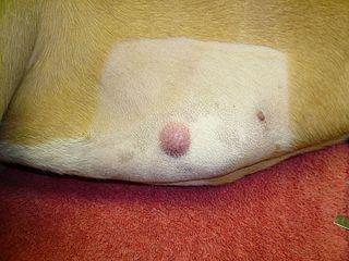
A mastocytoma in dogs is a neoplasm (neoplasia) originating from mast cells in the domestic dog, which occurs mainly in the skin and subcutis. Mastocytoma are not only extremely common in dogs, but also tend to be much more malignant in them than in other animal species. The average survival time for malignant tumors is only four months, whereas for benign tumors it is over two years.

References

  1. 1 2 Choi, Charles Q. (2006-08-10). "Contagious Canine Cancer Spread by Parasites". LiveScience. Archived from the original on 2006-08-20. Retrieved 2006-08-11.
  2. Wang, Xuan; Zhou, Bo-Wen; Yang, Melinda A.; Yin, Ting-Ting; Chen, Fang-Liang; Ommeh, Sheila C.; Esmailizadeh, Ali; Turner, Melissa M.; Poyarkov, Andrei D.; Savolainen, Peter; Wang, Guo-Dong; Fu, Qiaomei; Zhang, Ya-Ping (3 June 2019). "Canine transmissible venereal tumor genome reveals ancient introgression from coyotes to pre-contact dogs in North America". Cell Research. 29 (7): 592–595. bioRxiv   10.1101/350512 . doi: 10.1038/s41422-019-0183-2 . PMC   6796869 . PMID   31160719.
  3. Ní Leathlobhair, Máire; Perri, Angela R; Irving-Pease, Evan K; Witt, Kelsey E; Linderholm, Anna; Haile, James; Lebrasseur, Ophelie; Ameen, Carly; Blick, Jeffrey; Boyko, Adam R; Brace, Selina; Cortes, Yahaira Nunes; Crockford, Susan J; Devault, Alison; Dimopoulos, Evangelos A; Eldridge, Morley; Enk, Jacob; Gopalakrishnan, Shyam; Gori, Kevin; Grimes, Vaughan; Guiry, Eric; Hansen, Anders J; Hulme-Beaman, Ardern; Johnson, John; Kitchen, Andrew; Kasparov, Aleksei K; Kwon, Young-Mi; Nikolskiy, Pavel A; Lope, Carlos Peraza; et al. (2018). "The evolutionary history of dogs in the Americas" (PDF). Science. 361 (6397): 81–85. Bibcode:2018Sci...361...81N. doi: 10.1126/science.aao4776 . PMC   7116273 . PMID   29976825.
  4. 1 2 Rebbeck CA, Thomas R, Breen M, Leroi AM, Burt A (2009). "Origins and Evolution of a Transmissible Cancer". Evolution. 63 (9): 2340–2349. doi: 10.1111/j.1558-5646.2009.00724.x . PMID   19453727.
  5. Strakova, Andrea; Murchison, Elizabeth P (2015). "The cancer which survived: Insights from the genome of an 11000 year-old cancer". Current Opinion in Genetics & Development. 30: 49–55. doi:10.1016/j.gde.2015.03.005. PMID   25867244. S2CID   21195930.
  6. 1 2 3 Murgia, C; Pritchard JK; Kim SY; Fassati A; Weiss RA (2006-08-11). "Clonal Origin and Evolution of a Transmissible Cancer". Cell. 126 (3): 477–87. doi:10.1016/j.cell.2006.05.051. PMC   2593932 . PMID   16901782.
  7. 1 2 Mello Martins, M.I.; de Souza, F. Ferreira; Gobello, C. (2005). "Canine transmissible venereal tumor: Etiology, pathology, diagnosis and treatment". Recent Advances in Small Animal Reproduction. Retrieved 2006-05-25.
  8. Morrison, Wallace B. (1998). Cancer in Dogs and Cats (1st ed.). Williams and Wilkins. ISBN   978-0-683-06105-5.
  9. 1 2 Hasler A, Weber W (2000). "Theriogenology question of the month. Transmissible venereal tumor (TVT)". J. Am. Vet. Med. Assoc. 216 (10): 1557–9. doi: 10.2460/javma.2000.216.1557 . PMID   10825939.
  10. Papazoglou L, Koutinas A, Plevraki A, Tontis D (2001). "Primary intranasal transmissible venereal tumour in the dog: a retrospective study of six spontaneous cases". Journal of Veterinary Medicine, Series A. 48 (7): 391–400. doi:10.1046/j.1439-0442.2001.00361.x. PMID   11599677.
  11. Dingli, D; Nowak, MA (2006). "Cancer biology: infectious tumour cells". Nature. 443 (7107): 35–6. Bibcode:2006Natur.443...35D. doi:10.1038/443035a. PMC   2711443 . PMID   16957717.
  12. Vonholdt, B. M; Ostrander, E. A (2006). "The singular history of a canine transmissible tumor". Cell. 126 (3): 445–7. doi: 10.1016/j.cell.2006.07.016 . PMID   16901777.
  13. Mukaratirwa S, Gruys E (2003). "Canine transmissible venereal tumour: cytogenetic origin, immunophenotype, and immunobiology. A review". The Veterinary Quarterly. 25 (3): 101–11. doi: 10.1080/01652176.2003.9695151 . PMID   14535580.
  14. 1 2 Stettner N, Brenner O, Eilam R, Harmelin A (2005). "Pegylated liposomal doxorubicin as a chemotherapeutic agent for treatment of canine transmissible venereal tumor in murine models". J. Vet. Med. Sci. 67 (11): 1133–9. doi: 10.1292/jvms.67.1133 . PMID   16327225.
  15. Liao K, Hung S, Hsiao Y, Bennett M, Chu R (2003). "Canine transmissible venereal tumor cell depletion of B lymphocytes: molecule(s) specifically toxic for B cells". Vet. Immunol. Immunopathol. 92 (3–4): 149–62. doi:10.1016/S0165-2427(03)00032-1. PMID   12730015.
  16. "Canine Transmissible Venereal Tumor: Introduction". The Merck Veterinary Manual. 2006. Retrieved 2007-04-24.
  17. Arif, Syed Abdul; Das, Nilotpal; Goswami, Sushanta; Bhuyan, Manjyoti; Mahato, Gauranga; Pathak, Mamta; Das, Abhijit (2017-05-01). "Clinico-pathological study on metastatic form of canine Transmissible Veneral Tumour (TVT) and its therapeutic management". International Journal of Chemical Studies. 5 (3i): 593–595. doi:10.22271/chemi.2017.v5.i3i.08.
  18. 1 2 Rogers K, Walker M, Dillon H (1998). "Transmissible venereal tumor: a retrospective study of 29 cases". Journal of the American Animal Hospital Association . 34 (6): 463–70. doi:10.5326/15473317-34-6-463. PMID   9826280.
  19. Belov, Katherine; Jones, Elizabeth; Cheng, Yuanyuan (September 2015). "The origin, dynamics, and molecular evolution of transmissible cancers". Advances in Genomics and Genetics: 317. doi: 10.2147/AGG.S61298 .
  20. Frampton, D; Schwenzer, H; Marino, G; Butcher, LM; Pollara, G; Kriston-Vizi, J; Venturini, C; Austin, R; de Castro, KF; Ketteler, R; Chain, B; Goldstein, RA; Weiss, RA; Beck, S; Fassati, A (9 April 2018). "Molecular Signatures of Regression of the Canine Transmissible Venereal Tumor" (PDF). Cancer Cell. 33 (4): 620–633.e6. doi:10.1016/j.ccell.2018.03.003. PMC   5896242 . PMID   29634949.
  21. Ettinger, Stephen J.; Feldman, Edward C. (1995). Textbook of Veterinary Internal Medicine (4th ed.). W.B. Saunders Company. ISBN   978-0-7216-6795-9.
  22. Arif, S. A.; Das, T.; Deka, D.; Kachari, J.; Barman, U.; Changkija, B.; Patgiri, D. (2023-10-28). "Management of canine transmissible venereal tumour using autohaemotherapy: A vpromising approach" (PDF). INDIAN JOURNAL OF ANIMAL HEALTH. Online. doi:10.36062/ijah.2023.17222.