Urethra

Last updated
Urethra
Female and Male Urethra.jpg
The urethra transports urine from the bladder to the outside of the body. This image shows (a) a human female urethra and (b) a human male urethra.
Details
Precursor Urogenital sinus
Artery Inferior vesical artery
Middle rectal artery
Internal pudendal artery
Vein Inferior vesical vein
Middle rectal vein
Internal pudendal vein
Nerve Pudendal nerve
Pelvic splanchnic nerves
Inferior hypogastric plexus
Lymph Internal iliac lymph nodes
Deep inguinal lymph nodes
Identifiers
Latin urethra feminina (female); urethra masculina (male)
Greek οὐρήθρα
MeSH D014521
TA98 A08.4.01.001F
A08.5.01.001M
TA2 3426, 3442
FMA 19667
Anatomical terminology

The urethra (pl.: urethras or urethrae) is a tube that connects the mammalian urinary bladder to the urinary meatus in the glans penis or vulval vestibule. Male and female placental mammals release urine through the urethra during urination, but males also release semen through the urethra during ejaculation. [1]

Contents

The external urethral sphincter is a striated muscle that allows voluntary control over urination. [2] The internal sphincter, formed by the involuntary smooth muscles lining the bladder neck and urethra, receives its nerve supply by the sympathetic division of the autonomic nervous system. [3] The internal sphincter is present both in males and females. [4] [5] [6]

Structure

The urethra is a fibrous and muscular tube which connects the urinary bladder to the external urethral meatus. Its length differs between the sexes, because it passes through the penis in males.

Male

The human male urethra laid open on its anterior (upper) surface Gray1142.png
The human male urethra laid open on its anterior (upper) surface

In the human male, the urethra is on average 18 to 20 centimeters (7.1 to 7.9 inches) long and opens at the end of the external urethral meatus. [7]

The urethra is divided into four parts in men, named after the location: [7]

RegionDescription Epithelium
pre-prostatic urethra This is the intramural part of the urethra surrounded by the internal urethral sphincter and varies between 0.5 and 1.5 cm in length depending on the fullness of the bladder. Transitional
prostatic urethra Crosses through the prostate gland. There are several openings at the posterior wall : (1) the ejaculatory duct (2 lateral) receives sperm from the vas deferens and ejaculate fluid from the seminal vesicle, (2) prostatic sinus which has openings for several prostatic ducts where fluid from the prostate enters and contributes to the ejaculate, (3) the prostatic utricle, which is merely an indentation. These openings are collectively called the verumontanum (colliculus seminalis)

The prostatic urethra is a common site of obstruction to outflow of urine in BPH patients

Transitional
membranous urethra A small (1 or 2 cm) portion passing through the external urethral sphincter. This is the narrowest part of the urethra. It is located in the deep perineal pouch. The bulbourethral glands (Cowper's gland) are found posterior to this region but open in the spongy urethra. Pseudostratified columnar
spongy urethra (or penile urethra)Runs along the length of the penis on its ventral (underneath) surface. It is about 15 to 25 cm in length, [8] and travels through the corpus spongiosum. The ducts from the urethral gland (gland of Littré) enter here. The openings of the bulbourethral glands are also found here. [9] Some textbooks will subdivide the spongy urethra into two parts, the bulbous and pendulous urethra. The urethral lumen runs effectively parallel to the penis, except at the narrowest point, the external urethral meatus, where it is vertical. This produces a spiral stream of urine and has the effect of cleaning the external urethral meatus. The lack of an equivalent mechanism in the female urethra partly explains why urinary tract infections occur so much more frequently in females. Pseudostratified columnar – proximally, Stratified squamous – distally

There is inadequate data for the typical length of the male urethra; however, a study of 109 men showed an average length of 22.3 cm (SD = 2.4 cm), ranging from 15 cm to 29 cm. [10]

Female

In the human female, the urethra is about 4 cm long, [7] and exits the body between the clitoris and the vagina, extending from the internal to the external urethral orifice. The meatus is located below the clitoris. It is placed behind the symphysis pubis, embedded in the anterior wall of the vagina, and its direction is obliquely downward and forward; it is slightly curved with the concavity directed forward. The proximal two-thirds of the urethra is lined by transitional epithelial cells, while the distal third is lined by stratified squamous epithelial cells. [11]

Between the superior and inferior fascia of the urogenital diaphragm, the female urethra is surrounded by the urethral sphincter.

Microanatomy

The cells lining the urethra (the epithelium) start off as transitional cells as it exits the bladder, which are variable layers of flat to cuboidal cells that change shape depending on whether they are compressed by the contents of the urethra. [12] Further along the urethra there are pseudostratified columnar and stratified columnar epithelia. [12] The lining becomes multiple layers of flat cells near the end of the urethra, which is the same as the external skin around it. [12]

There are small mucus-secreting urethral glands, as well as bulbo-urethral glands of Cowper, that secrete mucous acting to lubricate the urethra. [12]

The urethra consists of three coats: muscular, erectile, and mucous, the muscular layer being a continuation of that of the bladder.

Blood and nerve supply and lymphatics

Somatic (conscious) innervation of the external urethral sphincter is supplied by the pudendal nerve.

Development

In the developing embryo, at the hind end lies a cloaca. This, over the fourth to the seventh week, divides into a urogenital sinus and the beginnings of the anal canal, with a wall forming between these two inpouchings called the urorectal septum. [13] The urogenital sinus divides into three parts, with the middle part forming the urethra; the upper part is largest and becomes the urinary bladder, and the lower part then changes depending on the biological sex of the embryo. [13] The cells lining the urethra (the epithelium) come from endoderm, whereas the connective tissue and smooth muscle parts are derived from mesoderm. [13]

After the third month, urethra also contributes to the development of associated structures depending on the biological sex of the embryo. In the male, the epithelium multiples to form the prostate. In the female, the upper part of the urethra forms the urethra and paraurethral glands. [13]

Function

Urination

The urethra is the vessel through which urine passes after leaving the bladder. During urination, the smooth muscle lining the urethra relaxes in concert with bladder contraction(s) to forcefully expel the urine in a pressurized stream. Following this, the urethra re-establishes muscle tone by contracting the smooth muscle layer, and the bladder returns to a relaxed, quiescent state. Urethral smooth muscle cells are mechanically coupled to each other to coordinate mechanical force and electrical signaling in an organized, unitary fashion. [14]

Ejaculation

The male urethra is the conduit for semen during orgasm. [1] Urine is removed before ejaculation by pre-ejaculate fluid called Cowper's fluid from the bulbourethral gland. [15] [16]

Clinical significance

Micrograph of urethral cancer (urothelial cell carcinoma), a rare problem of the urethra. Urethral urothelial cell carcinoma.jpg
Micrograph of urethral cancer (urothelial cell carcinoma), a rare problem of the urethra.

Infection of the urethra is urethritis, which often causes purulent urethral discharge. [17] It is most often due to a sexually transmitted infection such as gonorrhoea or chlamydia, and less commonly due to other bacteria such as ureaplasma or mycoplasma; trichomonas vaginalis; or the viruses herpes simplex virus and adenovirus. [17] Investigations such as a gram stain of the discharge might reveal the cause; nucleic acid testing based on the first urine sample passed in a day, or a swab of the urethra sent for bacterial culture and sensitivity may also be used. [17] Treatment usually involves antibiotics that treat both gonorrhoea and chlamydia, as these often occur together. [17] A person being treated for urethritis should not have sex until the infection is treated, so that they do not spread the infection to others. [17] Because of this spread, which may occur during an incubation period before a person gets symptoms, there is often contact tracing so that sexual partners of an affected person can be found and treatment offered. [17]

Cancer can also develop in the lining of the urethra. [18] When cancer is present, the most common symptom in an affected person is blood in the urine; a physical medical examination may be otherwise normal, except in late disease. [18] Cancer of the urethra is most often due to cancer of the cells lining the urethra, called transitional cell carcinoma, although it can more rarely occur as a squamous cell carcinoma if the type of cells lining the urethra have changed, such as due to a chronic schistosomiasis infection. [18] Investigations performed usually include collecting a sample of urine for an inspection for malignant cells under a microscope, called cytology, as well as examination with a flexible camera through the urethra, called urethroscopy. If a malignancy is found, a biopsy will be taken, and a CT scan will be performed of other body parts (a CT scan of the chest, abdomen and pelvis) to look for additional metastatic lesions. [18] After the cancer is staged, treatment may involve chemotherapy.[ citation needed ]

Injury

Passage of kidney stones through the urethra can be painful. Damage to the urethra, such as by kidney stones, chronic infection, cancer, or from catheterisation, can lead to narrowing, called a urethral stricture. [19] The location and structure of the narrowing can be investigated with a medical imaging scan in which dye is injected through the urinary meatus into the urethra, called a retrograde urethrogram. [20] Additional forms of imaging, such as ultrasound, computed tomography and magnetic resonance imaging may also be used to provide further details. [20]

Injuries to the urethra (e.g., from a pelvic fracture [21] )

Foreign bodies in the urethra are uncommon, but there have been medical case reports of self-inflicted injuries, a result of insertion of foreign bodies into the urethra such as an electrical wire. [22]

Other

Hypospadias and epispadias are forms of abnormal development of the urethra in the male, where the meatus is not located at the distal end of the penis (it occurs lower than normal with hypospadias, and higher with epispadias). In a severe chordee, the urethra can develop between the penis and the scrotum.

Catheterisation

A tube called a catheter can be inserted through the urethra to drain urine from the bladder, called an indwelling urinary catheter; or, to bypass the urethra, a catheter may be directly inserted through the abdominal wall into the bladder, called a suprapubic catheter. [23] This may be to relieve or bypass an obstruction, to monitor how much urine someone produces, or because a person has difficulty urinating, for example due to a neurological cause such as multiple sclerosis. [23] Complications that are associated with catheter insertion can include catheter-associated infections, injury to the urethra or nearby structures, or pain. [23]

Other animals

In all mammals, with the exception of monotremes, and in both sexes, the urethra serves primarily to drain and excrete urine, which in mammals, collects in the urinary bladder and is released from there into the urethra. In addition, the closing mechanisms of the urethra, together with immunoglobulins, largely prevent germs from penetrating the inside of the body. [24] In marsupials, the female's urethra empties into the urogenital sinus. [25]

History

The word "urethra" comes from the Ancient Greek οὐρήθρα – ourḗthrā. The stem "uro" relating to urination, with the structure described as early as the time of Hippocrates. [26] Confusingly however, at the time it was called "ureter". Thereafter, terms "ureter" and "urethra" were variably used to refer to each other thereafter for more than a millennia. [26] It was only in the 1550s that anatomists such as Bartolomeo Eustacchio and Jacques Dubois began to use the terms to specifically and consistently refer to what is in modern English called the ureter and the urethra. [26] Following this, in the 19th and 20th centuries, multiple terms relating to the structures such as urethritis and urethrography, were coined. [26]

Kidney stones have been identified and recorded about as long as written historical records exist. [27] The urinary tract as well as its function to drain urine from the kidneys, has been described by Galen in the second century AD. [28] Surgery to the urethra to remove kidney stones has been described since at least the first century AD by Aulus Cornelius Celsus. [28]

Additional images

See also

Related Research Articles

<span class="mw-page-title-main">Bladder</span> Organ in vertebrates that collects and stores urine from the kidneys before disposal

The bladder is a hollow organ in humans and other vertebrates that stores urine from the kidneys before disposal by urination. In placental mammals, urine enters the bladder via the ureters and exits via the urethra. In humans, the bladder is a distensible organ that sits on the pelvic floor. The typical adult human bladder will hold between 300 and 500 ml before the urge to empty occurs, but can hold considerably more.

<span class="mw-page-title-main">Prostate</span> Gland of the male reproductive system in most mammals

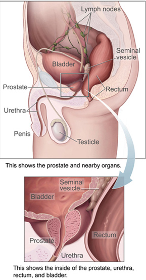
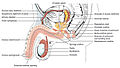
The prostate is both an accessory gland of the male reproductive system and a muscle-driven mechanical switch between urination and ejaculation. It is found in all male mammals. It differs between species anatomically, chemically, and physiologically. Anatomically, the prostate is found below the bladder, with the urethra passing through it. It is described in gross anatomy as consisting of lobes and in microanatomy by zone. It is surrounded by an elastic, fibromuscular capsule and contains glandular tissue, as well as connective tissue.

<span class="mw-page-title-main">Urinary system</span> Anatomical system consisting of the kidneys, ureters, urinary bladder, and the urethra

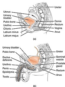
The human urinary system, also known as the urinary tract or renal system, consists of the kidneys, ureters, bladder, and the urethra. The purpose of the urinary system is to eliminate waste from the body, regulate blood volume and blood pressure, control levels of electrolytes and metabolites, and regulate blood pH. The urinary tract is the body's drainage system for the eventual removal of urine. The kidneys have an extensive blood supply via the renal arteries which leave the kidneys via the renal vein. Each kidney consists of functional units called nephrons. Following filtration of blood and further processing, wastes exit the kidney via the ureters, tubes made of smooth muscle fibres that propel urine towards the urinary bladder, where it is stored and subsequently expelled from the body by urination. The female and male urinary system are very similar, differing only in the length of the urethra.

<span class="mw-page-title-main">Cystoscopy</span> Medical procedure; endoscopy of the urinary bladder via the urethra

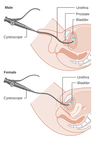
Cystoscopy is endoscopy of the urinary bladder via the urethra. It is carried out with a cystoscope.

<span class="mw-page-title-main">Urination</span> Release of urine from the urinary bladder

Urination is the release of urine from the bladder to the outside of the body. Urine is released from the urethra through the penis or vulva in placental mammals and through the cloaca in other vertebrates. It is the urinary system's form of excretion. It is also known medically as micturition, voiding, uresis, or, rarely, emiction, and known colloquially by various names including peeing, weeing, pissing, and euphemistically going number one. The process of urination is under voluntary control in healthy humans and other animals, but may occur as a reflex in infants, some elderly individuals, and those with neurological injury. It is normal for adult humans to urinate up to seven times during the day.

<span class="mw-page-title-main">Urinary incontinence</span> Uncontrolled leakage of urine

Urinary incontinence (UI), also known as involuntary urination, is any uncontrolled leakage of urine. It is a common and distressing problem, which may have a large impact on quality of life. It has been identified as an important issue in geriatric health care. The term enuresis is often used to refer to urinary incontinence primarily in children, such as nocturnal enuresis. UI is an example of a stigmatized medical condition, which creates barriers to successful management and makes the problem worse. People may be too embarrassed to seek medical help, and attempt to self-manage the symptom in secrecy from others.

<span class="mw-page-title-main">Ureter</span> Tubes used in the urinary system in most animals

The ureters are tubes composed of smooth muscle that transport urine from the kidneys to the urinary bladder. In an adult human, the ureters typically measure 20 to 30 centimeters in length and about 3 to 4 millimeters in diameter. They are lined with urothelial cells, a form of transitional epithelium, and feature an extra layer of smooth muscle in the lower third to aid in peristalsis. The ureters can be affected by a number of diseases, including urinary tract infections and kidney stone. Stenosis is when a ureter is narrowed, due to for example chronic inflammation. Congenital abnormalities that affect the ureters can include the development of two ureters on the same side or abnormally placed ureters. Additionally, reflux of urine from the bladder back up the ureters is a condition commonly seen in children.

<span class="mw-page-title-main">Hematuria</span> Medical condition

Hematuria or haematuria is defined as the presence of blood or red blood cells in the urine. "Gross hematuria" occurs when urine appears red, brown, or tea-colored due to the presence of blood. Hematuria may also be subtle and only detectable with a microscope or laboratory test. Blood that enters and mixes with the urine can come from any location within the urinary system, including the kidney, ureter, urinary bladder, urethra, and in men, the prostate. Common causes of hematuria include urinary tract infection (UTI), kidney stones, viral illness, trauma, bladder cancer, and exercise. These causes are grouped into glomerular and non-glomerular causes, depending on the involvement of the glomerulus of the kidney. But not all red urine is hematuria. Other substances such as certain medications and foods can cause urine to appear red. Menstruation in women may also cause the appearance of hematuria and may result in a positive urine dipstick test for hematuria. A urine dipstick test may also give an incorrect positive result for hematuria if there are other substances in the urine such as myoglobin, a protein excreted into urine during rhabdomyolysis. A positive urine dipstick test should be confirmed with microscopy, where hematuria is defined by three or more red blood cells per high power field. When hematuria is detected, a thorough history and physical examination with appropriate further evaluation can help determine the underlying cause.

<span class="mw-page-title-main">Urinary retention</span> Inability to completely empty the bladder

Urinary retention is an inability to completely empty the bladder. Onset can be sudden or gradual. When of sudden onset, symptoms include an inability to urinate and lower abdominal pain. When of gradual onset, symptoms may include loss of bladder control, mild lower abdominal pain, and a weak urine stream. Those with long-term problems are at risk of urinary tract infections.

<span class="mw-page-title-main">External sphincter muscle of male urethra</span>

The external sphincter muscle of male urethra, also sphincter urethrae membranaceae, sphincter urethrae externus, surrounds the whole length of the membranous urethra, and is enclosed in the fascia of the urogenital diaphragm.

In urology, voiding cystourethrography (VCUG) is a frequently performed technique for visualizing a person's urethra and urinary bladder while the person urinates (voids). It is used in the diagnosis of vesicoureteral reflux, among other disorders. The technique consists of catheterizing the person in order to fill the bladder with a radiocontrast agent, typically diatrizoic acid. Under fluoroscopy the radiologist watches the contrast enter the bladder and looks at the anatomy of the patient. If the contrast moves into the ureters and back into the kidneys, the radiologist makes the diagnosis of vesicoureteral reflux, and gives the degree of severity a score. The exam ends when the person voids while the radiologist is watching under fluoroscopy. Consumption of fluid promotes excretion of contrast media after the procedure. It is important to watch the contrast during voiding, because this is when the bladder has the most pressure, and it is most likely this is when reflux will occur. Despite this detailed description of the procedure, at least as of 2016 the technique had not been standardized across practices.

<span class="mw-page-title-main">Neurogenic bladder dysfunction</span> Medical condition

Neurogenic bladder dysfunction, often called by the shortened term neurogenic bladder, refers to urinary bladder problems due to disease or injury of the central nervous system or peripheral nerves involved in the control of urination. There are multiple types of neurogenic bladder depending on the underlying cause and the symptoms. Symptoms include overactive bladder, urinary urgency, frequency, incontinence or difficulty passing urine. A range of diseases or conditions can cause neurogenic bladder including spinal cord injury, multiple sclerosis, stroke, brain injury, spina bifida, peripheral nerve damage, Parkinson's disease, multiple system atrophy or other neurodegenerative diseases. Neurogenic bladder can be diagnosed through a history and physical as well as imaging and more specialized testing. In addition to symptomatic treatment, treatment depends on the nature of the underlying disease and can be managed with behavioral changes, medications, surgeries, or other procedures. The symptoms of neurogenic bladder, especially incontinence, can severely degrade a person's quality of life.

<span class="mw-page-title-main">Urethral cancer</span> Medical condition

Urethral cancer is a rare cancer originating from the urethra. The disease has been classified by the TNM staging system and the World Health Organization.

<span class="mw-page-title-main">Urinary meatus</span> Orifice of the urethra

The urinary meatus, also known as the external urethral orifice, is the opening where urine exits the male and female urethra. It is where semen also exits the male urethra. The meatus has varying degrees of sensitivity to touch.

<span class="mw-page-title-main">Urethral sphincters</span> Muscles keeping urine in the bladder

The urethral sphincters are two muscles used to control the exit of urine in the urinary bladder through the urethra. The two muscles are either the male or female external urethral sphincter and the internal urethral sphincter. When either of these muscles contracts, the urethra is sealed shut.

<span class="mw-page-title-main">Internal urethral sphincter</span> Urethral sphincter muscle which constricts the internal urethral orifice

The internal urethral sphincter is a urethral sphincter muscle which constricts the internal urethral orifice. It is located at the junction of the urethra with the urinary bladder and is continuous with the detrusor muscle, but anatomically and functionally fully independent from it. It is composed of smooth muscle, so it is under the control of the autonomic nervous system, specifically the sympathetic nervous system.

<span class="mw-page-title-main">Retrograde urethrogram</span> Medical imaging of the urethra

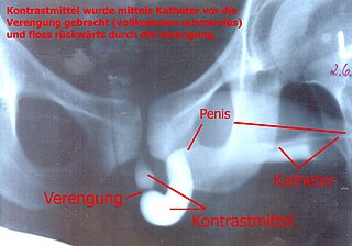
A retrograde urethrography is a routine radiologic procedure used to image the integrity of the urethra. Hence a retrograde urethrogram is essential for diagnosis of urethral injury, or urethral stricture.

Urologic diseases or conditions include urinary tract infections, kidney stones, bladder control problems, and prostate problems, among others. Some urologic conditions do not affect a person for that long and some are lifetime conditions. Kidney diseases are normally investigated and treated by nephrologists, while the specialty of urology deals with problems in the other organs. Gynecologists may deal with problems of incontinence in women.

<span class="mw-page-title-main">Diphallia</span> Genital medical condition

Diphallia, penile duplication (PD), diphallic terata, or diphallasparatus is an extremely rare developmental abnormality in which a male is born with two penises. The first reported case was by Johannes Jacob Wecker in 1609. Its occurrence is 1 in 5.5 million boys in the United States.

<span class="mw-page-title-main">Ureteral cancer</span> Medical condition

Ureteral cancer is cancer of the ureters, muscular tubes that propel urine from the kidneys to the urinary bladder. It is also known as ureter cancer, renal pelvic cancer, and rarely ureteric cancer or uretal cancer. Cancer in this location is rare. Ureteral cancer becomes more likely in older adults, usually ages 70–80, who have previously been diagnosed with bladder cancer.

References

  1. 1 2 Marvalee H. Wake (15 September 1992). Hyman's Comparative Vertebrate Anatomy. University of Chicago Press. pp. 583–. ISBN   978-0-226-87013-7 . Retrieved 6 May 2013.
  2. Legato, Marianne J.; Bilezikian, John P., eds. (2004). "109: The Evaluation and Treatment of Urinary Incontinence". Principles of Gender-specific Medicine. Vol. 1. Gulf Professional Publishing. p. 1187.
  3. Chancellor, Michael B; Yoshimura, Naoki (2004). "Neurophysiology of Stress Urinary Incontinence". Reviews in Urology. 6 (Suppl 3): S19–S28. PMC   1472861 . PMID   16985861.
  4. Jung, Junyang; Anh, Hyo Kwang; Huh, Youngbuhm (September 2012). "Clinical and Functional Anatomy of the Urethral Sphincter". International Neurourology Journal. 16 (3): 102–106. doi: 10.5213/inj.2012.16.3.102 . PMC   3469827 . PMID   23094214.
  5. Karam, I.; Moudouni, S.; Droupy, S.; Abd-Almasad, I.; Uhl, J. F.; Delmas, V. (April 2005). "The structure and innervation of the male urethra: histological and immunohistochemical studies with three-dimensional reconstruction". Journal of Anatomy. 206 (4): 395–403. doi: 10.1111/j.1469-7580.2005.00402.x . PMC   1571491 . PMID   15817107.
  6. Ashton-Miller, J. A.; DeLancey, J. O. (April 2007). "Functional anatomy of the female pelvic floor". Annals of the New York Academy of Sciences. 1101 (1): 266–296. Bibcode:2007NYASA1101..266A. doi:10.1196/annals.1389.034. hdl: 2027.42/72597 . PMID   17416924. S2CID   6310287 . Archived from the original on Aug 22, 2023 via Deep Blue Documents.
  7. 1 2 3 Standring, Susan, ed. (2016). "Bladder, prostate and urethra". Gray's Anatomy: The Anatomical Basis of Clinical Practice (41st ed.). Philadelphia: Elsevier Limited. pp. 1261–1266. ISBN   9780702052309. OCLC   920806541.
  8. "Male Urethra Function & Urethra Anatomy Pictures". Center For Reconstructive Urology. Archived from the original on Sep 26, 2023.
  9. Atlas of Human Anatomy 5th Edition, Netter.
  10. Kohler TS, Yadven M, Manvar A, Liu N, Monga M (2008). "The length of the male urethra". International Brazilian Journal of Urology. 34 (4): 451–4, discussion 455–6. doi: 10.1590/s1677-55382008000400007 . PMID   18778496.
  11. Manual of Obstetrics. (3rd ed.). Elsevier. pp. 1-16. ISBN   9788131225561.
  12. 1 2 3 4 Young, Barbara; O'Dowd, Geraldine; Woodford, Phillip (2013). "Male reproductive system". Wheater's functional histology: a text and colour atlas (6th ed.). Philadelphia: Elsevier. p. 349. ISBN   9780702047473.
  13. 1 2 3 4 Sadley, TW (2019). "Bladder and urethra". Langman's medical embryology (14th ed.). Philadelphia: Wolters Kluwer. pp. 263–66. ISBN   9781496383907.
  14. Kyle BD (Aug 2014). "Ion Channels of the Mammalian Urethra". Channels. 8 (5): 393–401. doi:10.4161/19336950.2014.954224. PMC   4594508 . PMID   25483582.
  15. Killick, Stephen R.; Leary, Christine; Trussell, James; Guthrie, Katherine A. (15 December 2010). "Sperm content of pre-ejaculatory fluid". Human Fertility. 14 (1): 48–52. doi:10.3109/14647273.2010.520798. PMC   3564677 . PMID   21155689.
  16. Chughtai B, Sawas A, O'Malley RL, Naik RR, Ali Khan S, Pentyala S (April 2005). "A neglected gland: a review of Cowper's gland". Int. J. Androl. 28 (2): 74–7. doi: 10.1111/j.1365-2605.2005.00499.x . PMID   15811067. S2CID   32553227.
  17. 1 2 3 4 5 6 Ralston, Stuart H.; Penman, Ian D.; Strachan, Mark W.; Hobson, Richard P. (eds.) (2018). "Urethral discharge". Davidson's principles and practice of medicine (23rd ed.). Elsevier. p. 333. ISBN   978-0-7020-7028-0.{{cite book}}: |first4= has generic name (help)
  18. 1 2 3 4 Ralston, Stuart H.; Penman, Ian D.; Strachan, Mark W.; Hobson, Richard P. (eds.) (2018). "Urothelial tumours". Davidson's principles and practice of medicine (23rd ed.). Elsevier. pp. 435–6. ISBN   978-0-7020-7028-0.{{cite book}}: |first4= has generic name (help)
  19. "Urethral stricture". Mayo Clinic. 12 December 2018. Retrieved 15 May 2020.
  20. 1 2 Maciejewski, Conrad; Rourke, Keith (2015-12-02). "Imaging of urethral stricture disease". Translational Andrology and Urology. 4 (1): 2–9. doi:10.3978/j.issn.2223-4683.2015.02.03. ISSN   2223-4691. PMC   4708283 . PMID   26816803.
  21. Stein DM, Santucci RA (July 2015). "An update on urotrauma". Current Opinion in Urology. 25 (4): 323–30. doi:10.1097/MOU.0000000000000184. PMID   26049876. S2CID   26994715.
  22. Stravodimos, Konstantinos G; Koritsiadis, Georgios; Koutalellis, Georgios (2009). "Electrical wire as a foreign body in a male urethra: a case report". Journal of Medical Case Reports. 3: 49. doi: 10.1186/1752-1947-3-49 . PMC   2649937 . PMID   19192284.
  23. 1 2 3 "Urinary catheters - NHS". nhs.uk. 26 February 2020. Retrieved 2 July 2020.
  24. Wilson, Michaeltitle=Microbial Inhabitants of Humans: Their Ecology and Role in Health and Disease (2005). Microbial Inhabitants of Humans: Their Ecology and Role in Health and Disease. Cambridge University Press. p. 195. ISBN   978-0-521-84158-0.
  25. Hugh Tyndale-Biscoe; Marilyn Renfree (30 January 1987). Reproductive Physiology of Marsupials. Cambridge University Press. pp. 172–. ISBN   978-0-521-33792-2.
  26. 1 2 3 4 Marx, Franz Josef; Karenberg, Axel (2010). "Uro-words making history: Ureter and urethra". The Prostate. 70 (9): 952–958. doi: 10.1002/pros.21129 . PMID   20166127. S2CID   32778667.
  27. Tefekli, Ahmet; Cezayirli, Fatin (2013). "The History of Urinary Stones: In Parallel with Civilization". The Scientific World Journal. 2013: 423964. doi: 10.1155/2013/423964 . PMC   3856162 . PMID   24348156.
  28. 1 2 Nahon, Irmina; Waddington, Gordon; Dorey, Grace; Adams, Roger (2011). "The History of Urologic Surgery: From Reeds to Robotics". Urologic Nursing. 31 (3): 173–180. doi:10.7257/1053-816X.2011.31.3.173. PMID   21805756.