Additional Images
 Medullary rays
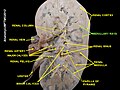 Medullary rays
| Medullary ray | |
|---|---|
| Details | |
| System | Urinary system |
| Identifiers | |
| Latin | radii medullares |
| TA98 | A08.1.01.018 |
| TA2 | 3378 |
| FMA | 74299 |
| Anatomical terminology | |
In anatomy, a medullary ray (Ferrein's pyramid) is the middle part of a cortical lobule (or renal lobule). Each consists of a group of nephrons in the renal cortex. [1] Their name is potentially misleading, as "medullary" refers to their destination, not their location. They travel perpendicular to the capsule, and extend from the cortex to the medulla. They may be visualised during urography. [1]


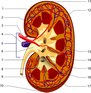
The kidneys are two reddish-brown bean-shaped organs found in vertebrates. They are located on the left and right in the retroperitoneal space, and in adult humans are about 12 centimetres in length. They receive blood from the paired renal arteries; blood exits into the paired renal veins. Each kidney is attached to a ureter, a tube that carries excreted urine to the bladder.

The collecting duct system of the kidney consists of a series of tubules and ducts that physically connect nephrons to a minor calyx or directly to the renal pelvis. The collecting duct system is the last part of nephron and participates in electrolyte and fluid balance through reabsorption and excretion, processes regulated by the hormones aldosterone and vasopressin.
The renal calyces are chambers of the kidney through which urine passes. The minor calyces surround the apex of the renal pyramids. Urine formed in the kidney passes through a renal papilla at the apex into the minor calyx; two or three minor calyces converge to form a major calyx, through which urine passes before continuing through the renal pelvis into the ureter.

The renal medulla is the innermost part of the kidney. The renal medulla is split up into a number of sections, known as the renal pyramids. Blood enters into the kidney via the renal artery, which then splits up to form the segmental arteries which then branch to form interlobar arteries. The interlobar arteries each in turn branch into arcuate arteries, which in turn branch to form interlobular arteries, and these finally reach the glomeruli. At the glomerulus the blood reaches a highly disfavourable pressure gradient and a large exchange surface area, which forces the serum portion of the blood out of the vessel and into the renal tubules. Flow continues through the renal tubules, including the proximal tubule, the Loop of Henle, through the distal tubule and finally leaves the kidney by means of the collecting duct, leading to the renal pelvis, the dilated portion of the ureter.

In the kidney, the macula densa is an area of closely packed specialized cells lining the wall of the distal tubule, at the point where the thick ascending limb of the Loop of Henle meets the distal convoluted tubule. The macula densa is the thickening where the distal tubule touches the glomerulus.
Podocytes are cells in Bowman's capsule in the kidneys that wrap around capillaries of the glomerulus. Podocytes make up the epithelial lining of Bowman's capsule, the third layer through which filtration of blood takes place. Bowman's capsule filters the blood, retaining large molecules such as proteins while smaller molecules such as water, salts, and sugars are filtered as the first step in the formation of urine. Although various viscera have epithelial layers, the name visceral epithelial cells usually refers specifically to podocytes, which are specialized epithelial cells that reside in the visceral layer of the capsule. One type of specialized epithelial cell is podocalyxin.
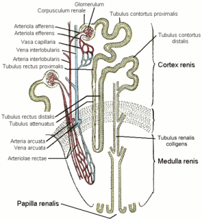
The vasa recta of the kidney, are the straight arterioles, and the straight venules of the kidney, – a series of blood vessels in the blood supply of the kidney that enter the medulla as the straight arterioles, and leave the medulla to ascend to the cortex as the straight venules.. They lie parallel to the loop of Henle.
Pyelogram is a form of imaging of the renal pelvis and ureter.
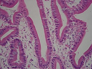
A brush border is the microvilli-covered surface of simple cuboidal and simple columnar epithelium found in different parts of the body. Microvilli are approximately 100 nanometers in diameter and their length varies from approximately 100 to 2,000 nanometers. Because individual microvilli are so small and are tightly packed in the brush border, individual microvilli can only be resolved using electron microscopes; with a light microscope they can usually only be seen collectively as a fuzzy fringe at the surface of the epithelium. This fuzzy appearance gave rise to the term brush border, as early anatomists noted that this structure appeared very much like the bristles of a paintbrush.
The renal hilum or renal pedicle is the hilum of the kidney, that is, its recessed central fissure where its vessels, nerves and ureter pass. The medial border of the kidney is concave in the center and convex toward either extremity; it is directed forward and a little downward. Its central part presents a deep longitudinal fissure, bounded by prominent overhanging anterior and posterior lips. This fissure is a hilum that transmits the vessels, nerves, and ureter. From anterior to posterior, the renal vein exits, the renal artery enters, and the renal pelvis exits the kidney.
The stellate veins join to form the interlobular veins, which pass inward between the rays, receive branches from the plexuses around the convoluted tubules, and, having arrived at the bases of the renal pyramids, join with the venae rectae.
Cortical radial arteries, formerly known as interlobular arteries, are renal blood vessels given off at right angles from the side of the arcuate arteries looking toward the cortical substance. The interlobular arteries pass directly outward between the medullary rays to reach the fibrous tunic, where they end in the capillary network of this part.
The stellate veins are veins that lie beneath the fibrous tunic of the kidney. They are stellate in arrangement and are derived from the capillary network, into which the terminal branches of the interlobular arteries break up. These join to form the interlobular veins, which pass inward between the rays.
The renal lobe is a portion of a kidney consisting of a renal pyramid and the renal cortex above it. In humans, on average there are 7 to 18 renal lobes.
A cortical lobule is a part of a renal lobe. It consists of the nephrons grouped around a single medullary ray, and draining into a single collecting duct. Its near identical parallel is the rectal lobe, which is present in the majority of mammals.

The arcuate arteries of the kidney, also known as arciform arteries, are vessels of the renal circulation. They are located at the border of the renal cortex and renal medulla.
The arcuate vein is a vessel of the renal circulation. It is located at the border of the renal cortex and renal medulla.
The interlobar arteries are vessels of the renal circulation which supply the renal lobes. The interlobar arteries branch from the lobar arteries which branch from the segmental arteries, from the renal artery. They give rise to arcuate arteries.
Within the nephron of the kidney, the ascending limb of the loop of Henle is a segment of the heterogenous loop of Henle downstream of the descending limb, after the sharp bend of the loop. This part of the renal tubule is divided into a thin and thick ascending limb; the thick portion is also known as the distal straight tubule, in contrast with the distal convoluted tubule downstream.
A computed tomography urography is a computed tomography scan that examines the urinary tract after contrast dye is injected into a vein.
This article includes a list of references, related reading or external links, but its sources remain unclear because it lacks inline citations .(May 2015) |
![]() This article incorporates text in the public domain from the 20th edition of Gray's Anatomy (1918)
This article incorporates text in the public domain from the 20th edition of Gray's Anatomy (1918)
| | This article related to the genitourinary system is a stub. You can help Wikipedia by expanding it. |