Median umbilical ligament

Last updated
Median umbilical ligament
Gray1036.png
Posterior view of the anterior abdominal wall in its lower half. The peritoneum is in place, and the various cords are shining through. Median umbilical ligament is not labeled, but it is located just underneath the median umbilical fold, seen in the center of the diagram
Details
FromApex of urinary bladder
To umbilicus
Identifiers
Latin ligamentum umbilicale medianum,
ligamentum suspensorium vesicae urinariae
TA98 A08.3.01.007
TA2 4322
FMA 16568
Anatomical terminology

In human anatomy, the median umbilical ligament is an unpaired midline ligamentous structure upon the lower inner surface of the anterior abdominal wall. [1] It is covered by the median umbilical fold.[ citation needed ]

Contents

The median umbilical ligament represents the remnant of the fetal urachus. [1] [2] It extends from the apex of the bladder to the umbilicus, on the deep surface of the anterior abdominal wall. [2]

The median umbilical ligament represents one of the five ligaments of the internal anterior abdominal wall inferior to the umbilicus; laterally on either side of it are one medial umbilical ligament and finally one lateral umbilical ligament. [1]

Development

The median umbilical ligament begins as the allantois in the embryonic period. It then becomes the urachus in the fetus. [3] [4] This later develops into the median umbilical ligament at birth. [3] It is also formed from the cloaca in utero. [2]

Function

The median umbilical ligament has no known function. [2]

Clinical significance

The median umbilical ligament may be used as a landmark for surgeons who are performing laparoscopy, such as laparoscopic inguinal hernia repair. Other than this, it has no function in a born human and may be cut or removed with impunity.

It contains the urachus, which is the obliterated form of the allantois. The allantois forms a communication between the cloaca (terminal part of hindgut) and the amniotic sac during embryonic development. If the urachus fails to close during fetal life, it can result in anatomical abnormalities such as a urachal cyst, urachal fistula, urachal diverticulum or urachal sinus.

In very rare cases the urachus can fail to close fully. This can lead to a condition known as a patent urachus (also urachal fistula). Although it is mainly immediately diagnosed after birth it can occur at any stages of life. In this condition there is a functional tubular connection between the umbilicus and the bladder which can lead to urine draining through the umbilicus. Patients often present with umbilical wetness or recurring infections in the area. Treatment options include laparoscopic surgery, excision of the umbilicus or conservative therapy. Due to limited risks conservative therapy, while still being chosen rarely, has been gaining some traction in the last years. [5]

Society and culture

The median umbilical ligament was jokingly referred to as "Xander's ligament" in a YouTube video published by Dr. Alexander R. Toftness (aka. ARTexplains on YouTube), and this name was subsequently used on Wikipedia for a short time. [6] This term was later used in a paper published in Mayo Clinic Proceedings. [6] [7] [8] The term has also appeared in a medical textbook of obstetrics and gynaecology. [9]

Related Research Articles

<span class="mw-page-title-main">Inguinal canal</span> Human abdominal anatomy

The inguinal canal is a passage in the anterior abdominal wall on each side of the body, which in males, convey the spermatic cords and in females, the round ligament of the uterus. The inguinal canals are larger and more prominent in males.

<span class="mw-page-title-main">Umbilical artery</span> Artery in the abdominal and pelvic regions

The umbilical artery is a paired artery that is found in the abdominal and pelvic regions. In the fetus, it extends into the umbilical cord.

<span class="mw-page-title-main">Allantois</span> Embryonic structure

The allantois is a hollow sac-like structure filled with clear fluid that forms part of a developing amniote's conceptus. It helps the embryo exchange gases and handle liquid waste.

<span class="mw-page-title-main">Inguinal ligament</span> Band running from the pubic tubercle to the anterior superior iliac spine

The inguinal ligament, also known as Poupart's ligament or groin ligament, is a band running from the pubic tubercle to the anterior superior iliac spine. It forms the base of the inguinal canal through which an indirect inguinal hernia may develop.

<span class="mw-page-title-main">Transverse abdominal muscle</span> Muscle of the abdominal area

The transverse abdominal muscle (TVA), also known as the transverse abdominis, transversalis muscle and transversus abdominis muscle, is a muscle layer of the anterior and lateral abdominal wall, deep to the internal oblique muscle. It is thought by most fitness instructors to be a significant component of the core.

<span class="mw-page-title-main">Internal iliac artery</span> Main artery of the pelvis

The internal iliac artery is the main artery of the pelvis.

<span class="mw-page-title-main">Inferior epigastric artery</span> Blood vessel

In human anatomy, the inferior epigastric artery is an artery that arises from the external iliac artery. It is accompanied by the inferior epigastric vein; inferiorly, these two inferior epigastric vessels together travel within the lateral umbilical fold The inferior epigastric artery then traverses the arcuate line of rectus sheath to enter the rectus sheath, then anastomoses with the superior epigastric artery within the rectus sheath.

<span class="mw-page-title-main">Conjoint tendon</span> Medial part of the posterior wall of the inguinal canal

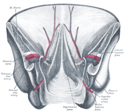
The conjoint tendon is a sheath of connective tissue formed from the lower part of the common aponeurosis of the abdominal internal oblique muscle and the transversus abdominis muscle, joining the muscle to the pelvis. It forms the medial part of the posterior wall of the inguinal canal.

<span class="mw-page-title-main">Round ligament of uterus</span> Ligament connecting the uterus to the labia majora

The round ligament of the uterus is a ligament that connects the uterus to the labia majora. It originates at the junction of the uterus and uterine tube. It passes through the inguinal canal to insert at the labium majus.

<span class="mw-page-title-main">Falciform ligament</span> Ligament attaching the liver to the front body wall

In human anatomy, the falciform ligament is a ligament that attaches the liver to the front body wall and divides the liver into the left lobe and right lobe. The falciform ligament is a broad and thin fold of peritoneum, its base being directed downward and backward and its apex upward and forward. It droops down from the hilum of the liver.

<span class="mw-page-title-main">Transversalis fascia</span> Aponeurosis between the transverse abdominal muscle and the extraperitoneal fat

The transversalis fascia is the fascial lining of the anterolateral abdominal wall situated between the inner surface of the transverse abdominal muscle, and the preperitoneal fascia. It is directly continuous with the iliac fascia, the internal spermatic fascia, and pelvic fascia.

<span class="mw-page-title-main">Round ligament of liver</span> Attaches the liver to the abdominal wall

The round ligament of the liver is a ligament that forms part of the free edge of the falciform ligament of the liver. It connects the liver to the umbilicus. It is the remnant of the left umbilical vein. The round ligament divides the left part of the liver into medial and lateral sections.

<span class="mw-page-title-main">Lateral umbilical fold</span>

The lateral umbilical fold is an elevation of the peritoneum lining the inner/posterior surface of the lower anterior abdominal wall formed by the underlying inferior epigastric artery and inferior epigastric vein which the peritoneum covers. Superiorly, the lateral umbilical fold ends where the vessels reach and enter the rectus sheath at the arcuate line of rectus sheath; in spite of the name, the lateral umbilical folds do not extend as far superiorly as the umbilicus. Inferiorly, it extends to just medial to the deep inguinal ring.

<span class="mw-page-title-main">Urachus</span>

The urachus is a fibrous remnant of the allantois, a canal that drains the urinary bladder of the fetus that joins and runs within the umbilical cord. The fibrous remnant lies in the space of Retzius, between the transverse fascia anteriorly and the peritoneum posteriorly.

<span class="mw-page-title-main">Arcuate line of rectus sheath</span>

The arcuate line of rectus sheath is a line of demarcation corresponding to the free inferior margin of the posterior layer of the rectus sheath inferior to which only the anterior layer of the rectus sheath is present and the rectus abdominis muscle is therefore in direct contact with the transversalis fascia. The arcuate line is concave inferior-wards.

<span class="mw-page-title-main">Medial umbilical ligament</span>

The medial umbilical ligament is a paired structure found in human anatomy. It is on the deep surface of the anterior abdominal wall, and is covered by the medial umbilical folds. It is different from the median umbilical ligament, a structure that represents the remnant of the embryonic urachus.

A urachal cyst is a sinus remaining from the allantois during embryogenesis. It is a cyst which occurs in the remnants between the umbilicus and bladder. This is a type of cyst occurring in a persistent portion of the urachus, presenting as an extraperitoneal mass in the umbilical region. It is characterized by abdominal pain, and fever if infected. It may rupture, leading to peritonitis, or it may drain through the umbilicus. Urachal cysts are usually silent clinically until infection, calculi or adenocarcinoma develop.

<span class="mw-page-title-main">Umbilical folds</span>

Related to the urinary bladder, anteriorly there are the following folds:

A urachal fistula is a congenital disorder caused by the persistence of the allantois, the structure that connects an embryo's bladder to the yolk sac. Normally, the urachus closes off to become the median umbilical ligament; however, if it remains open, urine can drain from the bladder to an opening by the umbilicus.

A urachal diverticulum is a congenital disorder caused by the partial persistence of the allantois. The allantois, which later becomes the urachus, connects an embryo's bladder to the yolk sac. Normally, the urachus closes off to become the median umbilical ligament; however, if it does not seal close to the bladder, a blind pouch connected to the bladder remains. This is usually asymptomatic but can lead to recurrent urinary tract infections. If the urachus is wholly patent, urine can drain from the bladder to an opening by the umbilicus, a condition known as urachal fistula.

References

  1. 1 2 3 Flynn, William; Vickerton, Paula (2022), "Anatomy, Abdomen and Pelvis, Abdominal Wall", StatPearls, Treasure Island (FL): StatPearls Publishing, PMID   31869113 , retrieved 2022-12-17
  2. 1 2 3 4 Wilson, Anthony L.; Gandhi, Jason; Seyam, Omar; Rahmani, Benjamin; Patel, Shrey; Joshi, Gunjan; Smith, Noel L.; Khan, Sardar Ali (2019-09-01). "Urachal anomalies: A review of pathological conditions, diagnosis, and management". Translational Research in Anatomy. 16: 100041. doi: 10.1016/j.tria.2019.100041 . ISSN   2214-854X.
  3. 1 2 Tan C, Simon MA, Dolin N, Gesner L (August 2020). "Incidental vesicourachal diverticulum in a young female". Radiology Case Reports. 15 (8): 1305–1308. doi:10.1016/j.radcr.2020.05.046. PMC   7322240 . PMID   32612730.
  4. Mrad Daly K, Ben Rhouma S, Zaghbib S, Oueslati A, Gharbi M, Nouira Y (September 2019). "Infected urachal cyst in an adult: A case report". Urology Case Reports. 26: 100976. doi:10.1016/j.eucr.2019.100976. PMC   6661533 . PMID   31380223.
  5. Briggs, Kayla B.; Rentea, Rebecca M. (2023), "Patent Urachus", StatPearls, Treasure Island (FL): StatPearls Publishing, PMID   32491655 , retrieved 2023-09-22
  6. 1 2 Wilson C. "This YouTuber Made Up A Name For A Body Part. It Ended Up In A Peer-Reviewed Medical Journal". BuzzFeed. Retrieved 2021-01-23.
  7. Giridhar KV, Kohli M (October 2017). "Management of Muscle-Invasive Urothelial Cancer and the Emerging Role of Immunotherapy in Advanced Urothelial Cancer". Mayo Clinic Proceedings. 92 (10): 1564–1582. doi: 10.1016/j.mayocp.2017.07.010 . PMID   28982487.
  8. Ledger T, Toftness A (August 2020). "The Xander ligament? Caution advised when using online encyclopaedias". Journal of Anatomy. 237 (2): 391. doi:10.1111/joa.13195. PMC   7369185 . PMID   32255503.
  9. DeRuiter MC, Kleinrensink GJ, de Bakker BS (2019). "Chapter 4: Anatomy of the pelvis, pelvic organs and reproductive system". In Steegers E, Fauser B, Hilders C, Jaddoe V, van der Post J, Schoenmakers S (eds.). Textbook of Obstetrics and Gynaecology: A Life Course Approach. bohn stafleu van loghum. pp. 51–84. doi:10.1007/978-90-368-2131-5. ISBN   978-90-368-2130-8. S2CID   59223366.