Collecting duct system

Last updated
Collecting duct system
Gray1128.png
Scheme of renal tubule and its vascular supply.
Details
Location Kidney
Identifiers
Latin tubulus renalis colligens
MeSH D007685
FMA 265239
Anatomical terminology

The collecting duct system of the kidney consists of a series of tubules and ducts that physically connect nephrons to a minor calyx or directly to the renal pelvis. The collecting duct participates in electrolyte and fluid balance through reabsorption and excretion, processes regulated by the hormones aldosterone and vasopressin (antidiuretic hormone).

Contents

There are several components of the collecting duct system, including the connecting tubules, cortical collecting ducts, and medullary collecting ducts.

Structure

Segments

Simple columnar epithelium and simple cuboidal epithelium in the collecting ducts of the pig kidney. The walls of the large and small connecting tubules (a and b respectively), the circular structures, are formed by simple columnar epithelium (a) and simple cuboidal epithelium (b). Gray1133.png
Simple columnar epithelium and simple cuboidal epithelium in the collecting ducts of the pig kidney. The walls of the large and small connecting tubules (a and b respectively), the circular structures, are formed by simple columnar epithelium (a) and simple cuboidal epithelium (b).

The segments of the system are as follows:

SegmentDescription
connecting tubule Connects distal convoluted tubule to the cortical collecting duct
initial collecting tubuleBefore convergence of nephrons
cortical collecting ductsReceives filtrate from the initial collecting tubules, and descends into the renal medulla, forming medullary collecting ducts
medullary collecting ducts
papillary ducts

Connecting tubule

With respect to the renal corpuscle, the connecting tubule (CNT, or junctional tubule, or arcuate renal tubule) is the most proximal part of the collecting duct system. It is adjacent to the distal convoluted tubule, the most distal segment of the renal tubule. Connecting tubules from several adjacent nephrons merge to form cortical collecting tubules, and these may join to form cortical collecting ducts (CCD). [1] Connecting tubules of some juxtamedullary nephrons may arch upward, forming an arcade. It is this "arcuate" feature which gives the tubule its alternate name.

The connecting tubule derives from the metanephric blastema, but the rest of the system derives from the ureteric bud. [2] Because of this, some sources group the connecting tubule as part of the nephron, rather than grouping it with the collecting duct system.

The initial collecting tubule is a segment with a constitution similar as the collecting duct, but before the convergence with other tubules.

The "cortical collecting ducts" receive filtrate from multiple initial collecting tubules and descend into the renal medulla to form medullary collecting ducts.

It participates in the regulation of water and electrolytes, including sodium, and chloride. [3] The CNT is sensitive to both isoprotenerol (more so than the cortical collecting ducts) and antidiuretic hormone (less so than the cortical collecting ducts), the latter largely determining its function in water reabsorption.

Medullary collecting duct

"Medullary collecting ducts" are divided into outer and inner segments, the latter reaching more deeply into the medulla. The variable reabsorption of water and, depending on fluid balances and hormonal influences, the reabsorption or secretion of sodium, potassium, hydrogen and bicarbonate ion continues here. Urea passively transports out of duct here and creates 500mOsm gradient.

The outer segment of the medullary collecting duct follows the cortical collecting duct. It reaches the level of the renal medulla where the thin descending limb of loop of Henle borders with the thick ascending limb of loop of Henle [4] :837

The inner segment is the part of the collecting duct system between the outer segment and the papillary ducts.

Papillary duct

Papillary (collecting) ducts are anatomical structures of the kidneys, previously known as the ducts of Bellini . Papillary ducts represent the most distal portion of the collecting duct. They receive renal filtrate (precursor to urine) from several medullary collecting ducts and empty into a minor calyx. Papillary ducts continue the work of water reabsorption and electrolyte balance initiated in the collecting tubules. [5]

Medullary collecting ducts converge to form a central (papillary) duct near the apex of each renal pyramid. This "papillary duct" exits the renal pyramid at the renal papillae. The renal filtrate it carries drains into a minor calyx as urine. [6]

The cells that comprise the duct itself are similar to rest of the collecting system. The duct is lined by a layer of simple columnar epithelium resting on a thin basement membrane. The epithelium is composed primarily of principal cells and α-intercalated cells. [7] The simple columnar epithelium of the collecting duct system transitions into urothelium near the junction of a papillary duct and a minor calyx. [6]

These cells work in tandem to reabsorb water, sodium, and urea and secrete acid and potassium. The amount of reabsorption or secretion that occurs is related to needs of the body at any given time. These processes are mediated by hormones (aldosterone, vasopressin) and the osmolarity (concentration of electrically charged chemicals) of the surrounding medulla. Hormones regulate how permeable the papillary duct is to water and electrolytes. In the medullary collecting duct specifically, vasopressin upregulates urea transporter A1. This increases the concentration of urea in the surrounding interstitium and increases the osmolarity. Osmolarity influences the strength of the force that pulls (reabsorbs) water from the papillary duct into the medullary interstitium. This is especially important in the papillary ducts. Osmolarity increases from the base of the renal pyramid to the apex. It is highest at the renal apex (up to 1200 mOsm). Thus the force driving the reabsorption of water from the collecting system is the greatest in the papillary duct. [8]

Cells

Each component of the collecting duct system contains two cell types, intercalated cells and a segment-specific cell type:

Principal cells

The principal cell mediates the collecting duct's influence on sodium and potassium balance via sodium channels and potassium channels located on the cell's apical membrane. Aldosterone determines expression of sodium channels (especially the ENaC on the collecting tubule). Increases in aldosterone increase expression of luminal sodium channels. [9] Aldosterone also increases the number of Na⁺/K⁺-ATPase pumps [10] :949 that allow increased sodium reabsorption and potassium excretion. [10] :336 Vasopressin determines the expression of aquaporin channels that provide a physical pathway for water to pass through the principal cells. [11] Together, aldosterone and vasopressin let the principal cell control the quantity of water that is reabsorbed.

Intercalated cells

Image depicting an a intercalated cell Alpha Intercalated Cell Cartoon.svg
Image depicting an α intercalated cell

Intercalated cells come in α, β, and non-α non-β varieties and participate in acid–base homeostasis. [12] [13]

Type of cellSecretesReabsorbs
α-intercalated cells acid (via an apical H+-ATPase and H+/K+ exchanger) in the form of hydrogen ions bicarbonate (via band 3, a basolateral Cl/HCO3 exchanger) [14]
β-intercalated cellsbicarbonate (via pendrin a specialised apical Cl/HCO3)acid (via a basal H+-ATPase)
non-α non-β intercalated cells acid (via an apical H+-ATPase and H+/K+ exchanger) and bicarbonate (via pendrin) [15] [16] -

For their contribution to acid–base homeostasis, the intercalated cells play important roles in the kidney's response to acidosis and alkalosis. Damage to the α-intercalated cell's ability to secrete acid can result in distal renal tubular acidosis (RTA type I, classical RTA)(reference). The intercalated cell population is also extensively modified in response to chronic lithium treatment, including the addition of a largely uncharacterized cell type which expressed markers for both intercalated and principal cells. [17] [18]

Function

Diagram outlining movement of ions in nephron, with the collecting ducts on the right. Kidney nephron molar transport diagram.svg
Diagram outlining movement of ions in nephron, with the collecting ducts on the right.

The collecting duct system is the final component of the kidney to influence the body's electrolyte and fluid balance. In humans, the system accounts for 4–5% of the kidney's reabsorption of sodium and 5% of the kidney's reabsorption of water. At times of extreme dehydration, over 24% of the filtered water may be reabsorbed in the collecting duct system.

The wide variation in water reabsorption levels for the collecting duct system reflects its dependence on hormonal activation. The collecting ducts, in particular, the outer medullary and cortical collecting ducts, are largely impermeable to water without the presence of antidiuretic hormone (ADH, or vasopressin).

The collecting duct system participates in the regulation of other electrolytes, including chloride, potassium, hydrogen ions, and bicarbonate.

An extracellular protein called hensin (protein) mediates the regulation of secretion of acid by alpha cells in acidosis, and secretion of bicarbonate by beta cells in alkalosis. [19] [20]

Collecting duct carcinoma

Carcinoma of the collecting duct is a relatively rare subtype of renal cell carcinoma (RCC), accounting for less than 1% of all RCCs. [21] [22] Many reported cases have occurred in younger patients, often in the third, fourth, or fifth decade of life. [23] Collecting duct carcinomas are derived from the medulla, but many are infiltrative, and extension into the cortex is common. [24] Most reported cases have been high grade and advanced stage and have not responded to conventional therapies. [23] [25] Most patients are symptomatic at presentation. [26] Immunohistochemical and molecular analyses suggest that collecting duct RCC may resemble transitional cell carcinoma, and some patients with advanced collecting duct RCC have responded to cisplatin- or gemcitabine-based chemotherapy. [27] [28]

See also

Related Research Articles

<span class="mw-page-title-main">Kidney</span> Organ that filters blood and produces urine

In humans, the kidneys are two reddish-brown bean-shaped blood-filtering organs that are a multilobar, multipapillary form of mammalian kidneys, usually without signs of external lobulation. They are located on the left and right in the retroperitoneal space, and in adult humans are about 12 centimetres in length. They receive blood from the paired renal arteries; blood exits into the paired renal veins. Each kidney is attached to a ureter, a tube that carries excreted urine to the bladder.

<span class="mw-page-title-main">Urinary system</span> Human anatomical system consisting of the kidneys, ureters, urinary bladder, and the urethra

The urinary system, also known as the urinary tract or renal system, consists of the kidneys, ureters, bladder, and the urethra. The purpose of the urinary system is to eliminate waste from the body, regulate blood volume and blood pressure, control levels of electrolytes and metabolites, and regulate blood pH. The urinary tract is the body's drainage system for the eventual removal of urine. The kidneys have an extensive blood supply via the renal arteries which leave the kidneys via the renal vein. Each kidney consists of functional units called nephrons. Following filtration of blood and further processing, wastes exit the kidney via the ureters, tubes made of smooth muscle fibres that propel urine towards the urinary bladder, where it is stored and subsequently expelled from the body by urination. The female and male urinary system are very similar, differing only in the length of the urethra.

<span class="mw-page-title-main">Nephron</span> Microscopic structural and functional unit of the kidney

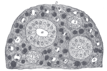
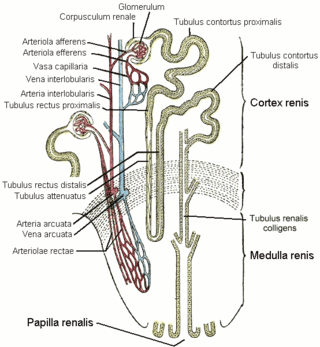
The nephron is the minute or microscopic structural and functional unit of the kidney. It is composed of a renal corpuscle and a renal tubule. The renal corpuscle consists of a tuft of capillaries called a glomerulus and a cup-shaped structure called Bowman's capsule. The renal tubule extends from the capsule. The capsule and tubule are connected and are composed of epithelial cells with a lumen. A healthy adult has 1 to 1.5 million nephrons in each kidney. Blood is filtered as it passes through three layers: the endothelial cells of the capillary wall, its basement membrane, and between the foot processes of the podocytes of the lining of the capsule. The tubule has adjacent peritubular capillaries that run between the descending and ascending portions of the tubule. As the fluid from the capsule flows down into the tubule, it is processed by the epithelial cells lining the tubule: water is reabsorbed and substances are exchanged ; first with the interstitial fluid outside the tubules, and then into the plasma in the adjacent peritubular capillaries through the endothelial cells lining that capillary. This process regulates the volume of body fluid as well as levels of many body substances. At the end of the tubule, the remaining fluid—urine—exits: it is composed of water, metabolic waste, and toxins.

<span class="mw-page-title-main">Renin–angiotensin system</span> Hormone system

The renin–angiotensin system (RAS), or renin–angiotensin–aldosterone system (RAAS), is a hormone system that regulates blood pressure, fluid and electrolyte balance, and systemic vascular resistance.

<span class="mw-page-title-main">Aldosterone</span> Mineralocorticoid steroid hormone

Aldosterone is the main mineralocorticoid steroid hormone produced by the zona glomerulosa of the adrenal cortex in the adrenal gland. It is essential for sodium conservation in the kidney, salivary glands, sweat glands, and colon. It plays a central role in the homeostatic regulation of blood pressure, plasma sodium (Na+), and potassium (K+) levels. It does so primarily by acting on the mineralocorticoid receptors in the distal tubules and collecting ducts of the nephron. It influences the reabsorption of sodium and excretion of potassium (from and into the tubular fluids, respectively) of the kidney, thereby indirectly influencing water retention or loss, blood pressure, and blood volume. When dysregulated, aldosterone is pathogenic and contributes to the development and progression of cardiovascular and kidney disease. Aldosterone has exactly the opposite function of the atrial natriuretic hormone secreted by the heart.

<span class="mw-page-title-main">Distal convoluted tubule</span> Feature of kidney anatomy

The distal convoluted tubule (DCT) is a portion of kidney nephron between the loop of Henle and the collecting tubule.

<span class="mw-page-title-main">Renal physiology</span> Study of the physiology of the kidney

Renal physiology is the study of the physiology of the kidney. This encompasses all functions of the kidney, including maintenance of acid-base balance; regulation of fluid balance; regulation of sodium, potassium, and other electrolytes; clearance of toxins; absorption of glucose, amino acids, and other small molecules; regulation of blood pressure; production of various hormones, such as erythropoietin; and activation of vitamin D.

<span class="mw-page-title-main">Loop of Henle</span> Part of kidney tissue

In the kidney, the loop of Henle is the portion of a nephron that leads from the proximal convoluted tubule to the distal convoluted tubule. Named after its discoverer, the German anatomist Friedrich Gustav Jakob Henle, the loop of Henle's main function is to create a concentration gradient in the medulla of the kidney.

<span class="mw-page-title-main">Renal medulla</span> Innermost part of the kidney

The renal medulla is the innermost part of the kidney. The renal medulla is split up into a number of sections, known as the renal pyramids. Blood enters into the kidney via the renal artery, which then splits up to form the segmental arteries which then branch to form interlobar arteries. The interlobar arteries each in turn branch into arcuate arteries, which in turn branch to form interlobular arteries, and these finally reach the glomeruli. At the glomerulus the blood reaches a highly disfavourable pressure gradient and a large exchange surface area, which forces the serum portion of the blood out of the vessel and into the renal tubules. Flow continues through the renal tubules, including the proximal tubule, the loop of Henle, through the distal tubule and finally leaves the kidney by means of the collecting duct, leading to the renal pelvis, the dilated portion of the ureter.

<span class="mw-page-title-main">Macula densa</span> A region of juxtaglomerular apparatus in nephron of kidney

In the kidney, the macula densa is an area of closely packed specialized cells lining the wall of the distal tubule where it touches the glomerulus. Specifically, the macula densa is found in the terminal portion of the distal straight tubule, after which the distal convoluted tubule begins.

<span class="mw-page-title-main">Potassium-sparing diuretic</span> Drugs that cause diuresis without causing potassium loss in the urine and leading to hyperkalemia

Potassium-sparing diuretics refers to drugs that cause diuresis without causing potassium loss in the urine. They are typically used as an adjunct in management of hypertension, cirrhosis, and congestive heart failure. The steroidal aldosterone antagonists can also be used for treatment of primary hyperaldosteronism. Spironolactone, a steroidal aldosterone antagonist, is also used in management of female hirsutism and acne from PCOS or other causes.

Fluid balance is an aspect of the homeostasis of organisms in which the amount of water in the organism needs to be controlled, via osmoregulation and behavior, such that the concentrations of electrolytes in the various body fluids are kept within healthy ranges. The core principle of fluid balance is that the amount of water lost from the body must equal the amount of water taken in; for example, in humans, the output must equal the input. Euvolemia is the state of normal body fluid volume, including blood volume, interstitial fluid volume, and intracellular fluid volume; hypovolemia and hypervolemia are imbalances. Water is necessary for all life on Earth. Humans can survive for 4 to 6 weeks without food but only for a few days without water.

<span class="mw-page-title-main">Vasa recta (kidney)</span>

The vasa recta of the kidney, are the straight arterioles, and the straight venules of the kidney, – a series of blood vessels in the blood supply of the kidney that enter the medulla as the straight arterioles, and leave the medulla to ascend to the cortex as the straight venules.. They lie parallel to the loop of Henle.

<span class="mw-page-title-main">Bartter syndrome</span> Medical condition

Bartter syndrome (BS) is a rare inherited disease characterised by a defect in the thick ascending limb of the loop of Henle, which results in low potassium levels (hypokalemia), increased blood pH (alkalosis), and normal to low blood pressure. There are two types of Bartter syndrome: neonatal and classic. A closely associated disorder, Gitelman syndrome, is milder than both subtypes of Bartter syndrome.

The actions of vasopressin are mediated by stimulation of tissue-specific G protein-coupled receptors (GPCRs) called vasopressin receptors that are classified into the V1 (V1A), V2, and V3 (V1B) receptor subtypes. These three subtypes differ in localization, function and signal transduction mechanisms.

In the physiology of the kidney, tubuloglomerular feedback (TGF) is a feedback system inside the kidneys. Within each nephron, information from the renal tubules is signaled to the glomerulus. Tubuloglomerular feedback is one of several mechanisms the kidney uses to regulate glomerular filtration rate (GFR). It involves the concept of purinergic signaling, in which an increased distal tubular sodium chloride concentration causes a basolateral release of adenosine from the macula densa cells. This initiates a cascade of events that ultimately brings GFR to an appropriate level.

<span class="mw-page-title-main">Ascending limb of loop of Henle</span>

Within the nephron of the kidney, the ascending limb of the loop of Henle is a segment of the heterogenous loop of Henle downstream of the descending limb, after the sharp bend of the loop. This part of the renal tubule is divided into a thin and thick ascending limb; the thick portion is also known as the distal straight tubule, in contrast with the distal convoluted tubule downstream.

Renal reabsorption of sodium (Na+) is a part of renal physiology. It uses Na-H antiport, Na-glucose symport, sodium ion channels (minor). It is stimulated by angiotensin II and aldosterone, and inhibited by atrial natriuretic peptide.

The rock dove, Columbia livia, has a number of special adaptations for regulating water uptake and loss.

<span class="mw-page-title-main">Mammalian kidney</span> Paired organ in the urinary system of mammals

The mammalian kidneys are a pair of excretory organs of the urinary system of mammals, a type of metanephric kidney. The kidneys in mammals are usually bean-shaped, located behind the peritoneum (retroperitoneally) on the back (dorsal) wall of the body. Each kidney consists of a renal capsule, peripheral cortex, internal medulla, calices, and renal pelvis, although the calices or renal pelvis may be absent in some species. Urine is excreted from the kidney through the ureter. The structure of the kidney may differ between species depending on the environment, in particular on its aridity. The cortex is responsible for filtering the blood, this part of the kidney is similar to the typical kidneys of less developed vertebrates. Nitrogen-containing waste products are excreted by the kidneys in mammals mainly in the form of urea.

References

  1. Imai M (1979). "The connecting tubule: a functional subdivision of the rabbit distal nephron segments". Kidney Int. 15 (4): 346–56. doi: 10.1038/ki.1979.46 . PMID   513494.
  2. Mitchell, B. S. (2009). Embryology: an illustrated colour text. Sharma, Ram, Britton, Robert. (2nd ed.). Edinburgh: Churchill Livingstone/Elsevier. pp. 50–51. ISBN   978-0-7020-5081-7. OCLC   787843894.
  3. Eaton, Douglas C.; Pooler, John P. (2004). Vander's Renal Physiology (6th ed.). Lange Medical Books/McGraw-Hill. ISBN   0-07-135728-9.
  4. Boron, Walter F. (2005). Medical Physiology: A Cellular and Molecular Approach (updated ed.). Philadelphia: Elsevier/Saunders. ISBN   1-4160-2328-3.
  5. Mescher, Anthony (2013). Junqueira's Basic Histology. McGraw-Hill. pp. 385–403. ISBN   9780071807203.
  6. 1 2 Mescher, Anthony (2013). Junqueira's Basic Histology. McGraw-Hill. p. 400. ISBN   9780071807203.
  7. Gartner, Leslie; Hiatt (2014). Color Atlas and Text of Histology. Baltimore, MD 21201: Lippincott & Wilkins. pp. 383–399. ISBN   9781451113433.{{cite book}}: CS1 maint: location (link)
  8. Costanzo, Linda (2011). Physiology. Baltimore, MD 21201: Wolters Kluwer Health. pp. 167–172. ISBN   9781451187953.{{cite book}}: CS1 maint: location (link)
  9. May, Anne; Puoti, Alessandro; Gaeggeler, Hans-Peter; Horisberger, Jean-Daniel; Rossier, Bernard C (1997). "Early Effect of Aldosterone on the Rate of Synthesis of the Epithelial Sodium Channel a Subunit in A6 Renal Cells" (PDF). Journal of the American Society of Nephrology. 8 (12): 1813–1822. doi: 10.1681/ASN.V8121813 . PMID   9402082 . Retrieved 21 November 2017.
  10. 1 2 Guyton, Arthur C.; John E. Hall (2006). Textbook of Medical Physiology (11 ed.). Philadelphia: Elsevier Saunders. ISBN   0-7216-0240-1.
  11. Schlatter, Eberhard; Schafer, James A. (1987). "Electrophysiological studies in principal cells of rat cortical collecting tubules ADH increases the apical membrane Na+-conductance". Pflügers Archiv: European Journal of Physiology. 409 (1–2): 81–92. doi:10.1007/BF00584753. PMID   2441357. S2CID   24655136.
  12. Alper, S. L.; Natale, J.; Gluck, S.; Lodish, H. F.; Brown, D. (1989-07-01). "Subtypes of intercalated cells in rat kidney collecting duct defined by antibodies against erythroid band 3 and renal vacuolar H+-ATPase". Proceedings of the National Academy of Sciences. 86 (14): 5429–5433. Bibcode:1989PNAS...86.5429A. doi: 10.1073/pnas.86.14.5429 . ISSN   0027-8424. PMC   297636 . PMID   2526338.
  13. Kim, J.; Kim, Y. H.; Cha, J. H.; Tisher, C. C.; Madsen, K. M. (January 1999). "Intercalated cell subtypes in connecting tubule and cortical collecting duct of rat and mouse". Journal of the American Society of Nephrology. 10 (1): 1–12. doi: 10.1681/ASN.V1011 . ISSN   1046-6673. PMID   9890303.
  14. Nosek, Thomas M. "Section 7/7ch07/7ch07p17". Essentials of Human Physiology. Archived from the original on 2016-03-24. – "Intercalated Cells"
  15. Kim, Young-Hee; Kwon, Tae-Hwan; Frische, Sebastian; Kim, Jin; Tisher, C. Craig; Madsen, Kirsten M.; Nielsen, Søren (2002-10-01). "Immunocytochemical localization of pendrin in intercalated cell subtypes in rat and mouse kidney". American Journal of Physiology. Renal Physiology. 283 (4): F744–F754. doi:10.1152/ajprenal.00037.2002. ISSN   1931-857X. PMID   12217866.
  16. Wall, Susan M.; Hassell, Kathryn A.; Royaux, Ines E.; Green, Eric D.; Chang, Judy Y.; Shipley, Gregory L.; Verlander, Jill W. (2003-01-01). "Localization of pendrin in mouse kidney". American Journal of Physiology. Renal Physiology. 284 (1): F229–F241. doi:10.1152/ajprenal.00147.2002. ISSN   1931-857X. PMID   12388426. S2CID   22831140.
  17. Christensen, Birgitte Mønster; Marples, David; Kim, Young-Hee; Wang, Weidong; Frøkiær, Jørgen; Nielsen, Søren (2004-04-01). "Changes in cellular composition of kidney collecting duct cells in rats with lithium-induced NDI" (PDF). American Journal of Physiology. Cell Physiology. 286 (4): C952–C964. doi:10.1152/ajpcell.00266.2003. ISSN   0363-6143. PMID   14613889. S2CID   20227998. Archived from the original (PDF) on 2019-02-19.
  18. Himmel, Nathaniel J.; Wang, Yirong; Rodriguez, Daniel A.; Sun, Michael A.; Blount, Mitsi A. (2018-04-18). "Chronic lithium treatment induces novel patterns of pendrin localization and expression". American Journal of Physiology. Renal Physiology. 315 (2): F313–F322. doi:10.1152/ajprenal.00065.2018. ISSN   1931-857X. PMC   6139525 . PMID   29667915.
  19. Harrison's principles of internal medicine. Jameson, J. Larry,, Kasper, Dennis L.,, Longo, Dan L. (Dan Louis), 1949-, Fauci, Anthony S., 1940-, Hauser, Stephen L.,, Loscalzo, Joseph (20th ed.). New York. 13 August 2018. p. 2097. ISBN   978-1-259-64403-0. OCLC   1029074059.{{cite book}}: CS1 maint: location missing publisher (link) CS1 maint: others (link)
  20. Takito, J; Hikita, C; Al-Awqati, Q (15 November 1996). "Hensin, a new collecting duct protein involved in the in vitro plasticity of intercalated cell polarity". The Journal of Clinical Investigation. 98 (10): 2324–31. doi:10.1172/JCI119044. PMC   507683 . PMID   8941650.
  21. Kennedy et al., 1990[ full citation needed ]
  22. Rumpelt et al., 1991[ full citation needed ]
  23. 1 2 Carter et al., 1992[ full citation needed ]
  24. Pickhardt et al., 2001[ full citation needed ]
  25. Chao et al., 2002b[ full citation needed ]
  26. Tokuda et al., 2004[ full citation needed ]
  27. Milowsky et al., 2002[ full citation needed ]
  28. Peyromaure et al., 2003[ full citation needed ]

PD-icon.svgThis article incorporates text in the public domain from page 1223 of the 20th edition of Gray's Anatomy (1918)