Aquaporin

Last updated

Aquaporin
Aquaporin-Sideview.png
Crystallographic structure of aquaporin 1 (AQP1) PDB 1j4n
Identifiers
SymbolAquaporin
Pfam PF00230
InterPro IPR000425
PROSITE PDOC00193
SCOP2 1fx8 / SCOPe / SUPFAM
TCDB 1.A.8
OPM superfamily 7
OPM protein 2zz9
Available protein structures:
Pfam   structures / ECOD  
PDB RCSB PDB; PDBe; PDBj
PDBsum structure summary

Aquaporins, also called water channels, are channel proteins from a larger family of major intrinsic proteins that form pores in the membrane of biological cells, mainly facilitating transport of water between cells. [1] The cell membranes of a variety of different bacteria, fungi, animal and plant cells contain aquaporins through which water can flow more rapidly into and out of the cell than by diffusing through the phospholipid bilayer. [2] Aquaporins have six membrane-spanning alpha helical domains with both carboxylic and amino terminals on the cytoplasmic side. Two hydrophobic loops contain conserved asparagineprolinealanine ("NPA motif") which form a barrel surrounding a central pore-like region that contains additional protein density. [3] Because aquaporins are usually always open and are prevalent in just about every cell type, this leads to a misconception that water readily passes through the cell membrane down its concentration gradient. Water can pass through the cell membrane through simple diffusion because it is a small molecule, and through osmosis, in cases where the concentration of water outside of the cell is greater than that of the inside. However, because water is a polar molecule this process of simple diffusion is relatively slow, and in tissues with high water permeability the majority of water passes through aquaporin. [4] [5]

Contents

The 2003 Nobel Prize in Chemistry was awarded jointly to Peter Agre for the discovery of aquaporins [6] and Roderick MacKinnon for his work on the structure and mechanism of potassium channels. [7]

Genetic defects involving aquaporin genes have been associated with several human diseases including nephrogenic diabetes insipidus and neuromyelitis optica. [8] [9] [10] [11]

History

The mechanism of facilitated water transport and the probable existence of water pores has attracted researchers since 1957. [12] In most cells, water moves in and out by osmosis through the lipid component of cell membranes. Due to the relatively high water permeability of some epithelial cells, it was long suspected that some additional mechanism for water transport across membranes must exist. Solomon and his co-workers performed pioneering work on water permeability across the cell membrane in the late 1950s. [13] [14] In the mid-1960s an alternative hypothesis (the "partition–diffusion model") sought to establish that the water molecules partitioned between the water phase and the lipid phase and then diffused through the membrane, crossing it until the next interphase where they left the lipid and returned to an aqueous phase. [15] [16] Studies by Parisi, Edelman, Carvounis et al. accented not only the importance of the presence of water channels but also the possibility to regulate their permeability properties. [17] [18] [19] In 1990, Verkman's experiments demonstrated functional expression of water channels, indicating that water channels are effectively proteins. [20] [21]

Discovery

It was not until 1992 that the first aquaporin, 'aquaporin-1' (originally known as CHIP 28), was reported by Peter Agre, of Johns Hopkins University. [22] In 1999, together with other research teams, Agre reported the first high-resolution images of the three-dimensional structure of an aquaporin, namely, aquaporin-1. [23] Further studies using supercomputer simulations identified the pathway of water as it moved through the channel and demonstrated how a pore can allow water to pass without the passage of small solutes. [24] The pioneering research and subsequent discovery of water channels by Agre and his colleagues won Agre the Nobel Prize in Chemistry in 2003. [7] Agre said he discovered aquaporins "by serendipity." He had been studying the Rh blood group antigens and had isolated the Rh molecule, but a second molecule, 28 kilodaltons in size (and therefore called 28K) kept appearing. At first they thought it was a Rh molecule fragment, or a contaminant, but it turned out to be a new kind of molecule with unknown function. It was present in structures such as kidney tubules and red blood cells, and related to proteins of diverse origins, such as in fruit fly brain, bacteria, the lens of the eye, and plant tissue. [23]

However the first report of protein-mediated water transport through membranes was by Gheorghe Benga and others in 1986, prior to Agre's first publication on the topic. [25] [26] This led to a controversy that Benga's work had not been adequately recognized either by Agre or by the Nobel Prize Committee. [27]

Function

Illustration of aquaporin molecule 173-Aquaporin 1fqy.jpg
Illustration of aquaporin molecule

Aquaporins are "the plumbing system for cells". Water moves through cells in an organized way, most rapidly in tissues that have aquaporin water channels. [28] For many years, scientists assumed that water leaked through the cell membrane, and some water does. However, this did not explain how water could move so quickly through some cells. [28]

Aquaporins selectively conduct water molecules in and out of the cell, while preventing the passage of ions and other solutes. Also known as water channels, aquaporins are integral membrane pore proteins. Some of them, known as aquaglyceroporins, also transport other small uncharged dissolved molecules including ammonia, CO2, glycerol, and urea. For example, the aquaporin 3 channel has a pore width of 8–10 Ångströms and allows the passage of hydrophilic molecules ranging between 150 and 200 Da. However, the water pores completely block ions including protons, essential to conserve the membrane's electrochemical potential difference. [29]

Water molecules traverse through the pore of the channel in single file. The presence of water channels increases membrane permeability to water. These are also essential for the water transport system in plants [30] and tolerance to drought and salt stresses. [31]

Structure

Schematic diagram of the 2D structure of aquaporin 1 (AQP1) depicting the six transmembrane alpha-helices and the five interhelical loop regions A-E AQP1.png
Schematic diagram of the 2D structure of aquaporin 1 (AQP1) depicting the six transmembrane alpha-helices and the five interhelical loop regions A-E
The 3D structure of aquaporin Z highlighting the 'hourglass'-shaped water channel that cuts through the center of the protein Aquaporin Z.png
The 3D structure of aquaporin Z highlighting the 'hourglass'-shaped water channel that cuts through the center of the protein

Aquaporin proteins are composed of a bundle of six transmembrane α-helices. They are embedded in the cell membrane. The amino and carboxyl ends face the inside of the cell. The amino and carboxyl halves resemble each other, apparently repeating a pattern of nucleotides. This may have been created by the doubling of a formerly half-sized gene. Between the helices are five regions (A – E) that loop into or out of the cell membrane, two of them hydrophobic (B, E), with an asparagine–proline–alanine ("NPA motif") pattern. They create a distinctive hourglass shape, making the water channel narrow in the middle and wider at each end. [29] [32]

Another and even narrower place in the AQP1 channel is the "ar/R selectivity filter", a cluster of amino acids enabling the aquaporin to selectively let through or block the passage of different molecules. [33]

Aquaporins form four-part clusters (tetramers) in the cell membrane, with each of the four monomers acting as a water channel. Different aquaporins have different sized water channels, the smallest types allowing nothing but water through. [29]

X-ray profiles show that aquaporins have two conical entrances. This hourglass shape could be the result of a natural selection process toward optimal permeability. [34] It has been shown that conical entrances with suitable opening angle can indeed provide a large increase of the hydrodynamic channel permeability. [34]

NPA motif

Aquaporin channels appear in simulations to allow only water to pass, as the molecules effectively queue up in single file. Guided by the aquaporin's local electric field, the oxygen in each water molecule faces forwards as it enters, turning around half way along and leaving with the oxygen facing backwards. [35] The arrangement of opposite-facing electrostatic potentials in the two halves of the channel prevents the flow of protons but permits water to pass freely. [36]

ar/R selectivity filter

Schematic depiction of water movement through the narrow selectivity filter of the aquaporin channel AQP-channel-EN.png
Schematic depiction of water movement through the narrow selectivity filter of the aquaporin channel

The aromatic/arginine or "ar/R" selectivity filter is a cluster of amino acids that help bind to water molecules and exclude other molecules that may try to enter the pore. It is the mechanism by which the aquaporin is able to selectively bind water molecules and so to allow them through, and to prevent other molecules from entering. The ar/R filter is made of two amino acid groups from helices B (HB) and E (HE) and two groups from loop E (LE1, LE2), from the two sides of the NPA motif. Its usual position is 8 Å on the outer side of the NPA motif; it is typically the tightest part of the channel. Its narrowness weakens the hydrogen bonds between water molecules, enabling the arginines, which carry a positive charge, to interact with the water molecules and to filter out undesirable protons. [37]

Taxonomic distribution

In mammals

There are thirteen known types of aquaporins in mammals; six of these are located in the kidney, [38] but the existence of many more is suspected. The most studied aquaporins are compared in the following table:

TypeLocation [39] Function [39]
Aquaporin 1 Water reabsorption
Aquaporin 2 Water reabsorption in response to ADH [40]
Aquaporin 3 Water reabsorption and glycerol permeability
Aquaporin 4 Water reabsorption

In plants

In plants, water is taken up from the soil through the roots, where it passes from the cortex into the vascular tissues. There are three routes for water to flow in these tissues, known as the apoplastic, symplastic and transcellular pathways. Specifically, aquaporins are found in the vacuolar membrane, in addition to the plasma membrane of plants; the transcellular pathway involves transport of water across the plasma and vacuolar membranes. [41] When plant roots are exposed to mercuric chloride, which is known to inhibit aquaporins, the flow of water is greatly reduced while the flow of ions is not, supporting the view that there exists a mechanism for water transport independent of the transport of ions: aquaporins. [42] Aquaporins can play a major role in extension growth by allowing an influx of water into expanding cells - a process necessary to sustain plant development. [41] Plant aquaporins are important for mineral nutrition and ion detoxification; these are both essential for the homeostasis of minerals such as boron. [43]

Aquaporins in plants are separated into four main homologous subfamilies, or groups: [44]

These five subfamilies have later been divided into smaller evolutionary subgroups based on their DNA sequence. PIPs cluster into two subgroups, PIP1 and PIP2, whilst TIPs cluster into 5 subgroups, TIP1, TIP2, TIP3, TIP4 and TIP5. Each subgroup is again split up into isoforms e.g. PIP1;1, PIP1;2. As isoforms nomenclature are historically based on functional parameters rather than evolutive ones, several novel propositions on plant aquaporines have been arisen with the study of the evolutionary relationships between the different aquaporins. [49] Within the various selection of aquaporin isoforms in plants, there are also unique patterns of cell- and tissue-specific expression. [41]

When plant aquaporins are silenced, the hydraulic conductance and photosynthesis of the leaf decrease. [50] When gating of plant aquaporins occurs, it stops the flow of water through the pore of the protein. This may happen for various reasons, for example when the plant contains low amounts of cellular water due to drought. [51] The gating of an aquaporin is carried out by an interaction between a gating mechanism and the aquaporin, which causes a 3D change in the protein so that it blocks the pore and, thus, disallows the flow of water through the pore. In plants, there are at least two forms of aquaporin gating: gating by the dephosphorylation of certain serine residues, in response to drought, and the protonation of specific histidine residues, in response to flooding. The phosphorylation of an aquaporin is involved in the opening and closing of petals in response to temperature. [52] [53]

In Heteroconts

Specific aquaporins called Large Intrinsic Proteins (LIP) [54] have been found in Heterokonts, including diatoms and brown algae. LIPs contain an NPM-motif in place of the second conserved NPA-motif typical of the majority of MIPs.

In other organisms

Aquaporins have been discovered in the fungi Saccharomyces cerevisiae (yeast), Dictyostelium , Candida and Ustilago and the protozoans Trypanosoma and Plasmodium . [30]

Clinical significance

There have been two clear examples of diseases identified as resulting from mutations in aquaporins: mutations in the aquaporin-2 gene cause hereditary nephrogenic diabetes insipidus in humans, [9] while mice homozygous for inactivating mutations in the aquaporin-0 gene develop congenital cataracts. [55] A small number of people have been identified with severe or total deficiency in aquaporin-1. They are, in general, healthy, but exhibit a defect in the ability to concentrate solutes in the urine and to conserve water when deprived of drinking water. [56] [57] Mice with targeted deletions in aquaporin-1 also exhibit a deficiency in water conservation due to an inability to concentrate solutes in the kidney medulla by countercurrent multiplication. [58] Aquaporins play a key role in acquired forms of nephrogenic diabetes insipidus, disorders that cause increased urine production. [59] Aquaporin 2 is regulated by vasopressin which, when bound to the cell-surface receptor, activates the cAMP signaling pathway. This results in aquaporin-2 containing vesicles to increase water uptake and return to circulation. Mutation of the aquaporin 2 vasopressin receptor is a cause of acquired diabetes insipidus. In rats, acquired nephrogenic diabetes insipidus can be caused by impaired regulation of aquaporin-2 due to administration of lithium salts, low potassium concentrations in the blood (hypokalemia) and high calcium concentrations in the blood (hypercalcemia). [60] [61] [62] Autoimmune reactions against aquaporin 4 in humans produce Devic's disease. [8] If aquaporin could be manipulated, that could potentially solve medical problems such as fluid retention in heart disease and brain edema after stroke. [28]

Related Research Articles

<span class="mw-page-title-main">Nuclear pore</span> Openings in nuclear envelope of eukaryotic cells

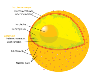
A nuclear pore is a channel as part of the nuclear pore complex (NPC), a large protein complex found in the nuclear envelope of eukaryotic cells. The nuclear envelope surrounds the cell nucleus containing DNA and facilitates the selective membrane transport of various molecules.

<span class="mw-page-title-main">Diabetes insipidus</span> Condition characterized by large amounts of dilute urine and increased thirst

Diabetes insipidus (DI), alternately called arginine vasopressin deficiency (AVP-D) or arginine vasopressin resistance (AVP-R), is a condition characterized by large amounts of dilute urine and increased thirst. The amount of urine produced can be nearly 20 liters per day. Reduction of fluid has little effect on the concentration of the urine. Complications may include dehydration or seizures.

<span class="mw-page-title-main">Semipermeable membrane</span> Membrane which will allow certain molecules or ions to pass through it by diffusion

Semipermeable membrane is a type of biological or synthetic, polymeric membrane that allows certain molecules or ions to pass through it by osmosis. The rate of passage depends on the pressure, concentration, and temperature of the molecules or solutes on either side, as well as the permeability of the membrane to each solute. Depending on the membrane and the solute, permeability may depend on solute size, solubility, properties, or chemistry. How the membrane is constructed to be selective in its permeability will determine the rate and the permeability. Many natural and synthetic materials which are rather thick are also semipermeable. One example of this is the thin film on the inside of an egg.

<span class="mw-page-title-main">Passive transport</span> Transport that does not require energy

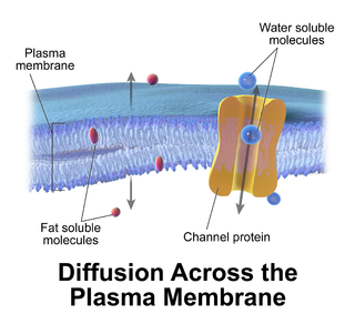
Passive transport is a type of membrane transport that does not require energy to move substances across cell membranes. Instead of using cellular energy, like active transport, passive transport relies on the second law of thermodynamics to drive the movement of substances across cell membranes. Fundamentally, substances follow Fick's first law, and move from an area of high concentration to an area of low concentration because this movement increases the entropy of the overall system. The rate of passive transport depends on the permeability of the cell membrane, which, in turn, depends on the organization and characteristics of the membrane lipids and proteins. The four main kinds of passive transport are simple diffusion, facilitated diffusion, filtration, and/or osmosis.

A membrane transport protein is a membrane protein involved in the movement of ions, small molecules, and macromolecules, such as another protein, across a biological membrane. Transport proteins are integral transmembrane proteins; that is they exist permanently within and span the membrane across which they transport substances. The proteins may assist in the movement of substances by facilitated diffusion, active transport, osmosis, or reverse diffusion. The two main types of proteins involved in such transport are broadly categorized as either channels or carriers. Examples of channel/carrier proteins include the GLUT 1 uniporter, sodium channels, and potassium channels. The solute carriers and atypical SLCs are secondary active or facilitative transporters in humans. Collectively membrane transporters and channels are known as the transportome. Transportomes govern cellular influx and efflux of not only ions and nutrients but drugs as well.

Nephrogenic diabetes insipidus,, is a form of diabetes insipidus primarily due to pathology of the kidney. This is in contrast to central or neurogenic diabetes insipidus, which is caused by insufficient levels of vasopressin. Nephrogenic diabetes insipidus is caused by an improper response of the kidney to vasopressin, leading to a decrease in the ability of the kidney to concentrate the urine by removing free water.

<span class="mw-page-title-main">Vasopressin receptor 2</span> Protein-coding gene in the species Homo sapiens

Vasopressin receptor 2 (V2R), or arginine vasopressin receptor 2, is a protein that acts as receptor for vasopressin. AVPR2 belongs to the subfamily of G-protein-coupled receptors. Its activity is mediated by the Gs type of G proteins, which stimulate adenylate cyclase.

The actions of vasopressin are mediated by stimulation of tissue-specific G protein-coupled receptors (GPCRs) called vasopressin receptors that are classified into the V1 (V1A), V2, and V3 (V1B) receptor subtypes. These three subtypes differ in localization, function and signal transduction mechanisms.

<span class="mw-page-title-main">Aquaporin-4</span> Protein-coding gene in the species Homo sapiens

Aquaporin-4, also known as AQP-4, is a water channel protein encoded by the AQP4 gene in humans. AQP-4 belongs to the aquaporin family of integral membrane proteins that conduct water through the cell membrane. A limited number of aquaporins are found within the central nervous system (CNS): AQP1, 3, 4, 5, 8, 9, and 11, but more exclusive representation of AQP1, 4, and 9 are found in the brain and spinal cord. AQP4 shows the largest presence in the cerebellum and spinal cord grey matter. In the CNS, AQP4 is the most prevalent aquaporin channel, specifically located at the perimicrovessel astrocyte foot processes, glia limitans, and ependyma. In addition, this channel is commonly found facilitating water movement near cerebrospinal fluid and vasculature.

<span class="mw-page-title-main">Aquaporin-2</span> Protein-coding gene in the species Homo sapiens

Aquaporin-2 (AQP-2) is found in the apical cell membranes of the kidney's collecting duct principal cells and in intracellular vesicles located throughout the cell. It is encoded by the AQP2 gene.

<span class="mw-page-title-main">Aquaporin-3</span> Protein-coding gene in the species Homo sapiens

Aquaporin 3 (AQP-3) is the protein product of the human AQP3 gene. It is found in the basolateral cell membrane of principal collecting duct cells and provides a pathway for water to exit these cells. Aquaporin-3 is also permeable to glycerol, ammonia, urea, and hydrogen peroxide. It is expressed in various tissues including the skin, respiratory tract, and kidneys as well as various types of cancers. In the kidney, aquaproin-3 is unresponsive to the antidiuretic hormone vasopressin, unlike aquaporin-2. This protein is also a determinant for the GIL blood group system.

<span class="mw-page-title-main">Aquaporin-1</span> Protein-coding gene in the species Homo sapiens

Aquaporin 1 (AQP-1) is a protein that in humans is encoded by the AQP1 gene.

<span class="mw-page-title-main">Major intrinsic proteins</span>

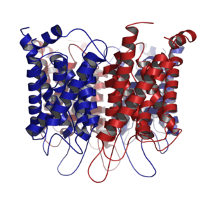
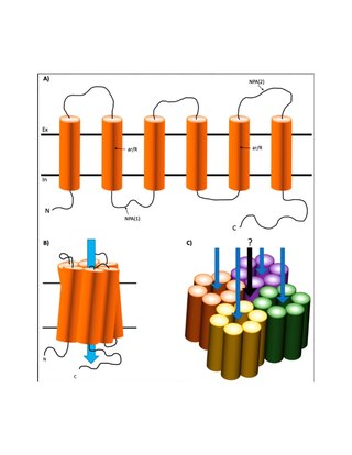
Major intrinsic proteins comprise a large superfamily of transmembrane protein channels that are grouped together on the basis of homology. The MIP superfamily includes three subfamilies: aquaporins, aquaglyceroporins and S-aquaporins.

  1. The aquaporins (AQPs) are water selective.
  2. The aquaglyceroporins are permeable to water, but also to other small uncharged molecules such as glycerol.
  3. The third subfamily, with little conserved amino acid sequences around the NPA boxes, include 'superaquaporins' (S-aquaporins).

A urea transporter is a membrane transport protein, transporting urea. Humans and other mammals have two types of urea transport proteins, UT-A and UT-B. The UT-A proteins are important for renal urea handling and are produced by alternative splicing of the SLC14A2 gene. Urea transport in the kidney is regulated by vasopressin.

<span class="mw-page-title-main">Lens fiber major intrinsic protein</span>

Lens fiber major intrinsic protein also known as aquaporin-0 is a protein that in humans is encoded by the MIP gene.

<span class="mw-page-title-main">Aquaporin-5</span> Protein-coding gene in the species Homo sapiens

Aquaporin-5 (AQP-5) is a protein that in humans is encoded by the AQP5 gene.

<span class="mw-page-title-main">Aquaporin-9</span> Protein-coding gene in the species Homo sapiens

Aquaporin-9 (AQP-9) is a protein that in humans is encoded by the AQP9 gene.

<span class="mw-page-title-main">Aquaporin-7</span> Protein-coding gene in the species Homo sapiens

Aquaporin-7 (AQP-7) is a protein that in humans is encoded by the AQP7 gene.

<span class="mw-page-title-main">Cell membrane</span> Biological membrane that separates the interior of a cell from its outside environment

The cell membrane is a biological membrane that separates and protects the interior of a cell from the outside environment. The cell membrane consists of a lipid bilayer, made up of two layers of phospholipids with cholesterols interspersed between them, maintaining appropriate membrane fluidity at various temperatures. The membrane also contains membrane proteins, including integral proteins that span the membrane and serve as membrane transporters, and peripheral proteins that loosely attach to the outer (peripheral) side of the cell membrane, acting as enzymes to facilitate interaction with the cell's environment. Glycolipids embedded in the outer lipid layer serve a similar purpose. The cell membrane controls the movement of substances in and out of a cell, being selectively permeable to ions and organic molecules. In addition, cell membranes are involved in a variety of cellular processes such as cell adhesion, ion conductivity, and cell signalling and serve as the attachment surface for several extracellular structures, including the cell wall and the carbohydrate layer called the glycocalyx, as well as the intracellular network of protein fibers called the cytoskeleton. In the field of synthetic biology, cell membranes can be artificially reassembled.

<span class="mw-page-title-main">Aquaporin-6</span> Protein-coding gene in the species Homo sapiens

Aquaporin-6, (AQP-6) also known as kidney-specific aquaporin is a protein in humans that is encoded by the AQP6 gene.

References

  1. Agre P (2006). "The aquaporin water channels". Proc Am Thorac Soc. 3 (1): 5–13. doi:10.1513/pats.200510-109JH. PMC   2658677 . PMID   16493146.
  2. Cooper G (2009). The Cell: A Molecular Approach. Washington, DC: ASM PRESS. p. 544. ISBN   978-0-87893-300-6.
  3. Verkman, AS (January 2000). "Structure and function of aquaporin water channels". Am J Physiol Renal Physiol. 278 (1): F13-28. doi:10.1152/ajprenal.2000.278.1.F13. PMID   10644652.
  4. Cooper, Geoffrey (2000). The Cell (2 ed.). MA: Sinauer Associates. Retrieved 23 April 2020.
  5. Lodish, Harvey; Berk, Arnold; Zipursky, S. Lawrence (2000). Molecular Cell Biology (4th ed.). New York: W. H. Freeman. ISBN   9781464183393 . Retrieved 20 May 2020.
  6. Knepper MA, Nielsen S (2004). "Peter Agre, 2003 Nobel Prize winner in chemistry". J. Am. Soc. Nephrol. 15 (4): 1093–5. doi: 10.1097/01.ASN.0000118814.47663.7D . PMID   15034115.
  7. 1 2 "The Nobel Prize in Chemistry 2003". Nobel Foundation. Retrieved 2008-01-23.
  8. 1 2 Lennon VA, Kryzer TJ, Pittock SJ, Verkman AS, Hinson SR (2005). "IgG marker of optic-spinal multiple sclerosis binds to the aquaporin-4 water channel". J. Exp. Med. 202 (4): 473–7. doi:10.1084/jem.20050304. PMC   2212860 . PMID   16087714.
  9. 1 2 Bichet DG (2006). "Nephrogenic diabetes insipidus" (PDF). Adv Chronic Kidney Dis. 13 (2): 96–104. doi:10.1053/j.ackd.2006.01.006. PMID   16580609. Archived from the original (PDF) on 2018-07-18.
  10. Agre P, Kozono D (2003). "Aquaporin water channels: molecular mechanisms for human diseases". FEBS Lett. 555 (1): 72–8. doi: 10.1016/S0014-5793(03)01083-4 . PMID   14630322. S2CID   35406097.
  11. Schrier RW (2007). "Aquaporin-related disorders of water homeostasis". Drug News Perspect. 20 (7): 447–53. doi:10.1358/dnp.2007.20.7.1138161. PMID   17992267.
  12. Parisi M, Dorr RA, Ozu M, Toriano R (December 2007). "From membrane pores to aquaporins: 50 years measuring water fluxes". J Biol Phys. 33 (5–6): 331–43. doi:10.1007/s10867-008-9064-5. PMC   2565768 . PMID   19669522.
  13. Paganelli CV, Solomon AK (November 1957). "The rate of exchange of tritiated water across the human red cell membrane". J. Gen. Physiol. 41 (2): 259–77. doi:10.1085/jgp.41.2.259. PMC   2194835 . PMID   13475690.
  14. Goldstein DA; Solomon AK (1960-09-01). "Determination of equivalent pore radius for human red cells by osmotic pressure measurement". The Journal of General Physiology. 44 (1): 1–17. doi:10.1085/jgp.44.1.1. PMC   2195086 . PMID   13706631.
  15. Dainty, J.; House, C. R. (1966-07-01). "An examination of the evidence for membrane pores in frog skin". The Journal of Physiology. 185 (1): 172–184. doi:10.1113/jphysiol.1966.sp007979. PMC   1395865 . PMID   5965891.
  16. Hanai T, Haydon DA (1966-08-01). "The permeability to water of bimolecular lipid membranes". Journal of Theoretical Biology. 11 (3): 370–382. Bibcode:1966JThBi..11..370H. doi:10.1016/0022-5193(66)90099-3. PMID   5967438.
  17. Parisi M, Bourguet J (1984-01-01). "Effects of cellular acidification on ADH-induced intramembrane particle aggregates". American Journal of Physiology. Cell Physiology. 246 (1): C157–C159. doi:10.1152/ajpcell.1984.246.1.c157. ISSN   0363-6143. PMID   6320654.
  18. Edelman, Isidore S. (25 May 1965). "Hydrogen-ion dependence of the antidiuretic action of vasopressin, oxytocin and deaminooxytocin". Biochimica et Biophysica Acta (BBA) - Biophysics Including Photosynthesis. 102 (1): 185–197. doi:10.1016/0926-6585(65)90212-8. PMID   5833400 via Elsevier Science Direct.
  19. Carvounis CP, Levine SD, Hays RM (1979-05-01). "pH-Dependence of water and solute transport in toad urinary bladder". Kidney International. 15 (5): 513–519. doi: 10.1038/ki.1979.66 . ISSN   0085-2538. PMID   39188.
  20. Zhang, RB; Logee, KA; Verkman, AS (1990-09-15). "Expression of mRNA coding for kidney and red cell water channels in Xenopus oocytes". The Journal of Biological Chemistry. 265 (26): 15375–15378. doi: 10.1016/S0021-9258(18)55405-3 . ISSN   0021-9258. PMID   2394728.
  21. Zhang, R; Alper, S L; Thorens, B; Verkman, A S (1991-11-01). "Evidence from oocyte expression that the erythrocyte water channel is distinct from band 3 and the glucose transporter". Journal of Clinical Investigation. 88 (5): 1553–1558. doi:10.1172/JCI115466. PMC   295670 . PMID   1939644.
  22. Agre P, Preston GM, Smith BL, Jung JS, Raina S, Moon C, Guggino WB, Nielsen S (1 October 1993). "Aquaporin CHIP: the archetypal molecular water channel". Am. J. Physiol. 265 (4 Pt 2): F463–76. doi:10.1152/ajprenal.1993.265.4.F463. PMID   7694481. S2CID   2685263.
  23. 1 2 Mitsuoka K, Murata K, Walz T, Hirai T, Agre P, Heymann JB, Engel A, Fujiyoshi Y (1999). "The structure of aquaporin-1 at 4.5-A resolution reveals short alpha-helices in the center of the monomer". J. Struct. Biol. 128 (1): 34–43. doi:10.1006/jsbi.1999.4177. PMID   10600556. S2CID   1076256.
  24. de Groot BL, Grubmüller H (2005). "The dynamics and energetics of water permeation and proton exclusion in aquaporins". Curr. Opin. Struct. Biol. 15 (2): 176–83. doi:10.1016/j.sbi.2005.02.003. hdl: 11858/00-001M-0000-0012-E99D-E . PMID   15837176.
  25. Benga G, Popescu O, Pop VI, Holmes RP (1986). "p-(Chloromercuri)benzenesulfonate binding by membrane proteins and the inhibition of water transport in human erythrocytes". Biochemistry. 25 (7): 1535–8. doi:10.1021/bi00355a011. PMID   3011064.
  26. Kuchel PW (2006). "The story of the discovery of aquaporins: convergent evolution of ideas--but who got there first?". Cell. Mol. Biol. (Noisy-le-grand). 52 (7): 2–5. PMID   17543213.
  27. Benga, G. "Gheorghe Benga". Ad Astra - Online project for the Romanian Scientific Community. Archived from the original on December 25, 2007. Retrieved 2008-04-05.
  28. 1 2 3 A Conversation With Peter Agre: Using a Leadership Role to Put a Human Face on Science, By Claudia Dreifus, New York Times, January 26, 2009
  29. 1 2 3 Gonen T, Walz T (2006). "The structure of aquaporins". Q. Rev. Biophys. 39 (4): 361–96. doi:10.1017/S0033583506004458. PMID   17156589. S2CID   40235608.
  30. 1 2 Kruse E, Uehlein N, Kaldenhoff R (2006). "The aquaporins". Genome Biol. 7 (2): 206. doi: 10.1186/gb-2006-7-2-206 . PMC   1431727 . PMID   16522221.
  31. Xu Y, et al. (2014). "A banana aquaporin gene". BMC Plant Biology. 14 (1): 59. doi: 10.1186/1471-2229-14-59 . PMC   4015420 . PMID   24606771.
  32. Fu D, Lu M (2007). "The structural basis of water permeation and proton exclusion in aquaporins (Review)". Molecular Membrane Biology. 24 (5–6): 366–74. doi:10.1080/09687680701446965. PMID   17710641. S2CID   343461.
  33. Sui, Haixin; Han, Bong-Gyoon; Lee, John K.; Walian, Peter; Jap, Bing K. (2001). "Structural basis of water-specific transport through the AQP1 water channel". Nature. 414 (6866): 872–878. doi:10.1038/414872a. PMID   11780053.
  34. 1 2 Gravelle S, Joly L, Detcheverry F, Ybert C, Cottin-Bizonne C, Bocquet L (2013). "Optimizing water permeability through the hourglass shape of aquaporins". PNAS. 110 (41): 16367–16372. arXiv: 1310.4309 . Bibcode:2013PNAS..11016367G. doi: 10.1073/pnas.1306447110 . PMC   3799357 . PMID   24067650.
  35. de Groot BL, Grubmüller H (2001). "Water permeation across biological membranes: mechanism and dynamics of aquaporin-1 and GlpF". Science. 294 (5550): 2353–2357. Bibcode:2001Sci...294.2353D. doi:10.1126/science.1062459. hdl: 11858/00-001M-0000-0014-61AF-6 . PMID   11743202. S2CID   446498.
  36. Tajkhorshid E, Nollert P, Jensen MØ, Miercke LJ, O'Connell J, Stroud RM, Schulten K (2002). "Control of the selectivity of the aquaporin water channel family by global orientational tuning". Science. 296 (5567): 525–30. Bibcode:2002Sci...296..525T. doi:10.1126/science.1067778. PMID   11964478. S2CID   22410850.
  37. Sui H, Han BG, Lee JK, Walian P, Jap BK (2001). "Structural basis of water-specific transport through the AQP1 water channel". Nature. 414 (6866): 872–878. doi:10.1038/414872a. PMID   11780053. S2CID   4315108.
  38. Nielsen S, Frøkiaer J, Marples D, Kwon TH, Agre P, Knepper MA (2002). "Aquaporins in the kidney: from molecules to medicine". Physiol. Rev. 82 (1): 205–44. doi:10.1152/physrev.00024.2001. PMID   11773613.
  39. 1 2 Unless else specified in table boxes, then ref is: Walter F. Boron (2005). Medical Physiology: A Cellular And Molecular Approaoch. Elsevier/Saunders. ISBN   978-1-4160-2328-9. Page 842
  40. Sands JM (2012). "Aquaporin 2: Not Just for Moving Water". Journal of the American Society of Nephrology. 23 (9): 1443–1444. doi:10.1681/ASN.2012060613. PMC   3431422 . PMID   22797179.
  41. 1 2 3 Johansson, I; Karlsson, M; Johanson, U; Larsson, C; Kjellbom, P (2000-05-01). "The role of aquaporins in cellular and whole plant water balance". Biochimica et Biophysica Acta (BBA) - Biomembranes. 1465 (1–2): 324–342. doi: 10.1016/S0005-2736(00)00147-4 . PMID   10748263.
  42. Chaumont, F; Tyerman, SD (2014-04-01). "Aquaporins: Highly Regulated Channels Controlling Plant Water Relations". Plant Physiology. 164 (4): 1600–1618. doi:10.1104/pp.113.233791. PMC   3982727 . PMID   24449709.
  43. Porcel, Rosa; Bustamante, Antonio; Ros, Roc; Serrano, Ramón; Mulet Salort, José M. (2018). "BvCOLD1: A novel aquaporin from sugar beet (Beta vulgaris L.) involved in boron homeostasis and abiotic stress". Plant, Cell & Environment. 41 (12): 2844–2857. doi:10.1111/pce.13416. hdl: 10251/145984 . PMID   30103284. S2CID   51974856.
  44. Kaldenhoff R, Bertl A, Otto B, Moshelion M, Uehlein N (2007). "Characterization of Plant Aquaporins". Osmosensing and Osmosignaling. Methods in Enzymology. Vol. 428. pp. 505–31. doi:10.1016/S0076-6879(07)28028-0. ISBN   978-0-12-373921-6. PMID   17875436.{{cite book}}: |journal= ignored (help)
  45. Kammerloher W, Fischer U, Piechottka GP, Schäffner AR (1994). "Water channels in the plant plasma membrane cloned by immunoselection from a mammalian expression system". Plant J. 6 (2): 187–99. doi: 10.1046/j.1365-313X.1994.6020187.x . PMID   7920711.
  46. Maeshima M (2001). "TONOPLAST TRANSPORTERS: Organization and Function". Annu Rev Plant Physiol Plant Mol Biol. 52 (1): 469–497. doi:10.1146/annurev.arplant.52.1.469. PMID   11337406.
  47. Wallace IS, Choi WG, Roberts DM (2006). "The structure, function and regulation of the nodulin 26-like intrinsic protein family of plant aquaglyceroporins". Biochim. Biophys. Acta. 1758 (8): 1165–75. doi: 10.1016/j.bbamem.2006.03.024 . PMID   16716251.
  48. Johanson U, Gustavsson S (2002). "A new subfamily of major intrinsic proteins in plants". Mol. Biol. Evol. 19 (4): 456–61. doi: 10.1093/oxfordjournals.molbev.a004101 . PMID   11919287.
  49. Johanson, Urban; Karlsson, Maria; Johansson, Ingela; Gustavsson, Sofia; Sjövall, Sara; Fraysse, Laure; Weig, Alfons R.; Kjellbom, Per (2001). "The Complete Set of Genes Encoding Major Intrinsic Proteins in Arabidopsis Provides a Framework for a New Nomenclature for Major Intrinsic Proteins in Plants". Plant Physiology. 126 (4): 1358–1369. doi:10.1104/pp.126.4.1358. PMC   117137 . PMID   11500536.
  50. Sade, N; Shatil-Cohen, A; Attia, Z; Maurel, C; Boursiac, Y; Kelly, G; Granot, D; Yaaran, A; Lerner, S (2014-11-01). "The Role of Plasma Membrane Aquaporins in Regulating the Bundle Sheath-Mesophyll Continuum and Leaf Hydraulics". Plant Physiology. 166 (3): 1609–1620. doi:10.1104/pp.114.248633. PMC   4226360 . PMID   25266632.
  51. Kaldenhoff R, Fischer M (2006). "Aquaporins in plants". Acta Physiol (Oxf). 187 (1–2): 169–76. doi:10.1111/j.1748-1716.2006.01563.x. PMID   16734753. S2CID   35656554.
  52. Azad AK, Sawa Y, Ishikawa T, Shibata H (2004). "Phosphorylation of plasma membrane aquaporin regulates temperature-dependent opening of tulip petals". Plant Cell Physiol. 45 (5): 608–17. doi: 10.1093/pcp/pch069 . PMID   15169943.
  53. Azad AK, Katsuhara M, Sawa Y, Ishikawa T, Shibata H (2008). "Characterization of four plasma membrane aquaporins in tulip petals: a putative homolog is regulated by phosphorylation". Plant Cell Physiology. 49 (8): 1196–208. doi: 10.1093/pcp/pcn095 . PMID   18567892.
  54. Khabudaev KV, Petrova DP, Grachev MA, Likhoshway YV (2014). "A new subfamily LIP of the major intrinsic proteins". BMC Genomics. 15 (1): 1–7. doi: 10.1186/1471-2164-15-173 . PMC   4022174 . PMID   24589353.
  55. Okamura T, Miyoshi I, Takahashi K, Mototani Y, Ishigaki S, Kon Y, Kasai N (2003). "Bilateral congenital cataracts result from a gain-of-function mutation in the gene for aquaporin-0 in mice". Genomics. 81 (4): 361–8. doi:10.1016/S0888-7543(03)00029-6. PMID   12676560.
  56. Radin, M. Judith; Yu, Ming-Jiun; Stoedkilde, Lene; Miller, R Lance; Hoffert, Jason D.; Frokiaer, Jorgen; Pisitkun, Trairak; Knepper, Mark A. (2017-03-06). "Aquaporin-2 Regulation in Health and Disease". Veterinary Clinical Pathology. 41 (4): 455–470. doi:10.1111/j.1939-165x.2012.00488.x. PMC   3562700 . PMID   23130944.
  57. King, Landon S; Choi, Michael; Fernandez, Pedro C; Cartron, Jean-Pierre; Agre, Peter (2001-07-19). "Defective Urinary Concentrating Ability Due to a Complete Deficiency of Aquaporin-1". New England Journal of Medicine. 345 (3): 175–179. doi: 10.1056/NEJM200107193450304 . PMID   11463012.
  58. Schnermann, Jurgen; Chou, Chung-Lin; Ma, Tonghui; Traynor, Timothy; Knepper, Mark A; Verkman, AS (1998-08-04). "Defective proximal tubular fluid reabsorption in transgenic aquaporin-1 null mice". Proceedings of the National Academy of Sciences of the United States of America. 95 (16): 9660–9664. Bibcode:1998PNAS...95.9660S. doi: 10.1073/pnas.95.16.9660 . PMC   21395 . PMID   9689137.
  59. Khanna A (2006). "Acquired nephrogenic diabetes insipidus". Semin. Nephrol. 26 (3): 244–8. doi:10.1016/j.semnephrol.2006.03.004. PMID   16713497.
  60. Christensen, S; Kusano, E; Yusufi, A N; Murayama, N; Dousa, TP (1985-06-01). "Pathogenesis of nephrogenic diabetes insipidus due to chronic administration of lithium in rats". Journal of Clinical Investigation. 75 (6): 1869–1879. doi:10.1172/JCI111901. PMC   425543 . PMID   2989335.
  61. Marples, D; Frøkiaer, J; Dørup, J; Knepper, M A; Nielsen, S (1996-04-15). "Hypokalemia-induced downregulation of aquaporin-2 water channel expression in rat kidney medulla and cortex". Journal of Clinical Investigation. 97 (8): 1960–1968. doi:10.1172/JCI118628. PMC   507266 . PMID   8621781.
  62. Marples, D; Christensen, S; Christensen, EI; Ottosen, P D; Nielsen, S (1995-04-01). "Lithium-induced downregulation of aquaporin-2 water channel expression in rat kidney medulla". Journal of Clinical Investigation. 95 (4): 1838–1845. doi:10.1172/JCI117863. PMC   295720 . PMID   7535800.