BK channel

Last updated
KCNMA1
BK-cartoon wp.jpg
The domain structure of BK channels
Identifiers
Symbol KCNMA1
Alt. symbolsSLO
NCBI gene 3778
HGNC 6284
OMIM 600150
RefSeq NM_002247
UniProt Q12791
Other data
Locus Chr. 10 q22
Search for
Structures Swiss-model
Domains InterPro
KCNMB1
Identifiers
Symbol KCNMB1
NCBI gene 3779
HGNC 6285
OMIM 603951
RefSeq NM_004137
UniProt Q16558
Other data
Locus Chr. 5 q34
Search for
Structures Swiss-model
Domains InterPro
KCNMB2
Identifiers
Symbol KCNMB2
NCBI gene 10242
HGNC 6286
OMIM 605214
RefSeq NM_181361
UniProt Q9Y691
Other data
Locus Chr. 3 q26.32
Search for
Structures Swiss-model
Domains InterPro
BK Channel Structure BK Channel Diagram.jpg
BK Channel Structure
KCNMB3
Identifiers
Symbol KCNMB3
Alt. symbolsKCNMB2, KCNMBL
NCBI gene 27094
HGNC 6287
OMIM 605222
RefSeq NM_171828
UniProt Q9NPA1
Other data
Locus Chr. 3 q26.3-q27
Search for
Structures Swiss-model
Domains InterPro
KCNMB3L
Identifiers
SymbolKCNMB3L
Alt. symbolsKCNMB2L, KCNMBLP
NCBI gene 27093
HGNC 6288
RefSeq NG_002679
Other data
Locus Chr. 22 q11.1
KCNMB4
Identifiers
Symbol KCNMB4
NCBI gene 27345
HGNC 6289
OMIM 605223
RefSeq NM_014505
UniProt Q86W47
Other data
Locus Chr. 12 q15
Search for
Structures Swiss-model
Domains InterPro
Calcium-activated BK potassium channel alpha subunit
Identifiers
SymbolBK_channel_a
Pfam PF03493
InterPro IPR003929
Available protein structures:
Pfam   structures / ECOD  
PDB RCSB PDB; PDBe; PDBj
PDBsum structure summary

BK channels (big potassium), are large conductance calcium-activated potassium channels, [1] also known as Maxi-K, slo1, or Kca1.1. BK channels are voltage-gated potassium channels that conduct large amounts of potassium ions (K+) across the cell membrane, hence their name, big potassium. These channels can be activated (opened) by either electrical means, or by increasing Ca2+ concentrations in the cell. [2] [3] BK channels help regulate physiological processes, such as circadian behavioral rhythms and neuronal excitability. [4] BK channels are also involved in many processes in the body, as it is a ubiquitous channel. They have a tetrameric structure that is composed of a transmembrane domain, voltage sensing domain, potassium channel domain, and a cytoplasmic C-terminal domain, with many X-ray structures for reference. Their function is to repolarize the membrane potential by allowing for potassium to flow outward, in response to a depolarization or increase in calcium levels.

Contents

Structure

Structurally, BK channels are homologous to voltage- and ligand-gated potassium channels, having a voltage sensor and pore as the membrane-spanning domain and a cytosolic domain for the binding of intracellular calcium and magnesium. [5] Each monomer of the channel-forming alpha subunit is the product of the KCNMA1 gene (also known as Slo1). The Slo1 subunit has three main structural domains, each with a distinct function:

The activation gate resides in the PGD, which is located at either the cytosolic side of S6 or the selectivity filter (selectivity is the preference of a channel to conduct a specific ion). [5] The voltage sensing domain and pore-gated domain are collectively referred as the membrane-spanning domains and are formed by transmembrane segments S1-S4 and S5-S6, respectively. Within the S4 helix contains a series of positively charged residues which serve as the primary voltage sensor. [6]

BK channels are quite similar to voltage gated K⁺ channels, however, in BK channels only one positively charged residue (Arg213) is involved in voltage sensing across the membrane. [5] Also unique to BK channels is an additional S0 segment, this segment is required for β subunit modulation. [7] [8] and voltage sensitivity. [9]

The Cytosolic domain is composed of two RCK (regulator of potassium conductance) domains, RCK1 and RCK2. These domains contain two high affinity Ca²⁺ binding sites: one in the RCK1 domain and the other in a region termed the Ca²⁺ bowl that consists of a series of Aspartic acid (Asp) residues that are located in the RCK2 domain. The Mg²⁺ binding site is located between the VSD and the cytosolic domain, which is formed by: Asp residues within the S0-S1 loop, Asparagine residues in the cytosolic end of S2, and Glutamine residues in RCK1. [5] In forming the Mg²⁺ binding site, two residues come from the RCK1 of one Slo1 subunit and the other two residues come from the VSD of the neighboring subunit. In order for these residues to coordinate the Mg²⁺ ion, the VSD and cytosolic domain from neighboring subunits must be in close proximity. [5] Modulatory beta subunits (encoded by KCNMB1, KCNMB2, KCNMB3, or KCNMB4) can associate with the tetrameric channel. There are four types of β subunits (β1-4), each of which have different expression patterns that modify the gating properties of the BK channel. The β1 subunit is primarily responsible for smooth muscle cell expression, both β2 and β3 subunits are neuronally expressed, while β4 is expressed within the brain. [5] The VSD associates with the PGD via three major interactions:

  1. Physical connection between the VSD and PGD through the S4-S5 linker.
  2. Interactions between the S4-S5 linker and the cytosolic side of S6.
  3. Interactions between S4 and S5 of a neighboring subunit.

Regulation

BK channels are associated and modulated by a wide variety of intra- and extracellular factors, such as auxiliary subunits (β, γ), Slobs (slo binding protein), phosphorylation, membrane voltage, chemical ligands (Ca²⁺, Mg²⁺), PKC, The BK α-subunits assemble 1:1 with four different auxiliary types of β-subunits (β1, β2, β3 or β4). [10]

Trafficking to and expression of BK channels in the plasma membrane has been found to be regulated by distinct splicing motifs located within the intracellular C-terminal RCK domains. In particular a splice variant that excluded these motifs prevented cell surface expression of BK channels and suggests that such a mechanism impacts physiology and pathophysiology. [10]

BK channels in the vascular system are modulated by agents naturally produced in the body, such as angiotensin II (Ang II), high glucose or arachidonic acid (AA) which is modulated in diabetes by oxidative stress (ROS). [10]

A weaker voltage sensitivity allows BK channels to function in a wide range of membrane potentials. This ensures that the channel can properly perform its physiological function. [11]

Inhibition of BK channel activity by phosphorylation of S695 by protein kinase C (PKC) is dependent on the phosphorylation of S1151 in C terminus of channel alpha-subunit. Only one of these phosphorylations in the tetrameric structure needs to occur for inhibition to be successful. Protein phosphatase 1 counteracts phosphorylation of S695. PKC decreases channel opening probability by shortening the channel open time and prolonging the closed state of the channel. PKC does not affect the single-channel conductance, voltage dependence, or the calcium sensitivity of BK channels. [11]

Activation mechanism

BK channels are synergistically activated through the binding of calcium and magnesium ions, but can also be activated via voltage dependence. [10] Ca²⁺ - dependent activation occurs when intracellular Ca²⁺ binds to two high affinity binding sites: one located in the C-terminus of the RCK2 domain (Ca²⁺ bowl), and the other located in the RCK1 domain. [5] The binding site within the RCK1 domain has somewhat of a lower affinity for calcium than the Ca²⁺ bowl, but is responsible for a larger portion of the Ca²⁺ sensitivity. [12] Voltage and calcium activate BK channels using two parallel mechanisms, with the voltage sensors and the Ca²⁺ bindings sites coupling to the activation gate independently, except for a weak interaction between the two mechanisms. The Ca²⁺ bowl accelerates activation kinetics at low Ca²⁺ concentrations while RCK1 site influences both activation and deactivation kinetics. [11] One mechanism model was originally proposed by Monod, Wyman, and Changeux, known as the MWC model. The MWC model for BK channels explains that a conformational change of the activation gate in channel opening is accompanied by a conformational change to the Ca²⁺ binding site, which increases the affinity of Ca²⁺ binding. [12]

Magnesium-dependent activation of BK channels activates via a low-affinity metal binding site that is independent from Ca²⁺-dependent activation. The Mg²⁺ sensor activates BK channels by shifting the activation voltage to a more negative range. Mg²⁺ activates the channel only when the voltage sensor domain stays in the activated state. The cytosolic tail domain (CTD) is a chemical sensor that has multiple binding sites for different ligands. The CTD activates the BK channel when bound with intracellular Mg²⁺ to allow for interaction with the voltage sensor domain (VSD). [11] Magnesium is predominantly coordinated by six oxygen atoms from the side chains of oxygen-containing residues, main chain carbonyl groups in proteins, or water molecules. [12] D99 at the C-terminus of the S0-S1 loop and N172 in the S2-S3 loop contain side chain oxygens in the voltage sensor domain that are essential for Mg²⁺ binding. Much like the Ca²⁺-dependent activation model, Mg²⁺-dependent activation can also be described by an allosteric MCW gating model. While calcium activates the channel largely independent of the voltage sensor, magnesium activates the channel by channel by an electrostatic interaction with the voltage sensor. [12] This is also known as the Nudging model, in which Magnesium activates the channel by pushing the voltage sensor via electrostatic interactions and involves the interactions among side chains in different structural domains. [5] Energy provided by voltage, Ca²⁺, and Mg²⁺ binding will propagate to the activation gate of BK channels to initiate ion conduction through the pore. [5]

Effects on the neuron, organ, body as a whole

Cellular level

BK channels help regulate both the firing of neurons and neurotransmitter release. [13] This modulation of synaptic transmission and electrical discharge at the cellular level is due to BK channel expression in conjunction with other potassium-calcium channels. [10] The opening of these channels causes a drive towards the potassium equilibrium potential and thus play a role in speeding up the repolarization of action potentials. [10] This would effectively allow for more rapid stimulation. [10] There is also a role played in shaping the general repolarization of cells, and thus after hyperpolarization (AHP) of action potentials. [14] The role that BK channels have in the fast phase of AHP has been studied extensively in the hippocampus. [14] It can also play a role in inhibiting the release of neurotransmitters. [15] There are many BK channels in Purkinje cells in the cerebellum, thus highlighting their role in motor coordination and function. [14] Furthermore, BK channels play a role in modulating the activity of dendrites as well as astrocytes and microglia. [15] They not only play a role in the CNS (central nervous system) but also in smooth muscle contractions, the secretion of endocrine cells, and the proliferation of cells. [13] Various γ subunits during early brain development are involved in neuronal excitability and in non-excitable cells they often are responsible as a driving force of calcium. [10] Therefore, these subunits can be targets for therapeutic treatments as BK channel activators. [10] There is further evidence that inhibiting BK channels would prevent the efflux of potassium and thus reduce the usage of ATP, in effect allowing for neuronal survival in low oxygen environments. [10] BK channels can also function as a neuronal protectant in terms such as limiting calcium entry into the cells through methionine oxidation. [10]

Organ level

BK channels also play a role in hearing. [14] This was found when the BK ɑ-subunit was knocked out in mice and progressive loss of cochlear hair cells, and thus hearing loss, was observed. [14] BK channels are not only involved in hearing, but also circadian rhythms. Slo binding proteins (Slobs) can modulate BK channels as a function of circadian rhythms in neurons. [10] BK channels are expressed in the suprachiasmatic nucleus (SCN), which is characterized to influence the pathophysiology of sleep. [14] BK channel openers can also have a protective effect on the cardiovascular system. [10] At a low concentration of calcium BK channels have a greater impact on vascular tone. [10] Furthermore, the signaling system of BK channels in the cardiovascular system have an influence on the functioning of coronary blood flow. [10] One of the functions of the β subunit in the brain includes inhibition of the BK channels, allowing for the slowing of channel properties as well as the ability to aid in prevention of seizures in the temporal lobe. [10]

Bodily function level

Mutations of BK channels, resulting in a lower amount of expression in mRNA, is more common in people who have mental disabilities (via hypofunction [15] ), schizophrenia or autism. [10] Moreover, increased repolarization caused by BK channel mutations may lead to dependency of alcohol initiation of dyskinesias, epilepsy or paroxysmal movement disorders. [10] Not only are BK channels important in many cellular processes in the adult it also is crucial for proper nutrition supply to a developing fetus. [10] Thus, estrogen can cause an increase in the density of BK channels in the uterus. [10] However, increased expression of BK channels have been found in tumor cells, and this could influence future cancer therapy, discussed more in the pharmacology section. [10] BK channels are ubiquitous throughout the body and thus have a large and vast impact on the body as a whole and at a more cellular level, as discussed.

Pharmacology

Potential issues

Several issues arise when there is a deficit in BK channels. Consequences of the malfunctioning BK channel can affect the functioning of a person in many ways, some more life-threatening than others. BK channels can be activated by exogenous pollutants and endogenous gasotransmitters carbon monoxide, [16] [17] nitric oxide, and hydrogen sulphide. [18] Mutations in the proteins involved with BK channels or genes encoding BK channels are involved in many diseases. A malfunction of BK channels can proliferate in many disorders such as: epilepsy, cancer, diabetes, asthma, and hypertension. [13] Specifically, β1 defect can increase blood pressure and hydrosaline retention in the kidney. [13] Both loss of function and gain of function mutations have been found to be involved in disorders such as epilepsy and chronic pain. [15] Furthermore, increases in BK channel activation, through gain-of-function mutants and amplification, has links to epilepsy and cancer. [13] Moreover, BK channels play a role in tumors as well as cancers. In certain cancers gBK, a variant ion channel called glioma BK channel, can be found. [14] It is known that BK channels do in some way influence the division of cells during replication, which when unregulated can lead to cancers and tumors. [14] Moreover, an aspect studied includes the migration of cancer cells and the role in which BK channels can facilitate this migration, though much is still unknown. [14] Another reason why BK channel understanding is important involves its role in organ transplant surgery. This is due to the activation of BK channels influencing repolarization of the resting membrane potential. [10] Thus, understanding is crucial for safety in effective transplantation.

Current developments

BK channels can be used as pharmacological targets for the treatment of several medical disorders including stroke [19] and overactive bladder. [20] There have been attempts to develop synthetic molecules targeting BK channels, [21] however their efforts have proven largely ineffective thus far. For instance, BMS-204352, a molecule developed by Bristol-Myers Squibb, failed to improve clinical outcome in stroke patients compared to placebo. [22] However, there have been some success from the agonist to BKCa channels, BMS-204352, in treating deficits observed in Fmr1 knockout mice, a model of Fragile X syndrome. [23] [24] BK channels also function as a blocker in ischemia and are a focus in investigating its use as a therapy for stroke. [10]

Future directions

There are many applications for therapeutic strategies involving BK channels. There has been research displaying that a blockage of BK channels results in an increase in neurotransmitter release, effectively indicating future therapeutic possibilities in cognition enhancement, improved memory, and relieving depression. [13] A behavioral response to alcohol is also modulated by BK channels, [10] therefore further understanding of this relationship can aid treatment in patients who are alcoholics. Oxidative stress on BK channels can lead to the negative impairments of lowering blood pressure through cardiovascular relaxation have on both aging and disease. [10] Thus, the signaling system can be involved in treating hypertension and atherosclerosis [10] through targeting of the ɑ subunit to prevent these detrimental effects. Furthermore, the known role that BK channels can play in cancer and tumors is limited. Thus, there is not a lot of current knowledge regarding specific aspects of BK channels that can influence tumors and cancers. [14] Further study is crucial, as this could lead to immense development in treatments for those with cancer and tumors. It is known that epilepsies are due to over-excitability of neurons, which BK channels have a large impact on controlling hyperexcitability. [4] Therefore, understanding could influence the treatment of epilepsy. Overall, BK channels are a target for future pharmacological agents that can be used for benevolent treatments of disease.

See also

Related Research Articles

<span class="mw-page-title-main">Ion channel</span> Pore-forming membrane protein

Ion channels are pore-forming membrane proteins that allow ions to pass through the channel pore. Their functions include establishing a resting membrane potential, shaping action potentials and other electrical signals by gating the flow of ions across the cell membrane, controlling the flow of ions across secretory and epithelial cells, and regulating cell volume. Ion channels are present in the membranes of all cells. Ion channels are one of the two classes of ionophoric proteins, the other being ion transporters.

Inorganic ions in animals and plants are ions necessary for vital cellular activity. In body tissues, ions are also known as electrolytes, essential for the electrical activity needed to support muscle contractions and neuron activation. They contribute to osmotic pressure of body fluids as well as performing a number of other important functions. Below is a list of some of the most important ions for living things as well as examples of their functions:

<span class="mw-page-title-main">Potassium channel</span> Ion channel that selectively passes K+

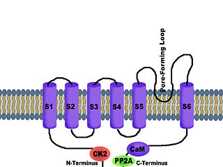
Potassium channels are the most widely distributed type of ion channel found in virtually all organisms. They form potassium-selective pores that span cell membranes. Potassium channels are found in most cell types and control a wide variety of cell functions.

<span class="mw-page-title-main">Voltage-gated ion channel</span> Type of ion channel transmembrane protein

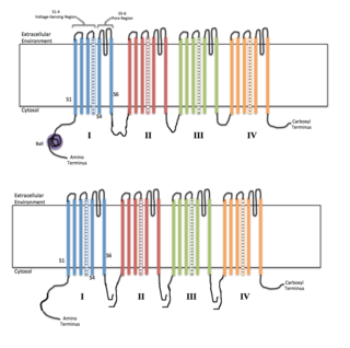
Voltage-gated ion channels are a class of transmembrane proteins that form ion channels that are activated by changes in the electrical membrane potential near the channel. The membrane potential alters the conformation of the channel proteins, regulating their opening and closing. Cell membranes are generally impermeable to ions, thus they must diffuse through the membrane through transmembrane protein channels. They have a crucial role in excitable cells such as neuronal and muscle tissues, allowing a rapid and co-ordinated depolarization in response to triggering voltage change. Found along the axon and at the synapse, voltage-gated ion channels directionally propagate electrical signals. Voltage-gated ion-channels are usually ion-specific, and channels specific to sodium (Na+), potassium (K+), calcium (Ca2+), and chloride (Cl) ions have been identified. The opening and closing of the channels are triggered by changing ion concentration, and hence charge gradient, between the sides of the cell membrane.

Voltage-gated calcium channels (VGCCs), also known as voltage-dependent calcium channels (VDCCs), are a group of voltage-gated ion channels found in the membrane of excitable cells (e.g., muscle, glial cells, neurons, etc.) with a permeability to the calcium ion Ca2+. These channels are slightly permeable to sodium ions, so they are also called Ca2+–Na+ channels, but their permeability to calcium is about 1000-fold greater than to sodium under normal physiological conditions.

<span class="mw-page-title-main">G protein-gated ion channel</span>

G protein-gated ion channels are a family of transmembrane ion channels in neurons and atrial myocytes that are directly gated by G proteins.

Sodium channels are integral membrane proteins that form ion channels, conducting sodium ions (Na+) through a cell's membrane. They belong to the superfamily of cation channels.

Calcium-activated potassium channels are potassium channels gated by calcium, or that are structurally or phylogenetically related to calcium gated channels. They were first discovered in 1958 by Gardos who saw that calcium levels inside of a cell could affect the permeability of potassium through that cell membrane. Then in 1970, Meech was the first to observe that intracellular calcium could trigger potassium currents. In humans they are divided into three subtypes: large conductance or BK channels, which have very high conductance which range from 100 to 300 pS, intermediate conductance or IK channels, with intermediate conductance ranging from 25 to 100 pS, and small conductance or SK channels with small conductances from 2-25 pS.

<span class="mw-page-title-main">Voltage-gated potassium channel</span> Class of transport proteins

Voltage-gated potassium channels (VGKCs) are transmembrane channels specific for potassium and sensitive to voltage changes in the cell's membrane potential. During action potentials, they play a crucial role in returning the depolarized cell to a resting state.

Two-pore channels (TPCs) are eukaryotic intracellular voltage-gated and ligand gated cation selective ion channels. There are two known paralogs in the human genome, TPC1s and TPC2s. In humans, TPC1s are sodium selective and TPC2s conduct sodium ions, calcium ions and possibly hydrogen ions. Plant TPC1s are non-selective channels. Expression of TPCs are found in both plant vacuoles and animal acidic organelles. These organelles consist of endosomes and lysosomes. TPCs are formed from two transmembrane non-equivalent tandem Shaker-like, pore-forming subunits, dimerized to form quasi-tetramers. Quasi-tetramers appear very similar to tetramers, but are not quite the same. Some key roles of TPCs include calcium dependent responses in muscle contraction(s), hormone secretion, fertilization, and differentiation. Disorders linked to TPCs include membrane trafficking, Parkinson's disease, Ebola, and fatty liver.

<span class="mw-page-title-main">KCNE1</span> Protein-coding gene in the species Homo sapiens

Potassium voltage-gated channel subfamily E member 1 is a protein that in humans is encoded by the KCNE1 gene.

<span class="mw-page-title-main">SK channel</span> Protein subfamily of calcium-activated potassium channels

SK channels are a subfamily of calcium-activated potassium channels. They are so called because of their small single channel conductance in the order of 10 pS. SK channels are a type of ion channel allowing potassium cations to cross the cell membrane and are activated (opened) by an increase in the concentration of intracellular calcium through N-type calcium channels. Their activation limits the firing frequency of action potentials and is important for regulating afterhyperpolarization in the neurons of the central nervous system as well as many other types of electrically excitable cells. This is accomplished through the hyperpolarizing leak of positively charged potassium ions along their concentration gradient into the extracellular space. This hyperpolarization causes the membrane potential to become more negative. SK channels are thought to be involved in synaptic plasticity and therefore play important roles in learning and memory.

T-type calcium channels are low voltage activated calcium channels that become inactivated during cell membrane hyperpolarization but then open to depolarization. The entry of calcium into various cells has many different physiological responses associated with it. Within cardiac muscle cell and smooth muscle cells voltage-gated calcium channel activation initiates contraction directly by allowing the cytosolic concentration to increase. Not only are T-type calcium channels known to be present within cardiac and smooth muscle, but they also are present in many neuronal cells within the central nervous system. Different experimental studies within the 1970s allowed for the distinction of T-type calcium channels from the already well-known L-type calcium channels. The new T-type channels were much different from the L-type calcium channels due to their ability to be activated by more negative membrane potentials, had small single channel conductance, and also were unresponsive to calcium antagonist drugs that were present. These distinct calcium channels are generally located within the brain, peripheral nervous system, heart, smooth muscle, bone, and endocrine system.

<span class="mw-page-title-main">L-type calcium channel</span> Family of transport proteins

The L-type calcium channel is part of the high-voltage activated family of voltage-dependent calcium channel. "L" stands for long-lasting referring to the length of activation. This channel has four isoforms: Cav1.1, Cav1.2, Cav1.3, and Cav1.4.

<span class="mw-page-title-main">Potassium channel tetramerisation domain</span>

K+ channel tetramerisation domain is the N-terminal, cytoplasmic tetramerisation domain (T1) of voltage-gated K+ channels. It defines molecular determinants for subfamily-specific assembly of alpha-subunits into functional tetrameric channels. It is distantly related to the BTB/POZ domain Pfam PF00651.

<span class="mw-page-title-main">Calcium-activated potassium channel subunit alpha-1</span> Voltage-gated potassium channel protein

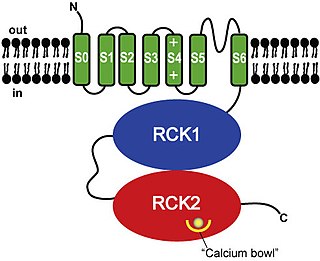
Calcium-activated potassium channel subunit alpha-1 also known as large conductance calcium-activated potassium channel, subfamily M, alpha member 1 (KCa1.1), or BK channel alpha subunit, is a voltage gated potassium channel encoded by the KCNMA1 gene and characterized by their large conductance of potassium ions (K+) through cell membranes.

<span class="mw-page-title-main">KCNMB1</span> Protein-coding gene in the species Homo sapiens

Calcium-activated potassium channel subunit beta-1 is a protein that in humans is encoded by the KCNMB1 gene.

<span class="mw-page-title-main">KCNB1</span> Protein-coding gene in the species Homo sapiens

Potassium voltage-gated channel, Shab-related subfamily, member 1, also known as KCNB1 or Kv2.1, is a protein that, in humans, is encoded by the KCNB1 gene.

<span class="mw-page-title-main">KCNMB2</span> Protein-coding gene in the species Homo sapiens

Calcium-activated potassium channel subunit beta-2 is a protein that in humans is encoded by the KCNMB2 gene.

<span class="mw-page-title-main">Ball and chain inactivation</span> Model in neuroscience

In neuroscience, ball and chain inactivation is a model to explain the fast inactivation mechanism of voltage-gated ion channels. The process is also called hinged-lid inactivation or N-type inactivation. A voltage-gated ion channel can be in three states: open, closed, or inactivated. The inactivated state is mainly achieved through fast inactivation, by which a channel transitions rapidly from an open to an inactivated state. The model proposes that the inactivated state, which is stable and non-conducting, is caused by the physical blockage of the pore. The blockage is caused by a "ball" of amino acids connected to the main protein by a string of residues on the cytoplasmic side of the membrane. The ball enters the open channel and binds to the hydrophobic inner vestibule within the channel. This blockage causes inactivation of the channel by stopping the flow of ions. This phenomenon has mainly been studied in potassium channels and sodium channels.

References

  1. Zang K, Zhang Y, Hu J, Wang Y (2018). "The Large Conductance Calcium- and Voltage-activated Potassium Channel (BK) and Epilepsy". CNS & Neurological Disorders Drug Targets. 17 (4): 248–254. doi:10.2174/1871527317666180404104055. PMID   29623857. S2CID   4663325.
  2. Miller, C. (2000). Genome Biology, 1(4), reviews0004.1. https://dx.doi.org/10.1186/gb-2000-1-4-reviews0004
  3. Yuan, P., Leonetti, M., Pico, A., Hsiung, Y., & MacKinnon, R. (2010). Structure of the Human BK Channel Ca2+-Activation Apparatus at 3.0 A Resolution. Science, 329(5988), 182-186. https://dx.doi.org/10.1126/science.1190414
  4. 1 2 N'Gouemo P (November 2011). "Targeting BK (big potassium) channels in epilepsy". Expert Opinion on Therapeutic Targets. 15 (11): 1283–95. doi:10.1517/14728222.2011.620607. PMC   3219529 . PMID   21923633.
  5. 1 2 3 4 5 6 7 8 9 Lee US, Cui J (September 2010). "BK channel activation: structural and functional insights". Trends in Neurosciences. 33 (9): 415–23. doi:10.1016/j.tins.2010.06.004. PMC   2929326 . PMID   20663573.
  6. Atkinson NS, Robertson GA, Ganetzky B (August 1991). "A component of calcium-activated potassium channels encoded by the Drosophila slo locus". Science. 253 (5019): 551–5. Bibcode:1991Sci...253..551A. doi:10.1126/science.1857984. PMID   1857984. S2CID   11317087.
  7. Morrow JP, Zakharov SI, Liu G, Yang L, Sok AJ, Marx SO (March 2006). "Defining the BK channel domains required for beta1-subunit modulation". Proceedings of the National Academy of Sciences of the United States of America. 103 (13): 5096–101. Bibcode:2006PNAS..103.5096M. doi: 10.1073/pnas.0600907103 . PMC   1458800 . PMID   16549765.
  8. Wallner M, Meera P, Toro L (December 1996). "Determinant for beta-subunit regulation in high-conductance voltage-activated and Ca(2+)-sensitive K+ channels: an additional transmembrane region at the N terminus". Proceedings of the National Academy of Sciences of the United States of America. 93 (25): 14922–7. Bibcode:1996PNAS...9314922W. doi: 10.1073/pnas.93.25.14922 . PMC   26238 . PMID   8962157.
  9. Koval OM, Fan Y, Rothberg BS (March 2007). "A role for the S0 transmembrane segment in voltage-dependent gating of BK channels". The Journal of General Physiology. 129 (3): 209–20. doi:10.1085/jgp.200609662. PMC   2151615 . PMID   17296928.
  10. 1 2 3 4 5 6 7 8 9 10 11 12 13 14 15 16 17 18 19 20 21 22 23 24 25 26 Hermann A, Sitdikova GF, Weiger TM (August 2015). "Oxidative Stress and Maxi Calcium-Activated Potassium (BK) Channels". Biomolecules. 5 (3): 1870–911. doi: 10.3390/biom5031870 . PMC   4598779 . PMID   26287261.
  11. 1 2 3 4 Yang H, Zhang G, Cui J (2015). "BK channels: multiple sensors, one activation gate". Frontiers in Physiology. 6: 29. doi: 10.3389/fphys.2015.00029 . PMC   4319557 . PMID   25705194.
  12. 1 2 3 4 Cui J, Yang H, Lee US (March 2009). "Molecular mechanisms of BK channel activation". Cellular and Molecular Life Sciences. 66 (5): 852–75. doi:10.1007/s00018-008-8609-x. PMC   2694844 . PMID   19099186.
  13. 1 2 3 4 5 6 Yu M, Liu SL, Sun PB, Pan H, Tian CL, Zhang LH (January 2016). "Peptide toxins and small-molecule blockers of BK channels". Acta Pharmacologica Sinica. 37 (1): 56–66. doi:10.1038/aps.2015.139. PMC   4722972 . PMID   26725735.
  14. 1 2 3 4 5 6 7 8 9 10 Bentzen BH, Olesen SP, Rønn LC, Grunnet M (2014). "BK channel activators and their therapeutic perspectives". Frontiers in Physiology. 5: 389. doi: 10.3389/fphys.2014.00389 . PMC   4191079 . PMID   25346695.
  15. 1 2 3 4 Contet C, Goulding SP, Kuljis DA, Barth AL (2016). "BK Channels in the Central Nervous System". Big on Bk - Current Insights into the Function of Large Conductance Voltage- and Ca2+ -Activated K+ Channels at the Molecular, Cellular and Systemic Levels. International Review of Neurobiology. Vol. 128. pp. 281–342. doi:10.1016/bs.irn.2016.04.001. ISBN   9780128036198. PMC   4902275 . PMID   27238267.
  16. Dubuis E, Potier M, Wang R, Vandier C (Feb 2005). "Continuous inhalation of carbon monoxide attenuates hypoxic pulmonary hypertension development presumably through activation of BKCa channels". Cardiovascular Research. 65 (3): 751–61. doi: 10.1016/j.cardiores.2004.11.007 . PMID   15664403.
  17. Hou S, Xu R, Heinemann SH, Hoshi T (Mar 2008). "The RCK1 high-affinity Ca2+ sensor confers carbon monoxide sensitivity to Slo1 BK channels". Proceedings of the National Academy of Sciences of the United States of America. 105 (10): 4039–43. Bibcode:2008PNAS..105.4039H. doi: 10.1073/pnas.0800304105 . PMC   2268785 . PMID   18316727.
  18. Sitdikova GF, Weiger TM, Hermann A (Feb 2010). "Hydrogen sulfide increases calcium-activated potassium (BK) channel activity of rat pituitary tumor cells". Pflügers Archiv. 459 (3): 389–97. doi:10.1007/s00424-009-0737-0. PMID   19802723. S2CID   23073556.
  19. Gribkoff VK, Starrett JE, Dworetzky SI (Apr 2001). "Maxi-K potassium channels: form, function, and modulation of a class of endogenous regulators of intracellular calcium". The Neuroscientist. 7 (2): 166–77. doi:10.1177/107385840100700211. PMID   11496927. S2CID   8791803.
  20. Layne JJ, Nausch B, Olesen SP, Nelson MT (Feb 2010). "BK channel activation by NS11021 decreases excitability and contractility of urinary bladder smooth muscle". American Journal of Physiology. Regulatory, Integrative and Comparative Physiology. 298 (2): R378–84. doi:10.1152/ajpregu.00458.2009. PMC   2828174 . PMID   19923353.
  21. Gribkoff VK, Winquist RJ (May 2005). "Voltage-gated cation channel modulators for the treatment of stroke". Expert Opinion on Investigational Drugs. 14 (5): 579–92. doi:10.1517/13543784.14.5.579. PMID   15926865. S2CID   10236998.
  22. Jensen BS (2002). "BMS-204352: a potassium channel opener developed for the treatment of stroke". CNS Drug Reviews. 8 (4): 353–60. doi:10.1111/j.1527-3458.2002.tb00233.x. PMC   6741660 . PMID   12481191.
  23. Laumonnier F, Roger S, Guérin P, Molinari F, M'rad R, Cahard D, Belhadj A, Halayem M, Persico AM, Elia M, Romano V, Holbert S, Andres C, Chaabouni H, Colleaux L, Constant J, Le Guennec JY, Briault S (2006). "Association of a functional deficit of the BKCa channel, a synaptic regulator of neuronal excitability, with autism and mental retardation". The American Journal of Psychiatry . 163 (9): 1622–1629. doi:10.1176/ajp.2006.163.9.1622. PMID   16946189. S2CID   25225269.
  24. Hébert B; Pietropaolo S; Même S; Laudier B; Laugeray A; Doisne N; Quartier A; Lefeuvre S; Got L; Cahard D; Laumonnier F; Crusio WE; Pichon J; Menuet A; Perche O; Briault S (2014). "Rescue of fragile X syndrome phenotypes in Fmr1 KO mice by a BKCa channel opener molecule". Orphanet Journal of Rare Diseases . 9: 124. doi: 10.1186/s13023-014-0124-6 . PMC   4237919 . PMID   25079250.

Further reading