Oxidative stress

Last updated
Oxidative stress mechanisms in tissue injury. Free radical toxicity induced by xenobiotics and the subsequent detoxification by cellular enzymes (termination). Free Radical Toxicity.svg
Oxidative stress mechanisms in tissue injury. Free radical toxicity induced by xenobiotics and the subsequent detoxification by cellular enzymes (termination).

Oxidative stress reflects an imbalance between the systemic manifestation of reactive oxygen species and a biological system's ability to readily detoxify the reactive intermediates or to repair the resulting damage. [1] Disturbances in the normal redox state of cells can cause toxic effects through the production of peroxides and free radicals that damage all components of the cell, including proteins, lipids, and DNA. Oxidative stress from oxidative metabolism causes base damage, as well as strand breaks in DNA. Base damage is mostly indirect and caused by the reactive oxygen species generated, e.g., O2 (superoxide radical), OH (hydroxyl radical) and H2O2 (hydrogen peroxide). [2] Further, some reactive oxidative species act as cellular messengers in redox signaling. Thus, oxidative stress can cause disruptions in normal mechanisms of cellular signaling.

Contents

In humans, oxidative stress is thought to be involved in the development of attention deficit hyperactivity disorder, [3] cancer, [4] Parkinson's disease, [5] Lafora disease, [6] Alzheimer's disease, [7] atherosclerosis, [8] heart failure, [9] myocardial infarction, [10] [11] fragile X syndrome, [12] sickle-cell disease, [13] lichen planus, [14] vitiligo, [15] autism, [16] infection, chronic fatigue syndrome, [17] and depression; [18] however, reactive oxygen species can be beneficial, as they are used by the immune system as a way to attack and kill pathogens. [19] Short-term oxidative stress may also be important in prevention of aging by induction of a process named mitohormesis, [20] and is required to initiate stress response processes in plants. [21]

Chemical and biological effects

Chemically, oxidative stress is associated with increased production of oxidizing species or a significant decrease in the effectiveness of antioxidant defenses, such as glutathione. [22] The effects of oxidative stress depend upon the size of these changes, with a cell being able to overcome small perturbations and regain its original state. However, more severe oxidative stress can cause cell death, and even moderate oxidation can trigger apoptosis, while more intense stresses may cause necrosis. [23]

Production of reactive oxygen species is a particularly destructive aspect of oxidative stress. Such species include free radicals and peroxides. Some of the less reactive of these species (such as superoxide) can be converted by oxidoreduction reactions with transition metals or other redox cycling compounds (including quinones) into more aggressive radical species that can cause extensive cellular damage. [24] Most long-term effects are caused by damage to DNA. [25] DNA damage induced by ionizing radiation is similar to oxidative stress, and these lesions have been implicated in aging and cancer. Biological effects of single-base damage by radiation or oxidation, such as 8-oxoguanine and thymine glycol, have been extensively studied. Recently the focus has shifted to some of the more complex lesions. Tandem DNA lesions are formed at substantial frequency by ionizing radiation and metal-catalyzed H2O2 reactions. Under anoxic conditions, the predominant double-base lesion is a species in which C8 of guanine is linked to the 5-methyl group of an adjacent 3'-thymine (G[8,5- Me]T). [26] Most of these oxygen-derived species are produced by normal aerobic metabolism. Normal cellular defense mechanisms destroy most of these. Repair of oxidative damages to DNA is frequent and ongoing, largely keeping up with newly induced damages. In rat urine, about 74,000 oxidative DNA adducts per cell are excreted daily. [27] There is also a steady state level of oxidative damages in the DNA of a cell. There are about 24,000 oxidative DNA adducts per cell in young rats and 66,000 adducts per cell in old rats. [27] Likewise, any damage to cells is constantly repaired. However, under the severe levels of oxidative stress that cause necrosis, the damage causes ATP depletion, preventing controlled apoptotic death and causing the cell to simply fall apart. [28] [29]

Polyunsaturated fatty acids, particularly arachidonic acid and linoleic acid, are primary targets for free radical and singlet oxygen oxidations. For example, in tissues and cells, the free radical oxidation of linoleic acid produces racemic mixtures of 13-hydroxy-9Z,11E-octadecadienoic acid, 13-hydroxy-9E,11E-octadecadienoic acid, 9-hydroxy-10E,12-E-octadecadienoic acid (9-EE-HODE), and 11-hydroxy-9Z,12-Z-octadecadienoic acid as well as 4-Hydroxynonenal while singlet oxygen attacks linoleic acid to produce (presumed but not yet proven to be racemic mixtures of) 13-hydroxy-9Z,11E-octadecadienoic acid, 9-hydroxy-10E,12-Z-octadecadienoic acid, 10-hydroxy-8E,12Z-octadecadienoic acid, and 12-hydroxy-9Z-13-E-octadecadienoic (see 13-Hydroxyoctadecadienoic acid and 9-Hydroxyoctadecadienoic acid). [30] [31] [32] Similar attacks on arachidonic acid produce a far larger set of products including various isoprostanes, hydroperoxy- and hydroxy- eicosatetraenoates, and 4-hydroxyalkenals. [31] [33] While many of these products are used as markers of oxidative stress, the products derived from linoleic acid appear far more predominant than arachidonic acid products and therefore easier to identify and quantify in, for example, atheromatous plaques. [34] Certain linoleic acid products have also been proposed to be markers for specific types of oxidative stress. For example, the presence of racemic 9-HODE and 9-EE-HODE mixtures reflects free radical oxidation of linoleic acid whereas the presence of racemic 10-hydroxy-8E,12Z-octadecadienoic acid and 12-hydroxy-9Z-13-E-octadecadienoic acid reflects singlet oxygen attack on linoleic acid. [32] [30] In addition to serving as markers, the linoleic and arachidonic acid products can contribute to tissue and/or DNA damage but also act as signals to stimulate pathways which function to combat oxidative stress. [31] [35] [36] [37] [38]

OxidantDescription
•O
2
, superoxide anion
One-electron reduction state of O
2
, formed in many autoxidation reactions and by the electron transport chain. Rather unreactive but can release Fe2+
from iron-sulfur proteins and ferritin. Undergoes dismutation to form H
2
O
2
spontaneously or by enzymatic catalysis and is a precursor for metal-catalyzed •OH formation.
H
2
O
2
, hydrogen peroxide
Two-electron reduction state, formed by dismutation of •O
2
or by direct reduction of O
2
. Lipid-soluble and thus able to diffuse across membranes.
•OH, hydroxyl radical Three-electron reduction state, formed by Fenton reaction and decomposition of peroxynitrite. Extremely reactive, will attack most cellular components
ROOH, organic hydroperoxide Formed by radical reactions with cellular components such as lipids and nucleobases.
RO•, alkoxy and ROO•, peroxy radicalsOxygen centred organic radicals. Lipid forms participate in lipid peroxidation reactions. Produced in the presence of oxygen by radical addition to double bonds or hydrogen abstraction.
HOCl, hypochlorous acid Formed from H
2
O
2
by myeloperoxidase. Lipid-soluble and highly reactive. Will readily oxidize protein constituents, including thiol groups, amino groups and methionine.
ONOO-, peroxynitrite Formed in a rapid reaction between •O
2
and NO•. Lipid-soluble and similar in reactivity to hypochlorous acid. Protonation forms peroxynitrous acid, which can undergo homolytic cleavage to form hydroxyl radical and nitrogen dioxide.

Table adapted from. [39] [40] [41]

Production and consumption of oxidants

One source of reactive oxygen under normal conditions in humans is the leakage of activated oxygen from mitochondria during oxidative phosphorylation. E. coli mutants that lack an active electron transport chain produce as much hydrogen peroxide as wild-type cells, indicating that other enzymes contribute the bulk of oxidants in these organisms. [42] One possibility is that multiple redox-active flavoproteins all contribute a small portion to the overall production of oxidants under normal conditions. [43] [44]

Other enzymes capable of producing superoxide are xanthine oxidase, NADPH oxidases and cytochromes P450. Hydrogen peroxide is produced by a wide variety of enzymes including several oxidases. Reactive oxygen species play important roles in cell signalling, a process termed redox signaling. Thus, to maintain proper cellular homeostasis, a balance must be struck between reactive oxygen production and consumption.

The best studied cellular antioxidants are the enzymes superoxide dismutase (SOD), catalase, and glutathione peroxidase. Less well studied (but probably just as important) enzymatic antioxidants are the peroxiredoxins and the recently discovered sulfiredoxin. Other enzymes that have antioxidant properties (though this is not their primary role) include paraoxonase, glutathione-S transferases, and aldehyde dehydrogenases.

The amino acid methionine is prone to oxidation, but oxidized methionine can be reversible. Oxidation of methionine is shown to inhibit the phosphorylation of adjacent Ser/Thr/Tyr sites in proteins. [45] This gives a plausible mechanism for cells to couple oxidative stress signals with cellular mainstream signaling such as phosphorylation.

Diseases

Oxidative stress is suspected to be important in neurodegenerative diseases including Lou Gehrig's disease (aka MND or ALS), Parkinson's disease, Alzheimer's disease, Huntington's disease, depression, and multiple sclerosis. [46] [47] It is also indicated in Neurodevelopmental conditions such as Autism Spectrum Disorder. [48] Indirect evidence via monitoring biomarkers such as reactive oxygen species, and reactive nitrogen species production indicates oxidative damage may be involved in the pathogenesis of these diseases, [49] [50] while cumulative oxidative stress with disrupted mitochondrial respiration and mitochondrial damage are related to Alzheimer's disease, Parkinson's disease, and other neurodegenerative diseases. [51]

Oxidative stress is thought to be linked to certain cardiovascular disease, since oxidation of LDL in the vascular endothelium is a precursor to plaque formation. Oxidative stress also plays a role in the ischemic cascade due to oxygen reperfusion injury following hypoxia. This cascade includes both strokes and heart attacks. Oxidative stress has also been implicated in chronic fatigue syndrome (ME/CFS). [52] Oxidative stress also contributes to tissue injury following irradiation and hyperoxia, as well as in diabetes. In hematological cancers, such as leukemia, the impact of oxidative stress can be bilateral. Reactive oxygen species can disrupt the function of immune cells, promoting immune evasion of leukemic cells. On the other hand, high levels of oxidative stress can also be selectively toxic to cancer cells. [53] [54]

Oxidative stress is likely to be involved in age-related development of cancer. The reactive species produced in oxidative stress can cause direct damage to the DNA and are therefore mutagenic, and it may also suppress apoptosis and promote proliferation, invasiveness and metastasis. [4] Infection by Helicobacter pylori which increases the production of reactive oxygen and nitrogen species in human stomach is also thought to be important in the development of gastric cancer. [55]

Oxidative stress can cause DNA damage in neurons. [56] In neuronal progenitor cells, DNA damage is associated with increased secretion of amyloid beta proteins Aβ40 and Aβ42. [56] This association supports the existence of a causal relationship between oxidative DNA damage and Aβ accumulation and suggests that oxidative DNA damage may contribute to Alzheimer’s disease (AD) pathology. [56] AD is associated with an accumulation of DNA damage (double-strand breaks) in vulnerable neuronal and glial cell populations from early stages onward, [57] and DNA double-strand breaks are increased in the hippocampus of AD brains compared to non-AD control brains. [58]

Antioxidants as supplements

The use of antioxidants to prevent some diseases is controversial. [59] In a high-risk group like smokers, high doses of beta carotene increased the rate of lung cancer since high doses of beta-carotene in conjunction of high oxygen tension due to smoking results in a pro-oxidant effect and an antioxidant effect when oxygen tension is not high. [60] [61] In less high-risk groups, the use of vitamin E appears to reduce the risk of heart disease. [62] However, while consumption of food rich in vitamin E may reduce the risk of coronary heart disease in middle-aged to older men and women, using vitamin E supplements also appear to result in an increase in total mortality, heart failure, and hemorrhagic stroke. The American Heart Association therefore recommends the consumption of food rich in antioxidant vitamins and other nutrients, but does not recommend the use of vitamin E supplements to prevent cardiovascular disease. [63] In other diseases, such as Alzheimer's, the evidence on vitamin E supplementation is also mixed. [64] [65] Since dietary sources contain a wider range of carotenoids and vitamin E tocopherols and tocotrienols from whole foods, ex post facto epidemiological studies can have differing conclusions than artificial experiments using isolated compounds. AstraZeneca's radical scavenging nitrone drug NXY-059 shows some efficacy in the treatment of stroke. [66]

Oxidative stress (as formulated in Denham Harman's free-radical theory of aging) is also thought to contribute to the aging process. While there is good evidence to support this idea in model organisms such as Drosophila melanogaster and Caenorhabditis elegans , [67] [68] recent evidence from Michael Ristow's laboratory suggests that oxidative stress may also promote life expectancy of Caenorhabditis elegans by inducing a secondary response to initially increased levels of reactive oxygen species. [69] The situation in mammals is even less clear. [70] [71] [72] Recent epidemiological findings support the process of mitohormesis, but a 2007 meta-analysis finds that in studies with a low risk of bias (randomization, blinding, follow-up), some popular antioxidant supplements (vitamin A, beta carotene, and vitamin E) may increase mortality risk (although studies more prone to bias reported the reverse). [73]

The USDA removed the table showing the Oxygen Radical Absorbance Capacity (ORAC) of Selected Foods Release 2 (2010) table due to the lack of evidence that the antioxidant level present in a food translated into a related antioxidant effect in the body. [74]

Metal catalysts

Metals such as iron, copper, chromium, vanadium, and cobalt are capable of redox cycling in which a single electron may be accepted or donated by the metal. This action catalyzes production of reactive radicals and reactive oxygen species. [75] The presence of such metals in biological systems in an uncomplexed form (not in a protein or other protective metal complex) can significantly increase the level of oxidative stress. These metals are thought to induce Fenton reactions and the Haber-Weiss reaction, in which hydroxyl radical is generated from hydrogen peroxide. [76] The hydroxyl radical then can modify amino acids. For example, meta-tyrosine and ortho-tyrosine form by hydroxylation of phenylalanine. Other reactions include lipid peroxidation and oxidation of nucleobases. Metal-catalyzed oxidations also lead to irreversible modification of arginine, lysine, proline, and threonine. Excessive oxidative-damage leads to protein degradation or aggregation. [77] [78]

The reaction of transition metals with proteins oxidated by reactive oxygen or nitrogen species can yield reactive products that accumulate and contribute to aging and disease. For example, in Alzheimer's patients, peroxidized lipids and proteins accumulate in lysosomes of the brain cells. [79]

Non-metal redox catalysts

Certain organic compounds in addition to metal redox catalysts can also produce reactive oxygen species. One of the most important classes of these is the quinones. Quinones can redox cycle with their conjugate semiquinones and hydroquinones, in some cases catalyzing the production of superoxide from dioxygen or hydrogen peroxide from superoxide.

Immune defense

The immune system uses the lethal effects of oxidants by making the production of oxidizing species a central part of its mechanism of killing pathogens; with activated phagocytes producing both reactive oxygen and nitrogen species. These include superoxide (•O
2
)
, nitric oxide (•NO) and their particularly reactive product, peroxynitrite (ONOO-). [80] Although the use of these highly reactive compounds in the cytotoxic response of phagocytes causes damage to host tissues, the non-specificity of these oxidants is an advantage since they will damage almost every part of their target cell. [41] This prevents a pathogen from escaping this part of immune response by mutation of a single molecular target.

Male infertility

Sperm DNA fragmentation appears to be an important factor in the cause of male infertility, since men with high DNA fragmentation levels have significantly lower odds of conceiving. [81] Oxidative stress is the major cause of DNA fragmentation in spermatozoa. [81] A high level of the oxidative DNA damage 8-oxo-2'-deoxyguanosine is associated with abnormal spermatozoa and male infertility. [82]

Aging

In a rat model of premature aging, oxidative stress induced DNA damage in the neocortex and hippocampus was substantially higher than in normally aging control rats. [83] Numerous studies have shown that the level of 8-oxo-2'-deoxyguanosine, a product of oxidative stress, increases with age in the brain and muscle DNA of the mouse, rat, gerbil and human. [84] Further information on the association of oxidative DNA damage with aging is presented in the article DNA damage theory of aging. However, it was recently shown that the fluoroquinolone antibiotic Enoxacin can diminish aging signals and promote lifespan extension in nematodes C. elegans by inducing oxidative stress. [85]

Origin of eukaryotes

The great oxygenation event began with the biologically induced appearance of oxygen in the Earth's atmosphere about 2.45 billion years ago. The rise of oxygen levels due to cyanobacterial photosynthesis in ancient microenvironments was probably highly toxic to the surrounding biota. Under these conditions, the selective pressure of oxidative stress is thought to have driven the evolutionary transformation of an archaeal lineage into the first eukaryotes. [86] Oxidative stress might have acted in synergy with other environmental stresses (such as ultraviolet radiation and/or desiccation) to drive this selection. Selective pressure for efficient repair of oxidative DNA damages may have promoted the evolution of eukaryotic sex involving such features as cell-cell fusions, cytoskeleton-mediated chromosome movements and emergence of the nuclear membrane. [86] Thus, the evolution of meiotic sex and eukaryogenesis may have been inseparable processes that evolved in large part to facilitate repair of oxidative DNA damages. [86] [87] [88]

COVID-19 and cardiovascular injury

It has been proposed that oxidative stress may play a major role in determining cardiac complications in COVID-19. [89] [90]

See also

Related Research Articles

Antioxidants are compounds that inhibit oxidation, a chemical reaction that can produce free radicals. Autoxidation leads to degradation of organic compounds, including living matter. Antioxidants are frequently added to industrial products, such as polymers, fuels, and lubricants, to extend their usable lifetimes. Foods are also treated with antioxidants to forestall spoilage, in particular the rancidification of oils and fats. In cells, antioxidants such as glutathione, mycothiol or bacillithiol, and enzyme systems like superoxide dismutase, can prevent damage from oxidative stress.

<span class="mw-page-title-main">Superoxide dismutase</span> Class of enzymes

Superoxide dismutase (SOD, EC 1.15.1.1) is an enzyme that alternately catalyzes the dismutation (or partitioning) of the superoxide (O
2
) radical into ordinary molecular oxygen (O2) and hydrogen peroxide (H
2
O
2
). Superoxide is produced as a by-product of oxygen metabolism and, if not regulated, causes many types of cell damage. Hydrogen peroxide is also damaging and is degraded by other enzymes such as catalase. Thus, SOD is an important antioxidant defense in nearly all living cells exposed to oxygen. One exception is Lactobacillus plantarum and related lactobacilli, which use a different mechanism to prevent damage from reactive O
2
.

<span class="mw-page-title-main">Xanthine oxidase</span> Class of enzymes

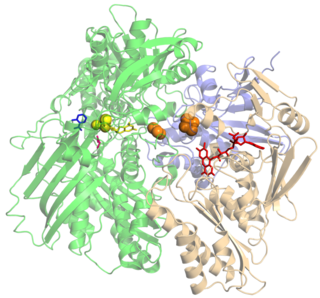
Xanthine oxidase is a form of xanthine oxidoreductase, a type of enzyme that generates reactive oxygen species. These enzymes catalyze the oxidation of hypoxanthine to xanthine and can further catalyze the oxidation of xanthine to uric acid. These enzymes play an important role in the catabolism of purines in some species, including humans.

The free radical theory of aging states that organisms age because cells accumulate free radical damage over time. A free radical is any atom or molecule that has a single unpaired electron in an outer shell. While a few free radicals such as melanin are not chemically reactive, most biologically relevant free radicals are highly reactive. For most biological structures, free radical damage is closely associated with oxidative damage. Antioxidants are reducing agents, and limit oxidative damage to biological structures by passivating them from free radicals.

<span class="mw-page-title-main">Reactive oxygen species</span> Highly reactive molecules formed from diatomic oxygen (O₂)

In chemistry and biology, reactive oxygen species (ROS) are highly reactive chemicals formed from diatomic oxygen (O2), water, and hydrogen peroxide. Some prominent ROS are hydroperoxide (O2H), superoxide (O2-), hydroxyl radical (OH.), and singlet oxygen. ROS are pervasive because they are readily produced from O2, which is abundant. ROS are important in many ways, both beneficial and otherwise. ROS function as signals, that turn on and off biological functions. They are intermediates in the redox behavior of O2, which is central to fuel cells. ROS are central to the photodegradation of organic pollutants in the atmosphere. Most often however, ROS are discussed in a biological context, ranging from their effects on aging and their role in causing dangerous genetic mutations.

Lipid peroxidation is the conversion of lipids to peroxide and hydroperoxide derivatives. These derivatives, known as lipid peroxides or lipid oxidation products (LOPs), are susceptible to further reactions that are relevant to "DNA and protein modification, radiation damage, aging..." Lipid peroxidation mainly applies to unsaturated fats, especially polyunsaturated fats such as those derived from linoleic acid.

Respiratory burst is the rapid release of the reactive oxygen species (ROS), superoxide anion and hydrogen peroxide, from different cell types.

NADPH oxidase is a membrane-bound enzyme complex that faces the extracellular space. It can be found in the plasma membrane as well as in the membranes of phagosomes used by neutrophil white blood cells to engulf microorganisms. Human isoforms of the catalytic component of the complex include NOX1, NOX2, NOX3, NOX4, NOX5, DUOX1, and DUOX2.

<span class="mw-page-title-main">Myricetin</span> Chemical compound

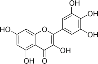
Myricetin is a member of the flavonoid class of polyphenolic compounds, with antioxidant properties. Common dietary sources include vegetables, fruits, nuts, berries, tea, and red wine.

Pro-oxidants are chemicals that induce oxidative stress, either by generating reactive oxygen species or by inhibiting antioxidant systems. The oxidative stress produced by these chemicals can damage cells and tissues, for example, an overdose of the analgesic paracetamol (acetaminophen) can fatally damage the liver, partly through its production of reactive oxygen species.

<span class="mw-page-title-main">Reactive nitrogen species</span>

Reactive nitrogen species (RNS) are a family of antimicrobial molecules derived from nitric oxide (•NO) and superoxide (O2•−) produced via the enzymatic activity of inducible nitric oxide synthase 2 (NOS2) and NADPH oxidase respectively. NOS2 is expressed primarily in macrophages after induction by cytokines and microbial products, notably interferon-gamma (IFN-γ) and lipopolysaccharide (LPS).

<span class="mw-page-title-main">Radical (chemistry)</span> Atom, molecule, or ion that has an unpaired valence electron; typically highly reactive

In chemistry, a radical, also known as a free radical, is an atom, molecule, or ion that has at least one unpaired valence electron. With some exceptions, these unpaired electrons make radicals highly chemically reactive. Many radicals spontaneously dimerize. Most organic radicals have short lifetimes.

<span class="mw-page-title-main">Pyocyanin</span> Chemical compound

Pyocyanin (PCN) is one of the many toxic compounds produced and secreted by the Gram negative bacterium Pseudomonas aeruginosa. Pyocyanin is a blue secondary metabolite, turning red below pH 4.9, with the ability to oxidise and reduce other molecules and therefore kill microbes competing against P. aeruginosa as well as mammalian cells of the lungs which P. aeruginosa has infected during cystic fibrosis. Since pyocyanin is a zwitterion at blood pH, it is easily able to cross the cell membrane. There are three different states in which pyocyanin can exist: oxidized (blue), monovalently reduced (colourless) or divalently reduced (red). Mitochondria play an important role in the cycling of pyocyanin between its redox states. Due to its redox-active properties, pyocyanin generates reactive oxygen species.

<span class="mw-page-title-main">Bacterial glutathione transferase</span>

Bacterial glutathione transferases are part of a superfamily of enzymes that play a crucial role in cellular detoxification. The primary role of GSTs is to catalyze the conjugation of glutathione (GSH) with the electrophilic centers of a wide variety of molecules. The most commonly known substrates of GSTs are xenobiotic synthetic chemicals. There are also classes of GSTs that utilize glutathione as a cofactor rather than a substrate. Often these GSTs are involved in reduction of reactive oxidative species toxic to the bacterium. Conjugation with glutathione receptors renders toxic substances more soluble, and therefore more readily exocytosed from the cell.

All living cells produce reactive oxygen species (ROS) as a byproduct of metabolism. ROS are reduced oxygen intermediates that include the superoxide radical (O2) and the hydroxyl radical (OH•), as well as the non-radical species hydrogen peroxide (H2O2). These ROS are important in the normal functioning of cells, playing a role in signal transduction and the expression of transcription factors. However, when present in excess, ROS can cause damage to proteins, lipids and DNA by reacting with these biomolecules to modify or destroy their intended function. As an example, the occurrence of ROS have been linked to the aging process in humans, as well as several other diseases including Alzheimer's, rheumatoid arthritis, Parkinson's, and some cancers. Their potential for damage also makes reactive oxygen species useful in direct protection from invading pathogens, as a defense response to physical injury, and as a mechanism for stopping the spread of bacteria and viruses by inducing programmed cell death.

Oxidation response is stimulated by a disturbance in the balance between the production of reactive oxygen species and antioxidant responses, known as oxidative stress. Active species of oxygen naturally occur in aerobic cells and have both intracellular and extracellular sources. These species, if not controlled, damage all components of the cell, including proteins, lipids and DNA. Hence cells need to maintain a strong defense against the damage. The following table gives an idea of the antioxidant defense system in bacterial system.

<span class="mw-page-title-main">Superoxide dismutase mimetics</span> Synthetic compounds

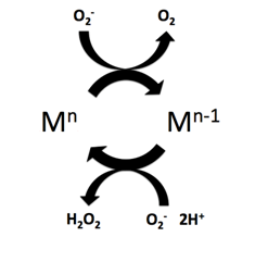
Superoxide dismutase (SOD) mimetics are synthetic compounds that mimic the native superoxide dismutase enzyme. SOD mimetics effectively convert the superoxide anion, a reactive oxygen species, into hydrogen peroxide, which is further converted into water by catalase. Reactive oxygen species are natural byproducts of cellular respiration and cause oxidative stress and cell damage, which has been linked to causing cancers, neurodegeneration, age-related declines in health, and inflammatory diseases. SOD mimetics are a prime interest in therapeutic treatment of oxidative stress because of their smaller size, longer half-life, and similarity in function to the native enzyme.

<span class="mw-page-title-main">9-Hydroxyoctadecadienoic acid</span> Chemical compound

9-Hydroxyoctadecadienoic acid (or 9-HODE) has been used in the literature to designate either or both of two stereoisomer metabolites of the essential fatty acid, linoleic acid: 9(S)-hydroxy-10(E),12(Z)-octadecadienoic acid (9(S)-HODE) and 9(R)-hydroxy-10(E),12(Z)-octadecadienoic acid (9(R)-HODE); these two metabolites differ in having their hydroxy residues in the S or R configurations, respectively. The accompanying figure gives the structure for 9(S)-HETE. Two other 9-hydroxy linoleic acid derivatives occur in nature, the 10E,12E isomers of 9(S)-HODE and 9(R)-HODE viz., 9(S)-hydroxy-10E,12E-octadecadienoic acid (9(S)-EE-HODE) and 9(R)-hydroxy-10E,12E-octadecadienoic acid (13(R)-EE-HODE); these two derivatives have their double bond at carbon 12 in the E or trans configuration as opposed to the Z or cis configuration. The four 9-HODE isomers, particularly under conditions of oxidative stress, may form together in cells and tissues; they have overlapping but not identical biological activities and significances. Because many studies have not distinguished between the S and R stereoisomers and, particularly in identifying tissue levels, the two EE isomers, 9-HODE is used here when the isomer studied is unclear.

Barry Halliwell is an English biochemist, chemist and university administrator, specialising in free radical metabolism in both animals and plants. His name is included in the "Foyer–Halliwell–Asada" pathway, a cellular process of hydrogen peroxide metabolism in plants and animals, named for the three principal discoverers, with Christine Foyer and Kozi Asada. He moved to Singapore in 2000, and served as Deputy President of the National University of Singapore (2006–15), where he continues to hold a Tan Chin Tuan Centennial professorship.

<span class="mw-page-title-main">Mitochondrial theory of ageing</span> Theory of ageing

The mitochondrial theory of ageing has two varieties: free radical and non-free radical. The first is one of the variants of the free radical theory of ageing. It was formulated by J. Miquel and colleagues in 1980 and was developed in the works of Linnane and coworkers (1989). The second was proposed by A. N. Lobachev in 1978.

References

  1. "Oxidative Stress". Handbook of Disease Burdens and Quality of Life Measures. New York, NY: Springer New York. 2010. p. 4278–4278. doi:10.1007/978-0-387-78665-0_6275. ISBN   978-0-387-78664-3. Definition: Imbalance between oxidants and antioxidants in favor of the oxidants.
  2. Birnboim HC (September 1986). "DNA strand breaks in human leukocytes induced by superoxide anion, hydrogen peroxide and tumor promoters are repaired slowly compared to breaks induced by ionizing radiation". Carcinogenesis. 7 (9): 1511–1517. doi:10.1093/carcin/7.9.1511. PMID   3017600.
  3. Joseph N, Zhang-James Y, Perl A, Faraone SV (November 2015). "Oxidative Stress and ADHD: A Meta-Analysis". Journal of Attention Disorders. 19 (11): 915–924. doi:10.1177/1087054713510354. PMC   5293138 . PMID   24232168.
  4. 1 2 Halliwell B (January 2007). "Oxidative stress and cancer: have we moved forward?". The Biochemical Journal. 401 (1): 1–11. doi:10.1042/BJ20061131. PMID   17150040.
  5. Hwang O (March 2013). "Role of oxidative stress in Parkinson's disease". Experimental Neurobiology. 22 (1): 11–17. doi:10.5607/en.2013.22.1.11. PMC   3620453 . PMID   23585717.
  6. Romá-Mateo C, Aguado C, García-Giménez JL, Ibáñez-Cabellos JS, Seco-Cervera M, Pallardó FV, et al. (2015). "Increased oxidative stress and impaired antioxidant response in Lafora disease". Molecular Neurobiology. 51 (3): 932–946. doi:10.1007/s12035-014-8747-0. hdl: 10261/123869 . PMID   24838580. S2CID   13096853.
  7. Valko M, Leibfritz D, Moncol J, Cronin MT, Mazur M, Telser J (2007). "Free radicals and antioxidants in normal physiological functions and human disease". The International Journal of Biochemistry & Cell Biology. 39 (1): 44–84. doi:10.1016/j.biocel.2006.07.001. PMID   16978905.
  8. Bonomini F, Tengattini S, Fabiano A, Bianchi R, Rezzani R (March 2008). "Atherosclerosis and oxidative stress". Histology and Histopathology. 23 (3): 381–390. doi:10.14670/HH-23.381. PMID   18072094.
  9. Singh N, Dhalla AK, Seneviratne C, Singal PK (1995). "Oxidative stress and heart failure". Molecular and Cellular Biochemistry. 147 (1–2): 77–81. doi:10.1007/BF00944786. PMID   7494558. S2CID   21662824.
  10. Ramond A, Godin-Ribuot D, Ribuot C, Totoson P, Koritchneva I, Cachot S, et al. (June 2013). "Oxidative stress mediates cardiac infarction aggravation induced by intermittent hypoxia". Fundamental & Clinical Pharmacology. 27 (3): 252–261. doi:10.1111/j.1472-8206.2011.01015.x. PMID   22145601. S2CID   40420948.
  11. Dean OM, van den Buuse M, Berk M, Copolov DL, Mavros C, Bush AI (July 2011). "N-acetyl cysteine restores brain glutathione loss in combined 2-cyclohexene-1-one and d-amphetamine-treated rats: relevance to schizophrenia and bipolar disorder". Neuroscience Letters. 499 (3): 149–153. doi:10.1016/j.neulet.2011.05.027. PMID   21621586. S2CID   32986064.
  12. de Diego-Otero Y, Romero-Zerbo Y, el Bekay R, Decara J, Sanchez L, Rodriguez-de Fonseca F, del Arco-Herrera I (March 2009). "Alpha-tocopherol protects against oxidative stress in the fragile X knockout mouse: an experimental therapeutic approach for the Fmr1 deficiency". Neuropsychopharmacology. 34 (4): 1011–1026. doi: 10.1038/npp.2008.152 . PMID   18843266.
  13. Amer J, Ghoti H, Rachmilewitz E, Koren A, Levin C, Fibach E (January 2006). "Red blood cells, platelets and polymorphonuclear neutrophils of patients with sickle cell disease exhibit oxidative stress that can be ameliorated by antioxidants". British Journal of Haematology. 132 (1): 108–113. doi: 10.1111/j.1365-2141.2005.05834.x . PMID   16371026.
  14. Aly DG, Shahin RS (2010). "Oxidative stress in lichen planus". Acta Dermatovenerologica Alpina, Pannonica, et Adriatica. 19 (1): 3–11. PMID   20372767.
  15. Arican O, Kurutas EB (March 2008). "Oxidative stress in the blood of patients with active localized vitiligo". Acta Dermatovenerologica Alpina, Pannonica, et Adriatica. 17 (1): 12–16. PMID   18454264.
  16. James SJ, Cutler P, Melnyk S, Jernigan S, Janak L, Gaylor DW, Neubrander JA (December 2004). "Metabolic biomarkers of increased oxidative stress and impaired methylation capacity in children with autism". The American Journal of Clinical Nutrition. 80 (6): 1611–1617. doi: 10.1093/ajcn/80.6.1611 . PMID   15585776.
  17. Kennedy G, Spence VA, McLaren M, Hill A, Underwood C, Belch JJ (September 2005). "Oxidative stress levels are raised in chronic fatigue syndrome and are associated with clinical symptoms". Free Radical Biology & Medicine. 39 (5): 584–589. doi:10.1016/j.freeradbiomed.2005.04.020. PMID   16085177.
  18. Jiménez-Fernández S, Gurpegui M, Díaz-Atienza F, Pérez-Costillas L, Gerstenberg M, Correll CU (December 2015). "Oxidative stress and antioxidant parameters in patients with major depressive disorder compared to healthy controls before and after antidepressant treatment: results from a meta-analysis". The Journal of Clinical Psychiatry. 76 (12): 1658–1667. doi:10.4088/JCP.14r09179. hdl: 10630/29937 . PMID   26579881.
  19. Segal AW (2005). "How neutrophils kill microbes". Annual Review of Immunology. 23: 197–223. doi:10.1146/annurev.immunol.23.021704.115653. PMC   2092448 . PMID   15771570.
  20. Gems D, Partridge L (March 2008). "Stress-response hormesis and aging: "that which does not kill us makes us stronger"". Cell Metabolism. 7 (3): 200–203. doi: 10.1016/j.cmet.2008.01.001 . PMID   18316025.
  21. Waszczak C, Carmody M, Kangasjärvi J (April 2018). "Reactive Oxygen Species in Plant Signaling". Annual Review of Plant Biology. 69 (1): 209–236. doi: 10.1146/annurev-arplant-042817-040322 . PMID   29489394.
  22. Schafer FQ, Buettner GR (June 2001). "Redox environment of the cell as viewed through the redox state of the glutathione disulfide/glutathione couple". Free Radical Biology & Medicine. 30 (11): 1191–1212. doi:10.1016/S0891-5849(01)00480-4. PMID   11368918.
  23. Lennon SV, Martin SJ, Cotter TG (March 1991). "Dose-dependent induction of apoptosis in human tumour cell lines by widely diverging stimuli". Cell Proliferation. 24 (2): 203–214. doi:10.1111/j.1365-2184.1991.tb01150.x. PMID   2009322. S2CID   37720004.
  24. Valko M, Morris H, Cronin MT (2005). "Metals, toxicity and oxidative stress". Current Medicinal Chemistry. 12 (10): 1161–1208. CiteSeerX   10.1.1.498.2796 . doi:10.2174/0929867053764635. PMID   15892631.
  25. Evans MD, Cooke MS (May 2004). "Factors contributing to the outcome of oxidative damage to nucleic acids". BioEssays. 26 (5): 533–542. doi:10.1002/bies.20027. PMID   15112233. S2CID   11714476.
  26. Colis LC, Raychaudhury P, Basu AK (August 2008). "Mutational specificity of gamma-radiation-induced guanine-thymine and thymine-guanine intrastrand cross-links in mammalian cells and translesion synthesis past the guanine-thymine lesion by human DNA polymerase eta". Biochemistry. 47 (31): 8070–8079. doi:10.1021/bi800529f. PMC   2646719 . PMID   18616294.
  27. 1 2 Helbock HJ, Beckman KB, Shigenaga MK, Walter PB, Woodall AA, Yeo HC, Ames BN (January 1998). "DNA oxidation matters: the HPLC-electrochemical detection assay of 8-oxo-deoxyguanosine and 8-oxo-guanine". Proceedings of the National Academy of Sciences of the United States of America. 95 (1): 288–293. Bibcode:1998PNAS...95..288H. doi: 10.1073/pnas.95.1.288 . PMC   18204 . PMID   9419368.
  28. Lelli JL, Becks LL, Dabrowska MI, Hinshaw DB (October 1998). "ATP converts necrosis to apoptosis in oxidant-injured endothelial cells". Free Radical Biology & Medicine. 25 (6): 694–702. doi:10.1016/S0891-5849(98)00107-5. PMID   9801070.
  29. Lee YJ, Shacter E (July 1999). "Oxidative stress inhibits apoptosis in human lymphoma cells". The Journal of Biological Chemistry. 274 (28): 19792–19798. doi: 10.1074/jbc.274.28.19792 . PMID   10391922.
  30. 1 2 Akazawa-Ogawa Y, Shichiri M, Nishio K, Yoshida Y, Niki E, Hagihara Y (February 2015). "Singlet-oxygen-derived products from linoleate activate Nrf2 signaling in skin cells". Free Radical Biology & Medicine. 79: 164–175. doi:10.1016/j.freeradbiomed.2014.12.004. PMID   25499849.
  31. 1 2 3 Riahi Y, Cohen G, Shamni O, Sasson S (December 2010). "Signaling and cytotoxic functions of 4-hydroxyalkenals". American Journal of Physiology. Endocrinology and Metabolism. 299 (6): E879–E886. doi:10.1152/ajpendo.00508.2010. PMID   20858748. S2CID   6062445.
  32. 1 2 Yoshida Y, Umeno A, Akazawa Y, Shichiri M, Murotomi K, Horie M (2015). "Chemistry of lipid peroxidation products and their use as biomarkers in early detection of diseases". Journal of Oleo Science. 64 (4): 347–356. doi: 10.5650/jos.ess14281 . PMID   25766928.
  33. Vigor C, Bertrand-Michel J, Pinot E, Oger C, Vercauteren J, Le Faouder P, et al. (August 2014). "Non-enzymatic lipid oxidation products in biological systems: assessment of the metabolites from polyunsaturated fatty acids" (PDF). Journal of Chromatography. B, Analytical Technologies in the Biomedical and Life Sciences. 964: 65–78. doi:10.1016/j.jchromb.2014.04.042. PMID   24856297.
  34. Waddington EI, Croft KD, Sienuarine K, Latham B, Puddey IB (March 2003). "Fatty acid oxidation products in human atherosclerotic plaque: an analysis of clinical and histopathological correlates". Atherosclerosis. 167 (1): 111–120. doi:10.1016/S0021-9150(02)00391-X. PMID   12618275.
  35. Cho KJ, Seo JM, Kim JH (July 2011). "Bioactive lipoxygenase metabolites stimulation of NADPH oxidases and reactive oxygen species". Molecules and Cells. 32 (1): 1–5. doi:10.1007/s10059-011-1021-7. PMC   3887656 . PMID   21424583.
  36. Galano JM, Mas E, Barden A, Mori TA, Signorini C, De Felice C, et al. (December 2013). "Isoprostanes and neuroprostanes: total synthesis, biological activity and biomarkers of oxidative stress in humans" (PDF). Prostaglandins & Other Lipid Mediators. 107: 95–102. doi:10.1016/j.prostaglandins.2013.04.003. PMID   23644158. S2CID   33638363.
  37. Cohen G, Riahi Y, Sunda V, Deplano S, Chatgilialoglu C, Ferreri C, et al. (December 2013). "Signaling properties of 4-hydroxyalkenals formed by lipid peroxidation in diabetes". Free Radical Biology & Medicine. 65: 978–987. doi:10.1016/j.freeradbiomed.2013.08.163. PMID   23973638.
  38. Speed N, Blair IA (December 2011). "Cyclooxygenase- and lipoxygenase-mediated DNA damage". Cancer and Metastasis Reviews. 30 (3–4): 437–447. doi:10.1007/s10555-011-9298-8. PMC   3237763 . PMID   22009064.
  39. Sies H (1985). "Oxidative stress: introductory remarks". In Sies H (ed.). Oxidative Stress. London: Academic Press. pp. 1–7.
  40. Docampo R (1995). "Antioxidant mechanisms". In Marr J, Müller M (eds.). Biochemistry and Molecular Biology of Parasites. London: Academic Press. pp. 147–160.
  41. 1 2 Rice-Evans CA, Gopinathan V (1995). "Oxygen toxicity, free radicals and antioxidants in human disease: biochemical implications in atherosclerosis and the problems of premature neonates". Essays in Biochemistry. 29: 39–63. PMID   9189713.
  42. Seaver LC, Imlay JA (November 2004). "Are respiratory enzymes the primary sources of intracellular hydrogen peroxide?". The Journal of Biological Chemistry. 279 (47): 48742–48750. doi: 10.1074/jbc.M408754200 . PMID   15361522.
  43. Messner KR, Imlay JA (November 2002). "Mechanism of superoxide and hydrogen peroxide formation by fumarate reductase, succinate dehydrogenase, and aspartate oxidase". The Journal of Biological Chemistry. 277 (45): 42563–42571. doi: 10.1074/jbc.M204958200 . PMID   12200425.
  44. Imlay JA (2003). "Pathways of oxidative damage". Annual Review of Microbiology. 57 (1): 395–418. doi:10.1146/annurev.micro.57.030502.090938. PMID   14527285.
  45. Hardin SC, Larue CT, Oh MH, Jain V, Huber SC (August 2009). "Coupling oxidative signals to protein phosphorylation via methionine oxidation in Arabidopsis". The Biochemical Journal. 422 (2): 305–312. doi:10.1042/BJ20090764. PMC   2782308 . PMID   19527223.
  46. Haider L, Fischer MT, Frischer JM, Bauer J, Höftberger R, Botond G, et al. (July 2011). "Oxidative damage in multiple sclerosis lesions". Brain. 134 (Pt 7): 1914–1924. doi:10.1093/brain/awr128. PMC   3122372 . PMID   21653539.
  47. Patel VP, Chu CT (March 2011). "Nuclear transport, oxidative stress, and neurodegeneration". International Journal of Clinical and Experimental Pathology. 4 (3): 215–229. PMC   3071655 . PMID   21487518.
  48. Hollis F, Kanellopoulos AK, Bagni C (August 2017). "Mitochondrial dysfunction in Autism Spectrum Disorder: clinical features and perspectives". Current Opinion in Neurobiology. 45: 178–187. doi:10.1016/j.conb.2017.05.018. PMID   28628841. S2CID   3617876.
  49. Nunomura A, Castellani RJ, Zhu X, Moreira PI, Perry G, Smith MA (July 2006). "Involvement of oxidative stress in Alzheimer disease". Journal of Neuropathology and Experimental Neurology. 65 (7): 631–641. doi: 10.1097/01.jnen.0000228136.58062.bf . PMID   16825950.
  50. Bošković M, Vovk T, Kores Plesničar B, Grabnar I (June 2011). "Oxidative stress in schizophrenia". Current Neuropharmacology. 9 (2): 301–312. doi:10.2174/157015911795596595. PMC   3131721 . PMID   22131939.
  51. Ramalingam M, Kim SJ (August 2012). "Reactive oxygen/nitrogen species and their functional correlations in neurodegenerative diseases". Journal of Neural Transmission. 119 (8): 891–910. doi:10.1007/s00702-011-0758-7. PMID   22212484. S2CID   2615132.
  52. Nijs J, Meeus M, De Meirleir K (August 2006). "Chronic musculoskeletal pain in chronic fatigue syndrome: recent developments and therapeutic implications". Manual Therapy. 11 (3): 187–191. doi:10.1016/j.math.2006.03.008. PMID   16781183.
  53. Domka K, Goral A, Firczuk M (2020). "cROSsing the Line: Between Beneficial and Harmful Effects of Reactive Oxygen Species in B-Cell Malignancies". Frontiers in Immunology. 11: 1538. doi: 10.3389/fimmu.2020.01538 . PMC   7385186 . PMID   32793211.
  54. Udensi UK, Tchounwou PB (December 2014). "Dual effect of oxidative stress on leukemia cancer induction and treatment". Journal of Experimental & Clinical Cancer Research. 33: 106. doi: 10.1186/s13046-014-0106-5 . PMC   4320640 . PMID   25519934.
  55. Handa O, Naito Y, Yoshikawa T (2011). "Redox biology and gastric carcinogenesis: the role of Helicobacter pylori". Redox Report. 16 (1): 1–7. doi: 10.1179/174329211X12968219310756 . PMC   6837368 . PMID   21605492.
  56. 1 2 3 Welty S, Thathiah A, Levine AS. DNA Damage Increases Secreted Aβ40 and Aβ42 in Neuronal Progenitor Cells: Relevance to Alzheimer's Disease. J Alzheimers Dis. 2022;88(1):177–190. doi: 10.3233/JAD-220030. PMID: 35570488; PMCID: PMC9277680
  57. Shanbhag NM, Evans MD, Mao W, Nana AL, Seeley WW, Adame A, Rissman RA, Masliah E, Mucke L. Early neuronal accumulation of DNA double strand breaks in Alzheimer's disease. Acta Neuropathol Commun. 2019 May 17;7(1):77. doi: 10.1186/s40478-019-0723-5. PMID: 31101070; PMCID: PMC6524256
  58. Thadathil N, Delotterie DF, Xiao J, Hori R, McDonald MP, Khan MM. DNA Double-Strand Break Accumulation in Alzheimer's Disease: Evidence from Experimental Models and Postmortem Human Brains. Mol Neurobiol. 2021 Jan;58(1):118–131. doi: 10.1007/s12035-020-02109-8. Epub 2020 Sep 8. PMID: 32895786
  59. Meyers DG, Maloley PA, Weeks D (May 1996). "Safety of antioxidant vitamins". Archives of Internal Medicine. 156 (9): 925–935. doi:10.1001/archinte.156.9.925. PMID   8624173.
  60. Ruano-Ravina A, Figueiras A, Freire-Garabal M, Barros-Dios JM (2006). "Antioxidant vitamins and risk of lung cancer". Current Pharmaceutical Design. 12 (5): 599–613. doi:10.2174/138161206775474396. PMID   16472151.
  61. Zhang P, Omaye ST (February 2001). "Antioxidant and prooxidant roles for beta-carotene, alpha-tocopherol and ascorbic acid in human lung cells". Toxicology in Vitro. 15 (1): 13–24. doi:10.1016/S0887-2333(00)00054-0. PMID   11259865.
  62. Pryor WA (January 2000). "Vitamin E and heart disease: basic science to clinical intervention trials". Free Radical Biology & Medicine. 28 (1): 141–164. doi:10.1016/S0891-5849(99)00224-5. PMID   10656300.
  63. Saremi A, Arora R (2010). "Vitamin E and cardiovascular disease". American Journal of Therapeutics. 17 (3): e56–e65. doi:10.1097/MJT.0b013e31819cdc9a. PMID   19451807. S2CID   25631305.
  64. Boothby LA, Doering PL (December 2005). "Vitamin C and vitamin E for Alzheimer's disease". The Annals of Pharmacotherapy. 39 (12): 2073–2080. doi:10.1345/aph.1E495. PMID   16227450. S2CID   46645284.
  65. Kontush K, Schekatolina S (December 2004). "Vitamin E in neurodegenerative disorders: Alzheimer's disease". Annals of the New York Academy of Sciences. 1031 (1): 249–262. Bibcode:2004NYASA1031..249K. doi:10.1196/annals.1331.025. PMID   15753151. S2CID   33556198.
  66. Fong JJ, Rhoney DH (March 2006). "NXY-059: review of neuroprotective potential for acute stroke". The Annals of Pharmacotherapy. 40 (3): 461–471. CiteSeerX   10.1.1.1001.6501 . doi:10.1345/aph.1E636. PMID   16507608. S2CID   38016035.
  67. Larsen PL (October 1993). "Aging and resistance to oxidative damage in Caenorhabditis elegans". Proceedings of the National Academy of Sciences of the United States of America. 90 (19): 8905–8909. Bibcode:1993PNAS...90.8905L. doi: 10.1073/pnas.90.19.8905 . PMC   47469 . PMID   8415630.
  68. Helfand SL, Rogina B (2003). "Genetics of aging in the fruit fly, Drosophila melanogaster". Annual Review of Genetics. 37 (1): 329–348. doi:10.1146/annurev.genet.37.040103.095211. PMID   14616064.
  69. Schulz TJ, Zarse K, Voigt A, Urban N, Birringer M, Ristow M (October 2007). "Glucose restriction extends Caenorhabditis elegans life span by inducing mitochondrial respiration and increasing oxidative stress". Cell Metabolism. 6 (4): 280–293. doi: 10.1016/j.cmet.2007.08.011 . PMID   17908557.
  70. Sohal RS, Mockett RJ, Orr WC (September 2002). "Mechanisms of aging: an appraisal of the oxidative stress hypothesis". Free Radical Biology & Medicine. 33 (5): 575–586. doi:10.1016/S0891-5849(02)00886-9. PMID   12208343.
  71. Sohal RS (July 2002). "Role of oxidative stress and protein oxidation in the aging process". Free Radical Biology & Medicine. 33 (1): 37–44. doi:10.1016/S0891-5849(02)00856-0. PMID   12086680.
  72. Rattan SI (December 2006). "Theories of biological aging: genes, proteins, and free radicals". Free Radical Research. 40 (12): 1230–1238. CiteSeerX   10.1.1.476.9259 . doi:10.1080/10715760600911303. PMID   17090411. S2CID   11125090.
  73. Bjelakovic G, Nikolova D, Gluud LL, Simonetti RG, Gluud C (February 2007). "Mortality in randomized trials of antioxidant supplements for primary and secondary prevention: systematic review and meta-analysis". JAMA. 297 (8): 842–857. doi:10.1001/jama.297.8.842. PMID   17327526.. See also the letter Archived 2008-07-24 at the Wayback Machine to JAMA by Philip Taylor and Sanford Dawsey and the reply Archived 2008-06-24 at the Wayback Machine by the authors of the original paper.
  74. "Oxygen Radical Absorbance Capacity (ORAC) of Selected Foods, Release 2 (2010)". USDA.
    • Pratviel G (2012). "Oxidative DNA Damage Mediated by Transition Metal Ions and Their Complexes". In Sigel A, Sigel H, Sigel RK (eds.). Interplay between Metal Ions and Nucleic Acids. Metal Ions in Life Sciences. Vol. 10. Springer. pp. 201–216. doi:10.1007/978-94-007-2172-2_7. ISBN   978-94-007-2171-5. PMID   22210340.
  75. Kodali V, Thrall BD (2015). "Oxidative Stress and Nanomaterial-Cellular Interactions". In Roberts SM, Kehrer JP, Klotz LO (eds.). Studies on Experimental Toxicology and Pharmacology. Oxidative Stress in Applied Basic Research and Clinical Practice. Cham: Springer International Publishing. pp. 347–367. doi:10.1007/978-3-319-19096-9_18. ISBN   978-3-319-19096-9.
  76. Dalle-Donne I, Aldini G, Carini M, Colombo R, Rossi R, Milzani A (2006). "Protein carbonylation, cellular dysfunction, and disease progression". Journal of Cellular and Molecular Medicine. 10 (2): 389–406. doi:10.1111/j.1582-4934.2006.tb00407.x. PMC   3933129 . PMID   16796807.
  77. Grimsrud PA, Xie H, Griffin TJ, Bernlohr DA (August 2008). "Oxidative stress and covalent modification of protein with bioactive aldehydes". The Journal of Biological Chemistry. 283 (32): 21837–21841. doi: 10.1074/jbc.R700019200 . PMC   2494933 . PMID   18445586.
  78. Devasagayam TP, Tilak JC, Boloor KK, Sane KS, Ghaskadbi SS, Lele RD (October 2004). "Free radicals and antioxidants in human health: current status and future prospects". The Journal of the Association of Physicians of India. 52: 794–804. PMID   15909857.
  79. Nathan C, Shiloh MU (August 2000). "Reactive oxygen and nitrogen intermediates in the relationship between mammalian hosts and microbial pathogens". Proceedings of the National Academy of Sciences of the United States of America. 97 (16): 8841–8848. Bibcode:2000PNAS...97.8841N. doi: 10.1073/pnas.97.16.8841 . PMC   34021 . PMID   10922044.
  80. 1 2 Wright C, Milne S, Leeson H (June 2014). "Sperm DNA damage caused by oxidative stress: modifiable clinical, lifestyle and nutritional factors in male infertility". Reproductive Biomedicine Online. 28 (6): 684–703. doi: 10.1016/j.rbmo.2014.02.004 . PMID   24745838.
  81. Guz J, Gackowski D, Foksinski M, Rozalski R, Zarakowska E, Siomek A, et al. (2013). "Comparison of oxidative stress/DNA damage in semen and blood of fertile and infertile men". PLOS ONE. 8 (7): e68490. Bibcode:2013PLoSO...868490G. doi: 10.1371/journal.pone.0068490 . PMC   3709910 . PMID   23874641.
  82. Sinha JK, Ghosh S, Swain U, Giridharan NV, Raghunath M (June 2014). "Increased macromolecular damage due to oxidative stress in the neocortex and hippocampus of WNIN/Ob, a novel rat model of premature aging". Neuroscience. 269: 256–264. doi:10.1016/j.neuroscience.2014.03.040. PMID   24709042. S2CID   9934178.
  83. Bernstein H, Payne CM, Bernstein C, Garewal H, Dvorak K (2008). "Cancer and Aging as Consequences of Un-Repaired DNA Damage". In Kimura H, Suzuki A (eds.). New Research on DNA Damages. New York: Nova Science Publishers, Inc. pp. 1–47. ISBN   978-1-60456-581-2. Archived from the original on 2014-10-25.
  84. Pinto S, Sato VN, De-Souza EA, Ferraz RC, Camara H, Pinca AP, et al. (September 2018). "Enoxacin extends lifespan of C. elegans by inhibiting miR-34-5p and promoting mitohormesis". Redox Biology. 18: 84–92. doi:10.1016/j.redox.2018.06.006. PMC   6037660 . PMID   29986212.
  85. 1 2 3 Gross J, Bhattacharya D (August 2010). "Uniting sex and eukaryote origins in an emerging oxygenic world". Biology Direct. 5: 53. doi: 10.1186/1745-6150-5-53 . PMC   2933680 . PMID   20731852.
  86. Bernstein H, Bernstein C. Sexual communication in archaea, the precursor to meiosis. pp. 103–117 in Biocommunication of Archaea (Guenther Witzany, ed.) 2017. Springer International Publishing ISBN   978-3-319-65535-2 DOI 10.1007/978-3-319-65536-9
  87. Hörandl E, Speijer D (February 2018). "How oxygen gave rise to eukaryotic sex". Proceedings. Biological Sciences. 285 (1872): 20172706. doi:10.1098/rspb.2017.2706. PMC   5829205 . PMID   29436502.
  88. Majumder N, Deepak V, Hadique S, Aesoph D, Velayutham M, Ye Q, et al. (October 2022). "Redox imbalance in COVID-19 pathophysiology". Redox Biology. 56: 102465. doi:10.1016/j.redox.2022.102465. PMC   9464257 . PMID   36116160.
  89. Loffredo L, Violi F (August 2020). "COVID-19 and cardiovascular injury: A role for oxidative stress and antioxidant treatment?". International Journal of Cardiology. 312: 136. doi: 10.1016/j.ijcard.2020.04.066 . PMC   7833193 . PMID   32505331.