Glutathione

Last updated

Contents

Glutathione [1]
Glutathione-skeletal.svg
Glutathione-from-xtal-3D-balls.png
Glutathione-3D-vdW.png
Names
IUPAC name
γ-Glutamylcysteinylglycine
Systematic IUPAC name
(2S)-2-Amino-5-({(2R)-1-[(carboxymethyl)amino]-1-oxo-3-sulfanylpropan-2-yl}amino)-5-oxopentanoic acid
Other names
γ-L-Glutamyl-L-cysteinylglycine
(2S)-2-Amino-4-({(1R)-1-[(carboxymethyl)carbamoyl]-2-sulfanylethyl}carbamoyl)butanoic acid
Identifiers
3D model (JSmol)
AbbreviationsGSH
ChEBI
ChEMBL
ChemSpider
DrugBank
ECHA InfoCard 100.000.660 OOjs UI icon edit-ltr-progressive.svg
KEGG
MeSH Glutathione
PubChem CID
UNII
  • InChI=1S/C10H17N3O6S/c11-5(10(18)19)1-2-7(14)13-6(4-20)9(17)12-3-8(15)16/h5-6,20H,1-4,11H2,(H,12,17)(H,13,14)(H,15,16)(H,18,19)/t5-,6-/m0/s1 Yes check.svgY
    Key: RWSXRVCMGQZWBV-WDSKDSINSA-N Yes check.svgY
  • C(CC(=O)N[C@@H](CS)C(=O)NCC(=O)O)[C@@H](C(=O)O)N
Properties
C10H17N3O6S
Molar mass 307.32 g·mol−1
Melting point 195 °C (383 °F; 468 K) [1]
Freely soluble [1]
Solubility in methanol, diethyl ether Insoluble [1]
Pharmacology
V03AB32 ( WHO )
Except where otherwise noted, data are given for materials in their standard state (at 25 °C [77 °F], 100 kPa).
Yes check.svgY  verify  (what is  Yes check.svgYX mark.svgN ?)

Glutathione (GSH, /ˌɡltəˈθn/ ) is an organic compound with the chemical formula HOCOCH(NH2)CH2CH2CONHCH(CH2SH)CONHCH2COOH. It is an antioxidant in plants, animals, fungi, and some bacteria and archaea. Glutathione is capable of preventing damage to important cellular components caused by sources such as reactive oxygen species, free radicals, peroxides, lipid peroxides, and heavy metals. [2] It is a tripeptide with a gamma peptide linkage between the carboxyl group of the glutamate side chain and cysteine. The carboxyl group of the cysteine residue is attached by normal peptide linkage to glycine.

Biosynthesis and occurrence

Glutathione biosynthesis involves two adenosine triphosphate-dependent steps:

While all animal cells are capable of synthesizing glutathione, glutathione synthesis in the liver has been shown to be essential. GCLC knockout mice die within a month of birth due to the absence of hepatic GSH synthesis. [4] [5]

The unusual gamma amide linkage in glutathione protects it from hydrolysis by peptidases. [6]

Occurrence

Glutathione is the most abundant non-protein thiol (R−SH-containing compound) in animal cells, ranging from 0.5 to 10 mmol/L. It is present in the cytosol and the organelles. [6] In healthy cells and tissue, more than 90% of the total glutathione pool is in the reduced form (GSH), with the remainder in the disulfide form (GSSG). [7] 80-85% of cellular GSH is in the cytosol and 10-15% is in the mitochondria. [8]

Human beings synthesize glutathione, but a few eukaryotes do not, including some members of Fabaceae, Entamoeba , and Giardia . The only known archaea that make glutathione are halobacteria. Some bacteria, such as "Cyanobacteria" and Pseudomonadota, can biosynthesize glutathione. [9] [10]

Systemic availability of orally consumed glutathione has poor bioavailability because the tripeptide is the substrate of proteases (peptidases) of the alimentary canal, and due to the absence of a specific carrier of glutathione at the level of cell membrane. [11] [12] The administration of N-acetylcysteine (NAC), a cysteine prodrug, helps replenish intracellular GSH levels. [13] The patented compound RiboCeine has been studied as a supplement that enhances production of glutathione which helps mitigate hyperglycemia. [14] [15]

Biochemical function

Glutathione exists in reduced (GSH) and oxidized (GSSG) states. [16] The ratio of reduced glutathione to oxidized glutathione within cells is a measure of cellular oxidative stress [17] [8] where increased GSSG-to-GSH ratio is indicative of greater oxidative stress.

In the reduced state, the thiol group of cysteinyl residue is a source of one reducing equivalent. Glutathione disulfide (GSSG) is thereby generated. The oxidized state is converted to the reduced state by NADPH. [18] This conversion is catalyzed by glutathione reductase:

NADPH + GSSG + H2O → 2 GSH + NADP+ + OH

Roles

Antioxidant

GSH protects cells by neutralising (reducing) reactive oxygen species. [19] [6] This conversion is illustrated by the reduction of peroxides:

2 GSH + R2O2 → GSSG + 2 ROH  (R = H, alkyl)

and with free radicals:

GSH + R1/2 GSSG + RH

Regulation

Aside from deactivating radicals and reactive oxidants, glutathione participates in thiol protection and redox regulation of cellular thiol proteins under oxidative stress by protein S-glutathionylation, a redox-regulated post-translational thiol modification. The general reaction involves formation of an unsymmetrical disulfide from the protectable protein (RSH) and GSH: [20]

RSH + GSH + [O] → GSSR + H2O

Glutathione is also employed for the detoxification of methylglyoxal and formaldehyde, toxic metabolites produced under oxidative stress. This detoxification reaction is carried out by the glyoxalase system. Glyoxalase I (EC 4.4.1.5) catalyzes the conversion of methylglyoxal and reduced glutathione to S-D-lactoylglutathione. Glyoxalase II (EC 3.1.2.6) catalyzes the hydrolysis of S-D-lactoylglutathione to glutathione and D-lactic acid.

It maintains exogenous antioxidants such as vitamins C and E in their reduced (active) states. [21] [22] [23]

Metabolism

Among the many metabolic processes in which it participates, glutathione is required for the biosynthesis of leukotrienes and prostaglandins. It plays a role in the storage of cysteine. Glutathione enhances the function of citrulline as part of the nitric oxide cycle. [24] It is a cofactor and acts on glutathione peroxidase. [25] Glutathione is used to produce S-sulfanylglutathione, which is part of hydrogen sulfide metabolism. [26]

Conjugation

Glutathione facilitates metabolism of xenobiotics. Glutathione S-transferase enzymes catalyze its conjugation to lipophilic xenobiotics, facilitating their excretion or further metabolism. [27] The conjugation process is illustrated by the metabolism of N-acetyl-p-benzoquinone imine (NAPQI). NAPQI is a reactive metabolite formed by the action of cytochrome P450 on paracetamol (acetaminophen). Glutathione conjugates to NAPQI, and the resulting ensemble is excreted.

In plants

In plants, glutathione is involved in stress management. It is a component of the glutathione-ascorbate cycle, a system that reduces poisonous hydrogen peroxide. [28] It is the precursor of phytochelatins, glutathione oligomers that chelate heavy metals such as cadmium. [29] Glutathione is required for efficient defence against plant pathogens such as Pseudomonas syringae and Phytophthora brassicae. [30] Adenylyl-sulfate reductase, an enzyme of the sulfur assimilation pathway, uses glutathione as an electron donor. Other enzymes using glutathione as a substrate are glutaredoxins. These small oxidoreductases are involved in flower development, salicylic acid, and plant defence signalling. [31]

Uses

Winemaking

The content of glutathione in must, the first raw form of wine, determines the browning, or caramelizing effect, during the production of white wine by trapping the caffeoyltartaric acid quinones generated by enzymic oxidation as grape reaction product. [32] Its concentration in wine can be determined by UPLC-MRM mass spectrometry. [33]

See also

Related Research Articles

<span class="mw-page-title-main">Protein disulfide-isomerase</span> Class of enzymes

Protein disulfide isomerase, or PDI, is an enzyme in the endoplasmic reticulum (ER) in eukaryotes and the periplasm of bacteria that catalyzes the formation and breakage of disulfide bonds between cysteine residues within proteins as they fold. This allows proteins to quickly find the correct arrangement of disulfide bonds in their fully folded state, and therefore the enzyme acts to catalyze protein folding.

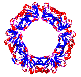
Thioredoxin reductases are enzymes that reduce thioredoxin (Trx). Two classes of thioredoxin reductase have been identified: one class in bacteria and some eukaryotes and one in animals. In bacteria TrxR also catalyzes the reduction of glutaredoxin like proteins known as NrdH. Both classes are flavoproteins which function as homodimers. Each monomer contains a FAD prosthetic group, a NADPH binding domain, and an active site containing a redox-active disulfide bond.

Drug metabolism is the metabolic breakdown of drugs by living organisms, usually through specialized enzymatic systems. More generally, xenobiotic metabolism is the set of metabolic pathways that modify the chemical structure of xenobiotics, which are compounds foreign to an organism's normal biochemistry, such as any drug or poison. These pathways are a form of biotransformation present in all major groups of organisms and are considered to be of ancient origin. These reactions often act to detoxify poisonous compounds. The study of drug metabolism is called pharmacokinetics.

<span class="mw-page-title-main">Glutathione disulfide</span> Chemical compound

Glutathione disulfide (GSSG) is a disulfide derived from two glutathione molecules.

<span class="mw-page-title-main">Glutathione reductase</span> Enzyme

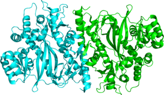
Glutathione reductase (GR) also known as glutathione-disulfide reductase (GSR) is an enzyme that in humans is encoded by the GSR gene. Glutathione reductase catalyzes the reduction of glutathione disulfide (GSSG) to the sulfhydryl form glutathione (GSH), which is a critical molecule in resisting oxidative stress and maintaining the reducing environment of the cell. Glutathione reductase functions as dimeric disulfide oxidoreductase and utilizes an FAD prosthetic group and NADPH to reduce one molar equivalent of GSSG to two molar equivalents of GSH:

<span class="mw-page-title-main">Sulfur assimilation</span> Incorporation of sulfur into living organisms

Sulfur assimilation is the process by which living organisms incorporate sulfur into their biological molecules. In plants, sulfate is absorbed by the roots and then be transported to the chloroplasts by the transipration stream where the sulfur are reduced to sulfide with the help of a series of enzymatic reactions. Furthermore, the reduced sulfur is incorporated into cysteine, an amino acid that is a precursor to many other sulfur-containing compounds. In animals, sulfur assimilation occurs primarily through the diet, as animals cannot produce sulfur-containing compounds directly. Sulfur is incorporated into amino acids such as cysteine and methionine, which are used to build proteins and other important molecules.

<span class="mw-page-title-main">Glutathione synthetase</span> Enzyme

Glutathione synthetase (GSS) is the second enzyme in the glutathione (GSH) biosynthesis pathway. It catalyses the condensation of gamma-glutamylcysteine and glycine, to form glutathione. Glutathione synthetase is also a potent antioxidant. It is found in many species including bacteria, yeast, mammals, and plants.

<span class="mw-page-title-main">Glutaredoxin</span>

Glutaredoxins are small redox enzymes of approximately one hundred amino-acid residues that use glutathione as a cofactor. In humans this oxidation repair enzyme is also known to participate in many cellular functions, including redox signaling and regulation of glucose metabolism. Glutaredoxins are oxidized by substrates, and reduced non-enzymatically by glutathione. In contrast to thioredoxins, which are reduced by thioredoxin reductase, no oxidoreductase exists that specifically reduces glutaredoxins. Instead, glutaredoxins are reduced by the oxidation of glutathione. Reduced glutathione is then regenerated by glutathione reductase. Together these components compose the glutathione system.

<span class="mw-page-title-main">Peroxiredoxin</span> Family of antioxidant enzymes

Peroxiredoxins are a ubiquitous family of antioxidant enzymes that also control cytokine-induced peroxide levels and thereby mediate signal transduction in mammalian cells. The family members in humans are PRDX1, PRDX2, PRDX3, PRDX4, PRDX5, and PRDX6. The physiological importance of peroxiredoxins is indicated by their relative abundance. Their function is the reduction of peroxides, specifically hydrogen peroxide, alkyl hydroperoxides, and peroxynitrite.

<span class="mw-page-title-main">NFE2L2</span> Human protein and coding gene

Nuclear factor erythroid 2-related factor 2 (NRF2), also known as nuclear factor erythroid-derived 2-like 2, is a transcription factor that in humans is encoded by the NFE2L2 gene. NRF2 is a basic leucine zipper (bZIP) protein that may regulate the expression of antioxidant proteins that protect against oxidative damage triggered by injury and inflammation, according to preliminary research. In vitro, NRF2 binds to antioxidant response elements (AREs) in the promoter regions of genes encoding cytoprotective proteins. NRF2 induces the expression of heme oxygenase 1 in vitro leading to an increase in phase II enzymes. NRF2 also inhibits the NLRP3 inflammasome.

Glutamate–cysteine ligase (GCL) EC 6.3.2.2), previously known as γ-glutamylcysteine synthetase (GCS), is the first enzyme of the cellular glutathione (GSH) biosynthetic pathway that catalyzes the chemical reaction:

The glyoxalase system is a set of enzymes that carry out the detoxification of methylglyoxal and the other reactive aldehydes that are produced as a normal part of metabolism. This system has been studied in both bacteria and eukaryotes. This detoxification is accomplished by the sequential action of two thiol-dependent enzymes; firstly glyoxalase І, which catalyzes the isomerization of the spontaneously formed hemithioacetal adduct between glutathione and 2-oxoaldehydes into S-2-hydroxyacylglutathione. Secondly, glyoxalase ІІ hydrolyses these thiolesters and in the case of methylglyoxal catabolism, produces D-lactate and GSH from S-D-lactoyl-glutathione.

<span class="mw-page-title-main">GLRX2</span> Protein-coding gene in the species Homo sapiens

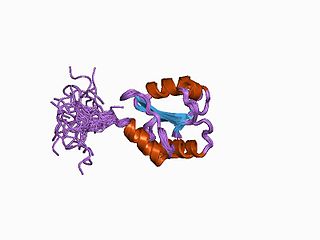
Glutaredoxin 2 (GLRX2) is an enzyme that in humans encoded by the GLRX2 gene. GLRX2, also known as GRX2, is a glutaredoxin family protein and a thiol-disulfide oxidoreductase that maintains cellular thiol homeostasis. This gene consists of four exons and three introns, spanned 10 kilobase pairs, and localized to chromosome 1q31.2–31.3.

γ-<small>L</small>-Glutamyl-<small>L</small>-cysteine Chemical compound

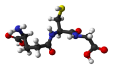
γ-L-Glutamyl-L-cysteine, also known as γ-glutamylcysteine (GGC), is a dipeptide found in animals, plants, fungi, some bacteria, and archaea. It has a relatively unusual γ-bond between the constituent amino acids, L-glutamic acid and L-cysteine and is a key intermediate in the γ-glutamyl cycle first described by Meister in the 1970s. It is the most immediate precursor to the antioxidant glutathione.

<span class="mw-page-title-main">RoGFP</span> Prodified GFP protein that exhibits different fluorescent properties when oxidized and reduced

The reduction-oxidation sensitive green fluorescent protein (roGFP) is a green fluorescent protein engineered to be sensitive to changes in the local redox environment. roGFPs are used as redox-sensitive biosensors.

The ascorbate-glutathione cycle, sometimes Foyer-Halliwell-Asada pathway, is a metabolic pathway that detoxifies hydrogen peroxide (H2O2), a reactive oxygen species that is produced as a waste product in metabolism. The cycle involves the antioxidant metabolites: ascorbate, glutathione and NADPH and the enzymes linking these metabolites.

<span class="mw-page-title-main">Bacterial glutathione transferase</span>

Bacterial glutathione transferases are part of a superfamily of enzymes that play a crucial role in cellular detoxification. The primary role of GSTs is to catalyze the conjugation of glutathione (GSH) with the electrophilic centers of a wide variety of molecules. The most commonly known substrates of GSTs are xenobiotic synthetic chemicals. There are also classes of GSTs that utilize glutathione as a cofactor rather than a substrate. Often these GSTs are involved in reduction of reactive oxidative species toxic to the bacterium. Conjugation with glutathione receptors renders toxic substances more soluble, and therefore more readily exocytosed from the cell.

Oxidation response is stimulated by a disturbance in the balance between the production of reactive oxygen species and antioxidant responses, known as oxidative stress. Active species of oxygen naturally occur in aerobic cells and have both intracellular and extracellular sources. These species, if not controlled, damage all components of the cell, including proteins, lipids and DNA. Hence cells need to maintain a strong defense against the damage. The following table gives an idea of the antioxidant defense system in bacterial system.

Thiol oxidoreductases are proteins that redox control by utilizing catalytic cysteine (Cys) residues for oxidation or reduction of their substrates. Examples of such proteins include thioredoxin, thioredoxin reductase, glutathione reductase, glutaredoxin, glutathione peroxidase, and peroxiredoxin.

<span class="mw-page-title-main">Danyelle Townsend</span> Biomedical scientist and academic

Danyelle M. Townsend is a biomedical scientist, and academic. She is a Professor and acting Department Chair of Drug Discovery and Biomedical Sciences at the Medical University of South Carolina (MUSC).

References

  1. 1 2 3 4 Haynes, William M., ed. (2016). CRC Handbook of Chemistry and Physics (97th ed.). CRC Press. p. 3.284. ISBN   9781498754293.
  2. Pompella A, Visvikis A, Paolicchi A, De Tata V, Casini AF (October 2003). "The changing faces of glutathione, a cellular protagonist". Biochemical Pharmacology. 66 (8): 1499–1503. doi:10.1016/S0006-2952(03)00504-5. PMID   14555227.
  3. White CC, Viernes H, Krejsa CM, Botta D, Kavanagh TJ (July 2003). "Fluorescence-based microtiter plate assay for glutamate-cysteine ligase activity". Analytical Biochemistry. 318 (2): 175–180. doi:10.1016/S0003-2697(03)00143-X. PMID   12814619.
  4. Chen Y, Yang Y, Miller ML, Shen D, Shertzer HG, Stringer KF, Wang B, Schneider SN, Nebert DW, Dalton TP (May 2007). "Hepatocyte-specific Gclc deletion leads to rapid onset of steatosis with mitochondrial injury and liver failure". Hepatology. 45 (5): 1118–1128. doi: 10.1002/hep.21635 . PMID   17464988. S2CID   25000753.
  5. Sies H (1999). "Glutathione and its role in cellular functions". Free Radical Biology & Medicine. 27 (9–10): 916–921. doi:10.1016/S0891-5849(99)00177-X. PMID   10569624.
  6. 1 2 3 Guoyao Wu; Yun-Zhong Fang; Sheng Yang; Joanne R. Lupton; Nancy D. Turner (2004). "Glutathione Metabolism and its Implications for Health". Journal of Nutrition. 134 (3): 489–492. doi: 10.1093/jn/134.3.489 . PMID   14988435.
  7. Halprin KM, Ohkawara A (1967). "The measurement of glutathione in human epidermis using glutathione reductase". The Journal of Investigative Dermatology. 48 (2): 149–152. doi: 10.1038/jid.1967.24 . PMID   6020678.
  8. 1 2 Lu SC (May 2013). "Glutathione synthesis". Biochimica et Biophysica Acta . 1830 (5): 3143–3153. doi:10.1016/j.bbagen.2012.09.008. PMC   3549305 . PMID   22995213.
  9. Copley SD, Dhillon JK (29 April 2002). "Lateral gene transfer and parallel evolution in the history of glutathione biosynthesis genes". Genome Biology. 3 (5): research0025. doi: 10.1186/gb-2002-3-5-research0025 . PMC   115227 . PMID   12049666.
  10. Wonisch W, Schaur RJ (2001). "Chapter 2: Chemistry of Glutathione". In Grill D, Tausz T, De Kok L (eds.). Significance of glutathione in plant adaptation to the environment. Springer. ISBN   978-1-4020-0178-9 via Google Books.
  11. Witschi A, Reddy S, Stofer B, Lauterburg BH (1992). "The systemic availability of oral glutathione". European Journal of Clinical Pharmacology. 43 (6): 667–669. doi:10.1007/bf02284971. PMID   1362956. S2CID   27606314.
  12. "Acetylcysteine Monograph for Professionals". Drugs.com.
  13. Atkuri, K. R.; Mantovani, J. J.; Herzenberg, L. A.; Herzenberg, L. A. (2007). "N-acetylcysteine - a safe antidote for cysteine/glutathione deficiency". Current Opinion in Pharmacology. 7 (4): 355–359. doi:10.1016/j.coph.2007.04.005. PMC   4540061 . PMID   17602868.
  14. Nagasawa, Herbert T. (27 November 2014). "COMPOSITIONS COMPRISING SUGAR-CYSTEINE PRODUCTS - US-20140348811-A1". ppubs.uspto.gov. United States Patent Office. p. 16. Retrieved 31 October 2023. 30. A method of increasing ATP and/or glutathione...
  15. Ukwenya VO, Alese MO, Ogunlade B, Folorunso IM, Omotuyi OI (2022). "Anacardium occidentale leaves extract and riboceine mitigate hyperglycemia through anti-oxidative effects and modulation of some selected genes associated with diabetes". J Diabetes Metab Disord. 22 (1): 455–468. doi:10.1007/s40200-022-01165-2. PMC   10225389 . PMID   37255827.
  16. Iskusnykh IY, Zakharova AA, Pathak D (January 2022). "Glutathione in Brain Disorders and Aging". Molecules. 27 (1): 324. doi: 10.3390/molecules27010324 . PMC   8746815 . PMID   35011559.
  17. Pastore A, Piemonte F, Locatelli M, Lo Russo A, Gaeta LM, Tozzi G, Federici G (August 2001). "Determination of blood total, reduced, and oxidized glutathione in pediatric subjects". Clinical Chemistry. 47 (8): 1467–1469. doi: 10.1093/clinchem/47.8.1467 . PMID   11468240.
  18. Couto N, Malys N, Gaskell SJ, Barber J (June 2013). "Partition and turnover of glutathione reductase from Saccharomyces cerevisiae: a proteomic approach" (PDF). Journal of Proteome Research. 12 (6): 2885–2894. doi:10.1021/pr4001948. PMID   23631642.
  19. Michael Brownlee (2005). "The pathobiology of diabetic complications: A unifying mechanism". Diabetes. 54 (6): 1615–1625. doi: 10.2337/diabetes.54.6.1615 . PMID   15919781.
  20. Dalle-Donne, Isabella; Rossi, Ranieri; Colombo, Graziano; Giustarini, Daniela; Milzani, Aldo (2009). "Protein S-glutathionylation: a regulatory device from bacteria to humans". Trends in Biochemical Sciences. 34 (2): 85–96. doi:10.1016/j.tibs.2008.11.002. PMID   19135374.
  21. Dringen R (December 2000). "Metabolism and functions of glutathione in brain". Progress in Neurobiology. 62 (6): 649–671. doi:10.1016/s0301-0082(99)00060-x. PMID   10880854. S2CID   452394.
  22. Scholz RW, Graham KS, Gumpricht E, Reddy CC (1989). "Mechanism of interaction of vitamin E and glutathione in the protection against membrane lipid peroxidation". Annals of the New York Academy of Sciences. 570 (1): 514–517. Bibcode:1989NYASA.570..514S. doi:10.1111/j.1749-6632.1989.tb14973.x. S2CID   85414084.
  23. Hughes RE (1964). "Reduction of dehydroascorbic acid by animal tissues". Nature. 203 (4949): 1068–1069. Bibcode:1964Natur.203.1068H. doi:10.1038/2031068a0. PMID   14223080. S2CID   4273230.
  24. Ha SB, Smith AP, Howden R, Dietrich WM, Bugg S, O'Connell MJ, Goldsbrough PB, Cobbett CS (June 1999). "Phytochelatin synthase genes from Arabidopsis and the yeast Schizosaccharomyces pombe". The Plant Cell. 11 (6): 1153–1164. doi:10.1105/tpc.11.6.1153. JSTOR   3870806. PMC   144235 . PMID   10368185.
  25. Grant CM (2001). "Role of the glutathione/glutaredoxin and thioredoxin systems in yeast growth and response to stress conditions". Molecular Microbiology. 39 (3): 533–541. doi: 10.1046/j.1365-2958.2001.02283.x . PMID   11169096. S2CID   6467802.
  26. Melideo, SL; Jackson, MR; Jorns, MS (22 July 2014). "Biosynthesis of a central intermediate in hydrogen sulfide metabolism by a novel human sulfurtransferase and its yeast ortholog". Biochemistry. 53 (28): 4739–53. doi:10.1021/bi500650h. PMC   4108183 . PMID   24981631.
  27. Hayes, John D.; Flanagan, Jack U.; Jowsey, Ian R. (2005). "Glutathione transferases". Annual Review of Pharmacology and Toxicology. 45: 51–88. doi:10.1146/annurev.pharmtox.45.120403.095857. PMID   15822171.
  28. Noctor G, Foyer CH (June 1998). "Ascorbate and Glutathione: Keeping Active Oxygen Under Control". Annual Review of Plant Physiology and Plant Molecular Biology. 49 (1): 249–279. doi:10.1146/annurev.arplant.49.1.249. PMID   15012235.
  29. Ha SB, Smith AP, Howden R, Dietrich WM, Bugg S, O'Connell MJ, Goldsbrough PB, Cobbett CS (June 1999). "Phytochelatin synthase genes from Arabidopsis and the yeast Schizosaccharomyces pombe". The Plant Cell. 11 (6): 1153–1164. doi:10.1105/tpc.11.6.1153. PMC   144235 . PMID   10368185.
  30. Parisy V, Poinssot B, Owsianowski L, Buchala A, Glazebrook J, Mauch F (January 2007). "Identification of PAD2 as a gamma-glutamylcysteine synthetase highlights the importance of glutathione in disease resistance of Arabidopsis" (PDF). The Plant Journal. 49 (1): 159–172. doi: 10.1111/j.1365-313X.2006.02938.x . PMID   17144898.
  31. Rouhier N, Lemaire SD, Jacquot JP (2008). "The role of glutathione in photosynthetic organisms: emerging functions for glutaredoxins and glutathionylation" (PDF). Annual Review of Plant Biology. 59 (1): 143–166. doi:10.1146/annurev.arplant.59.032607.092811. PMID   18444899.
  32. Rigaud J, Cheynier V, Souquet JM, Moutounet M (1991). "Influence of must composition on phenolic oxidation kinetics". Journal of the Science of Food and Agriculture. 57 (1): 55–63. Bibcode:1991JSFA...57...55R. doi:10.1002/jsfa.2740570107.
  33. Vallverdú-Queralt A, Verbaere A, Meudec E, Cheynier V, Sommerer N (January 2015). "Straightforward method to quantify GSH, GSSG, GRP, and hydroxycinnamic acids in wines by UPLC-MRM-MS". Journal of Agricultural and Food Chemistry. 63 (1): 142–149. doi:10.1021/jf504383g. PMID   25457918.