Quinolinic acid

Last updated
Quinolinic acid
2,3-Pyridine Dicarboxylic Acid V.2.svg
Names
Preferred IUPAC name
Pyridine-2,3-dicarboxylic acid
Other names
2,3-Pyridinedicarboxylic acid
Identifiers
3D model (JSmol)
ChEBI
ChEMBL
ChemSpider
ECHA InfoCard 100.001.704 OOjs UI icon edit-ltr-progressive.svg
EC Number
  • 201-874-8
KEGG
MeSH D017378
PubChem CID
UNII
  • InChI=1S/C7H5NO4/c9-6(10)4-2-1-3-8-5(4)7(11)12/h1-3H,(H,9,10)(H,11,12) X mark.svgN
    Key: GJAWHXHKYYXBSV-UHFFFAOYSA-N X mark.svgN
  • InChI=1/C7H5NO4/c9-6(10)4-2-1-3-8-5(4)7(11)12/h1-3H,(H,9,10)(H,11,12)
    Key: GJAWHXHKYYXBSV-UHFFFAOYAW
  • C1=CC(=C(N=C1)C(=O)O)C(=O)O
Properties
C7H5NO4
Molar mass 167.12 g/mol
Melting point 185 to 190 °C (365 to 374 °F; 458 to 463 K) (decomposes)
Hazards
Safety data sheet (SDS) External MSDS
Except where otherwise noted, data are given for materials in their standard state (at 25 °C [77 °F], 100 kPa).
X mark.svgN  verify  (what is  Yes check.svgYX mark.svgN ?)

Quinolinic acid (abbreviated QUIN or QA), also known as pyridine-2,3-dicarboxylic acid, is a dicarboxylic acid with a pyridine backbone. It is a colorless solid. It is the biosynthetic precursor to niacin. [1]

Contents

Quinolinic acid is a downstream product of the kynurenine pathway, which metabolizes the amino acid tryptophan. It acts as an NMDA receptor agonist. [2]

Quinolinic acid has a potent neurotoxic effect. Studies have demonstrated that quinolinic acid may be involved in many psychiatric disorders, neurodegenerative processes in the brain, as well as other disorders. Within the brain, quinolinic acid is only produced by activated microglia and macrophages. [3]

History

In 1949 L. Henderson was one of the earliest to describe quinolinic acid. Lapin followed up this research by demonstrating that quinolinic acid could induce convulsions when injected into mice brain ventricles. However, it was not until 1981 that Stone and Perkins showed that quinolinic acid activates the N-methyl-D-aspartate receptor (NMDAR). After this, Schwarcz demonstrated that elevated quinolinic acid levels could lead to axonal neurodegeneration. [4]

Synthesis

One of the earliest reported syntheses of this quinolinic acid was by Zdenko Hans Skraup, who found that methyl-substituted quinolines could be oxidized to quinolinic acid by potassium permanganate. [5]

This compound is commercially available. It is generally obtained by the oxidation of quinoline. Oxidants such as ozone, [6] hydrogen peroxide, [7] and potassium permanganate have been used. Electrolysis is able to perform the transformation as well. [8] [9]

Quinolinic acid may undergo further decarboxylation to nicotinic acid (niacin):

Synthesis Niacin I.svg

Biosynthesis

From aspartate

Oxidation of aspartate by the enzyme aspartate oxidase gives iminosuccinate, containing the two carboxylic acid groups that are found in quinolinic acid. Condensation of iminosuccinate with glyceraldehyde-3-phosphate, mediated by quinolinate synthase, affords quinolinic acid. [1]

Catabolism of tryptophan

The Kynurenine pathway, which connects quinolinic acid to tryptophan. The pathway is named for the first intermediate, Kynurenine, which is a precursor to kynurenic acid and 3-hydroxykynurenine. Kynurenine pathway wordless.svg
The Kynurenine pathway, which connects quinolinic acid to tryptophan. The pathway is named for the first intermediate, Kynurenine, which is a precursor to kynurenic acid and 3-hydroxykynurenine.

Quinolinic acid is a byproduct of the kynurenine pathway, which is responsible for catabolism of tryptophan in mammals. This pathway is important for its production of the coenzyme nicotinamide adenine dinucleotide (NAD+) and produces several neuroactive intermediates including quinolinic acid, kynurenine (KYN), kynurenic acid (KYNA), 3-hydroxykynurenine (3-HK), and 3-hydroxyanthranilic acid (3-HANA). [10] [11] Quinolinic acid's neuroactive and excitatory properties are a result of NMDA receptor agonism in the brain. [11] It also acts as a neurotoxin, gliotoxin, proinflammatory mediator, and pro-oxidant molecule. [10]

While quinolinic acid cannot pass the BBB, kynurenine, [12] tryptophan and 3-hydroxykynurenine do and subsequently act as precursors to the production of quinolinic acid in the brain. The quinolinic acid produced in microglia is then released and stimulates NMDA receptors, resulting in excitatory neurotoxicity. [11] While astrocytes do not produce quinolinic acid directly, they do produce KYNA, which when released from the astrocytes can be taken in by migroglia that can in turn increase quinolinic acid production. [10] [11]

Microglia and macrophages produce the vast majority of quinolinic acid present in the body. This production increases during an immune response. It is suspected that this is a result of activation of indoleamine dioxygenases (to be specific, IDO-1 and IDO-2) as well as tryptophan 2,3-dioxygenase (TDO) stimulation by inflammatory cytokines (mainly IFN-gamma, but also IFN-beta and IFN-alpha). [10]

IDO-1, IDO-2 and TDO are present in microglia and macrophages. Under inflammatory conditions and conditions of T cell activation, leukocytes are retained in the brain by cytokine and chemokine production, which can lead to the breakdown of the BBB, thus increasing the quinolinic acid that enters the brain. Furthermore, quinolinic acid has been shown to play a role in destabilization of the cytoskeleton within astrocytes and brain endothelial cells, contributing to the degradation of the BBB, which results in higher concentrations of quinolinic acid in the brain. [13]

Toxicity

Quinolinic acid is an excitotoxin in the CNS. It reaches pathological levels in response to inflammation in the brain, which activates resident microglia and macrophages. High levels of quinolinic acid can lead to hindered neuronal function or even apoptotic death. [10] Quinolinic acid produces its toxic effect through several mechanisms, primarily as its function as an NMDA receptor agonist, which triggers a chain of deleterious effects, but also through lipid peroxidation, and cytoskeletal destabilization. [10] The gliotoxic effects of quinolinic acid further amplify the inflammatory response. Quinolinic acid affects neurons located mainly in the hippocampus, striatum, and neocortex, due to the selectivity toward quinolinic acid by the specific NMDA receptors residing in those regions. [10]

When inflammation occurs, quinolinic acid is produced in excessive levels through the kynurenine pathway. This leads to over excitation of the NMDA receptor, which results in an influx of Ca2+ into the neuron. High levels of Ca2+ in the neuron trigger an activation of destructive enzymatic pathways including protein kinases, phospholipases, NO synthase, and proteases. [14] These enzymes will degenerate crucial proteins in the cell and increase NO levels, leading to an apoptotic response by the cell, which results in cell death.

In normal cell conditions, astrocytes in the neuron will provide a glutamate–glutamine cycle, which results in reuptake of glutamate from the synapse into the pre-synaptic cell to be recycled, keeping glutamate from accumulating to lethal levels inside the synapse. At high concentrations, quinolinic acid inhibits glutamine synthetase, a critical enzyme in the glutamate–glutamine cycle. In addition, It can also promote glutamate release and block its reuptake by astrocytes. All three of these actions result in increased levels of glutamate activity that could be neurotoxic. [10]

This results in a loss of function of the cycle, and results in an accumulation of glutamate. This glutamate further stimulates the NMDA receptors, thus acting synergistically with quinolinic acid to increase its neurotoxic effect by increasing the levels of glutamate, as well as inhibiting its uptake. In this way, quinolinic acid self-potentiates its own toxicity. [10] Furthermore, quinolinic acid results in changes of the biochemistry and structure of the astrocytes themselves, resulting in an apoptotic response. A loss of astrocytes results in a pro-inflammatory effect, further increasing the initial inflammatory response which initiates quinolinic acid production. [10]

Quinolinic acid can also exert neurotoxicity through lipid peroxidation, as a result of its pro-oxidant properties. Quinolinic acid can interact with Fe(II) to form a complex that induces a reactive oxygen and nitrogen species (ROS/RNS), notably the hydroxyl radical •OH. This free radical causes oxidative stress by further increasing glutamate release and inhibiting its reuptake, and results in the breakdown of DNA in addition to lipid peroxidation. [14] Quinolinic acid has also been noted to increase phosphorylation of proteins involved in cell structure, leading to destabilization of the cytoskeleton. [10]

Clinical implications

Psychiatric disorders

Mood disorders

The prefrontal cortices in the post-mortem brains of patients with major depression and bipolar depression contain increased quinolinic acid immunoreactivity compared to the brains of patients never having had depression. [15] The fact that NMDA receptor antagonists possess antidepressant properties suggests that increased levels of quinolinic acid in patients with depression may overactivate NMDA receptors. [11] By inducing increased levels of quinolinic acid in the cerebral spinal fluid with interferon α, researchers have demonstrated that increased quinolinic acid levels correlate with increased depressive symptoms. [16]

Increased levels of quinolinic acid might contribute to the apoptosis of astrocytes and certain neurons, resulting in decreased synthesis of neurotrophic factors. With less neurotrophic factors, the astrocyte-microglia-neuronal network is weaker and thus is more likely to be affected by environmental factors such as stress. In addition, increased levels of quinolinic acid could play a role in impairment of the glial-neuronal network, which could be associated with the recurrent and chronic nature of depression. [15]

Furthermore, studies have shown that unpredictable chronic mild stress (UCMS) can lead to the metabolism of quinolinic acid in the amygdala and striatum and a reduction in quinolinic acid pathway in the cingulate cortex. Experiments with mice demonstrate how quinolinic acid can affect behavior and act as endogenous anxiogens. For instance, when quinolinic acid levels are increased, mice socialize and groom for shorter periods of time. [16] There is also evidence that increased concentrations of quinolinic acid can play a role in adolescent depression. [15]

Schizophrenia

Quinolinic acid may be involved in schizophrenia; however, there has been no research done to examine the specific effects of quinolinic acid in schizophrenia. There are many studies that show that kynurenic acid (KYNA) plays a role in the positive symptoms of schizophrenia, and there has been some research to suggest that 3-hydroxykynurenine (OHK) plays a role in the disease as well. Because quinolinic acid is strongly associated with KYNA and OHK, it may too play a role in schizophrenia. [11] [15]

The cytotoxic effects of quinolinic acid elaborated upon in the toxicity section amplify cell death in neurodegenerative conditions.

Amyotrophic lateral sclerosis (ALS)

Quinolinic acid may contribute to the causes of amyotrophic lateral sclerosis (ALS). Researchers have found elevated levels of quinolinic acid in the cerebral spinal fluid (CSF), motor cortex, and spinal cord in ALS patients. These increased concentrations of quinolinic acid could lead to neurotoxicity. In addition, quinolinic acid is associated with overstimulating NMDA receptors on motor neurons. Studies have demonstrated that quinolinic acid leads to depolarization of spinal motor neurons by interacting with the NMDA receptors on those cells in rats. Also, quinolinic acid plays a role in mitochondrial dysfunction in neurons. All of these effects could contribute to ALS symptoms. [17]

Alzheimer's disease

Researchers have found a correlation between quinolinic acid and Alzheimer's disease. For example, studies have found in the post-mortem brains of Alzheimer's disease patients higher neuronal quinolinic acid levels and that quinolinic acid can associate with tau protein. [11] [18] Furthermore, researchers have demonstrated that quinolinic acid increases tau phosphorylation in vitro in human fetal neurons [11] [18] and induces ten neuronal genes including some known to correlate with Alzheimer's disease. [18] In immunoreactivity studies, researchers have found that quinolinic acid immunoreactivity is strongest in glial cells that are located close to amyloid plaques and that there is immunoreactivity with neurofibrillary tangles. [11]

Brain ischemia

Brain ischemia is characterized by insufficient blood flow to the brain. Studies with ischaemic gerbils indicate that, after a delay, levels of quinolinic acid significantly increase, which correlates with increased neuronal damage. [15] [19] In addition, researchers have found that, after transient global ischaemia, there are microglia containing quinolinic acid within the brain. Following cerebral ischaemia, delayed neuronal death may occur in part because of central microglia and macrophages, which possess and secrete quinolinic acid. This delayed neurodegeneration could be associated with chronic brain damage that follows a stroke. [19]

Human immunodeficiency virus (HIV) and Acquired immunodeficiency syndrome (AIDS)

Studies have found that there is a correlation between levels of quinolinic acid in cerebral spinal fluid (CSF) and HIV-associated neurocognitive disorder (HAND) severity. About 20% of HIV patients have this disorder. Concentrations of quinolinic acid in the CSF are associated to different stages of HAND. For example, raised levels of quinolinic acid after infection are correlated to perceptual-motor slowing in patients. Then, in later stages of HIV, increased concentrations of quinolinic acid in the CSF of HAND patients correlates with HIV encephalitis and cerebral atrophy. [20]

Quinolinic acid has also been found in HAND patients' brains. In fact, the amount of quinolinic acid found in the brain of HAND patients can be up to 300 times greater than that found in the CSF. [21] Neurons exposed to quinolinic acid for long periods of time can develop cytoskeletal abnormalities, vacuolization, and cell death. HAND patients' brains contain many of these defects. Furthermore, studies in rats have demonstrated that quinolinic acid can lead to neuronal death in brains structures that are affected by HAND, including the striatum, hippocampus, the substantia nigra, and non-limbic cortex. [20]

Levels of quinolinic acid in the CSF of AIDS patients with AIDS- dementia can be up to twenty times higher than normal. Similar to HIV patients, this increased quinolinic acid concentration correlates with cognitive and motor dysfunction. When patients were treated with zidovudine to decrease quinolinic acid levels, the amount of neurological improvement was related to the amount of quinolinic acid decreased. [21]

Huntington's disease

In the initial stages of Huntington's disease, patients have substantially increased quinolinic acid levels, in particular in the neostriatum and cortex. These areas of the brain that had the most damage at these stages. [17] [19] The increase in quinolinic acid correlates with the early activation of microglia and increased cerebral 3-hydroxykynurenine (3-HK) levels. Furthermore, these increased levels of quinolinic acid are great enough to produce excitotoxic neuronal damage. [11] Studies have demonstrated that activation of NMDA receptors by quinolinic acid leads to neuronal dysfunction and death of striatal GABAergic medium spiny neurons (MSN). [17]

Researchers utilize quinolinic acid in order to study Huntington's disease in many model organisms. Because injection of quinolinic acid into the striatum of rodents induces electrophysiological, neuropathological, and behavioral changes similar to those found in Huntington's disease, this is the most common method researchers use to produce a Huntington's disease phenotype. [15] [19] Neurological changes produced by quinolinic acid injections include altered levels of glutamate, GABA, and other amino acids. Lesions in the pallidum can suppress effects of quinolinic acid in monkeys injected with quinolinic acid into their striatum. In humans, such lesions can also diminish some of the effects of Huntington's disease and Parkinson's disease. [21]

Parkinson's disease

Quinolinic acid neurotoxicity is thought to play a role in Parkinson's disease. [17] [22] Studies show that quinolinic acid is involved in the degeneration of the dopaminergic neurons in the substantia nigra (SN) of Parkinson's disease patients. SN degeneration is one of the key characteristics of Parkinson's disease. Microglia associated with dopaminergic cells in the SN produce quinolinic acid at this location when scientists induce Parkinson's disease symptoms in macaques. Quinolinic acid levels are too high at these sites to be controlled by KYNA, causing neurotoxicity to occur. [17]

Other

Quinolinic acid levels are increased in the brains of children infected with a range of bacterial infections of the central nervous system (CNS), [19] [21] of poliovirus patients, [21] and of Lyme disease with CNS involvement patients. [15] [21] In addition, raised quinolinic acid levels have been found in traumatic CNS injury patients, patients with cognitive decline with ageing, hyperammonaemia patients, hypoglycaemia patients, and systemic lupus erythematosus patients. Also, it has been found that people with malaria and patients with olivopontocerebellar atrophy have raised quinolinic acid metabolism. [21]

Treatment focus

Reduction of the excitotoxic effects of quinolinic acid is the subject of on-going research. NMDAr antagonists have been shown to provide protection to motor neurons from excitotoxicity resulting from quinolinic acid production. [10] Kynurenic acid, another product of the kynurenine pathway acts as an NMDA receptor antagonist. [23]

Kynurenic acid thus acts as a neuroprotectant, by reducing the dangerous over-activation of the NMDA receptors. Manipulation of the kynurenine pathway away from quinolinic acid and toward kynurenic acid is therefore a major therapeutic focus. Nicotinylalanine has been shown to be an inhibitor of kynurenine hydroxylase, which results in a decreased production of quinolinic acid, thus favoring kynurenic acid production. [23] This change in balance has the potential to reduce hyperexcitability, and thus excitotoxic damage produced from elevated levels of quinolinic acid. [23] Therapeutic efforts are also focusing on antioxidants, which have been shown to provide protection against the pro-oxidant properties of quinolinic acid. [10]

Norharmane suppresses the production of quinolinic acid, 3-hydroxykynurenine and nitric oxide synthase, thereby acting as a neuroprotectant. [24] Natural phenols such as catechin hydrate, curcumin, and epigallocatechin gallate reduce the neurotoxicity of quinolinic acid, via anti-oxidant and possibly calcium influx mechanisms. [25] COX-2 inhibitors, such as licofelone have also demonstrated protective properties against the neurotoxic effects of quinolinic acid. COX-2 is upregulated in many neurotoxic disorders and is associated with increased ROS production. Inhibitors have demonstrated some evidence of efficacy in mental health disorders such as major depressive disorder, schizophrenia, and Huntington's disease. [23]

See also

Related Research Articles

<span class="mw-page-title-main">Neurotoxin</span> Toxin harmful to nervous tissue

Neurotoxins are toxins that are destructive to nerve tissue. Neurotoxins are an extensive class of exogenous chemical neurological insults that can adversely affect function in both developing and mature nervous tissue. The term can also be used to classify endogenous compounds, which, when abnormally contacted, can prove neurologically toxic. Though neurotoxins are often neurologically destructive, their ability to specifically target neural components is important in the study of nervous systems. Common examples of neurotoxins include lead, ethanol, glutamate, nitric oxide, botulinum toxin, tetanus toxin, and tetrodotoxin. Some substances such as nitric oxide and glutamate are in fact essential for proper function of the body and only exert neurotoxic effects at excessive concentrations.

<i>N</i>-Methyl-<small>D</small>-aspartic acid Amino acid derivative

N-methyl-D-aspartic acid or N-methyl-D-aspartate (NMDA) is an amino acid derivative that acts as a specific agonist at the NMDA receptor mimicking the action of glutamate, the neurotransmitter which normally acts at that receptor. Unlike glutamate, NMDA only binds to and regulates the NMDA receptor and has no effect on other glutamate receptors. NMDA receptors are particularly important when they become overactive during, for example, withdrawal from alcohol as this causes symptoms such as agitation and, sometimes, epileptiform seizures.

<span class="mw-page-title-main">NMDA receptor</span> Glutamate receptor and ion channel protein found in nerve cells

The N-methyl-D-aspartatereceptor (also known as the NMDA receptor or NMDAR), is a glutamate receptor and ion channel found in neurons. The NMDA receptor is one of three types of ionotropic glutamate receptors, the other two being AMPA and kainate receptors. Depending on its subunit composition, its ligands are glutamate and glycine (or D-serine). However, the binding of the ligands is typically not sufficient to open the channel as it may be blocked by Mg2+ ions which are only removed when the neuron is sufficiently depolarized. Thus, the channel acts as a “coincidence detector” and only once both of these conditions are met, the channel opens and it allows positively charged ions (cations) to flow through the cell membrane. The NMDA receptor is thought to be very important for controlling synaptic plasticity and mediating learning and memory functions.

<span class="mw-page-title-main">Excitatory synapse</span> Sort of synapse

An excitatory synapse is a synapse in which an action potential in a presynaptic neuron increases the probability of an action potential occurring in a postsynaptic cell. Neurons form networks through which nerve impulses travels, each neuron often making numerous connections with other cells of neurons. These electrical signals may be excitatory or inhibitory, and, if the total of excitatory influences exceeds that of the inhibitory influences, the neuron will generate a new action potential at its axon hillock, thus transmitting the information to yet another cell.

<span class="mw-page-title-main">Excitotoxicity</span> Process that kills nerve cells

In excitotoxicity, nerve cells suffer damage or death when the levels of otherwise necessary and safe neurotransmitters such as glutamate become pathologically high, resulting in excessive stimulation of receptors. For example, when glutamate receptors such as the NMDA receptor or AMPA receptor encounter excessive levels of the excitatory neurotransmitter, glutamate, significant neuronal damage might ensue. Excess glutamate allows high levels of calcium ions (Ca2+) to enter the cell. Ca2+ influx into cells activates a number of enzymes, including phospholipases, endonucleases, and proteases such as calpain. These enzymes go on to damage cell structures such as components of the cytoskeleton, membrane, and DNA. In evolved, complex adaptive systems such as biological life it must be understood that mechanisms are rarely, if ever, simplistically direct. For example, NMDA in subtoxic amounts induces neuronal survival of otherwise toxic levels of glutamate.

Neuroprotection refers to the relative preservation of neuronal structure and/or function. In the case of an ongoing insult the relative preservation of neuronal integrity implies a reduction in the rate of neuronal loss over time, which can be expressed as a differential equation. It is a widely explored treatment option for many central nervous system (CNS) disorders including neurodegenerative diseases, stroke, traumatic brain injury, spinal cord injury, and acute management of neurotoxin consumption. Neuroprotection aims to prevent or slow disease progression and secondary injuries by halting or at least slowing the loss of neurons. Despite differences in symptoms or injuries associated with CNS disorders, many of the mechanisms behind neurodegeneration are the same. Common mechanisms of neuronal injury include decreased delivery of oxygen and glucose to the brain, energy failure, increased levels in oxidative stress, mitochondrial dysfunction, excitotoxicity, inflammatory changes, iron accumulation, and protein aggregation. Of these mechanisms, neuroprotective treatments often target oxidative stress and excitotoxicity—both of which are highly associated with CNS disorders. Not only can oxidative stress and excitotoxicity trigger neuron cell death but when combined they have synergistic effects that cause even more degradation than on their own. Thus limiting excitotoxicity and oxidative stress is a very important aspect of neuroprotection. Common neuroprotective treatments are glutamate antagonists and antioxidants, which aim to limit excitotoxicity and oxidative stress respectively.

<span class="mw-page-title-main">Glutamate receptor</span> Cell-surface proteins that bind glutamate and trigger changes which influence the behavior of cells

Glutamate receptors are synaptic and non synaptic receptors located primarily on the membranes of neuronal and glial cells. Glutamate is abundant in the human body, but particularly in the nervous system and especially prominent in the human brain where it is the body's most prominent neurotransmitter, the brain's main excitatory neurotransmitter, and also the precursor for GABA, the brain's main inhibitory neurotransmitter. Glutamate receptors are responsible for the glutamate-mediated postsynaptic excitation of neural cells, and are important for neural communication, memory formation, learning, and regulation.

Glutamate transporters are a family of neurotransmitter transporter proteins that move glutamate – the principal excitatory neurotransmitter – across a membrane. The family of glutamate transporters is composed of two primary subclasses: the excitatory amino acid transporter (EAAT) family and vesicular glutamate transporter (VGLUT) family. In the brain, EAATs remove glutamate from the synaptic cleft and extrasynaptic sites via glutamate reuptake into glial cells and neurons, while VGLUTs move glutamate from the cell cytoplasm into synaptic vesicles. Glutamate transporters also transport aspartate and are present in virtually all peripheral tissues, including the heart, liver, testes, and bone. They exhibit stereoselectivity for L-glutamate but transport both L-aspartate and D-aspartate.

<span class="mw-page-title-main">Kynurenine</span> Chemical compound

l-Kynurenine is a metabolite of the amino acid l-tryptophan used in the production of niacin.

Gliosis is a nonspecific reactive change of glial cells in response to damage to the central nervous system (CNS). In most cases, gliosis involves the proliferation or hypertrophy of several different types of glial cells, including astrocytes, microglia, and oligodendrocytes. In its most extreme form, the proliferation associated with gliosis leads to the formation of a glial scar.

<span class="mw-page-title-main">Kynurenic acid</span> Chemical compound

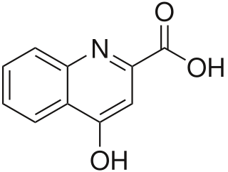
Kynurenic acid is a product of the normal metabolism of amino acid L-tryptophan. It has been shown that kynurenic acid possesses neuroactive activity. It acts as an antiexcitotoxic and anticonvulsant, most likely through acting as an antagonist at excitatory amino acid receptors. Because of this activity, it may influence important neurophysiological and neuropathological processes. As a result, kynurenic acid has been considered for use in therapy in certain neurobiological disorders. Conversely, increased levels of kynurenic acid have also been linked to certain pathological conditions.

<span class="mw-page-title-main">NMDA receptor antagonist</span> Class of anesthetics

NMDA receptor antagonists are a class of drugs that work to antagonize, or inhibit the action of, the N-Methyl-D-aspartate receptor (NMDAR). They are commonly used as anesthetics for animals and humans; the state of anesthesia they induce is referred to as dissociative anesthesia.

Gliotransmitters are chemicals released from glial cells that facilitate neuronal communication between neurons and other glial cells. They are usually induced from Ca2+ signaling, although recent research has questioned the role of Ca2+ in gliotransmitters and may require a revision of the relevance of gliotransmitters in neuronal signalling in general.

<span class="mw-page-title-main">Kynurenine 3-monooxygenase</span> Enzyme

In enzymology, a kynurenine 3-monooxygenase (EC 1.14.13.9) is an enzyme that catalyzes the chemical reaction

<span class="mw-page-title-main">GRIN3A</span> Protein-coding gene in the species Homo sapiens

Glutamate [NMDA] receptor subunit 3A is a protein that in humans is encoded by the GRIN3A gene.

<span class="mw-page-title-main">GRIN3B</span> Protein-coding gene in the species Homo sapiens

Glutamate [NMDA] receptor subunit 3B is a protein that in humans is encoded by the GRIN3B gene.

<span class="mw-page-title-main">Cystine/glutamate transporter</span> Protein found in humans

Cystine/glutamate transporter is an antiporter that in humans is encoded by the SLC7A11 gene.

<span class="mw-page-title-main">Hypertryptophanemia</span> Medical condition

Hypertryptophanemia is a rare autosomal recessive metabolic disorder that results in a massive buildup of the amino acid tryptophan in the blood, with associated symptoms and tryptophanuria.

<span class="mw-page-title-main">Kynurenine pathway</span> Metabolic pathway that produces the NAD coenzyme

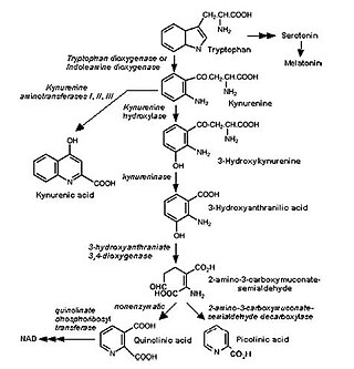
The kynurenine pathway is a metabolic pathway leading to the production of nicotinamide adenine dinucleotide (NAD+). Metabolites involved in the kynurenine pathway include tryptophan, kynurenine, kynurenic acid, xanthurenic acid, quinolinic acid, and 3-hydroxykynurenine. The kynurenine pathway is responsible for total catabolization of tryptophan about 95%. Disruption in the pathway is associated with certain genetic and psychiatric disorders.

Immuno-psychiatry, according to Pariante, is a discipline that studies the connection between the brain and the immune system. It differs from psychoneuroimmunology by postulating that behaviors and emotions are governed by peripheral immune mechanisms. Depression, for instance, is seen as malfunctioning of the immune system.

References

  1. 1 2 Hiroshi Ashihara, Alan Crozier, Atsushi Komamine "Nicotine Biosynthesis" in Plant Metabolism and Biotechnology, Tsubasa Shoji, Takashi Hashimoto Eds. Wiley-VCH, Weinheim, 2011. {{DOI: 10.1002/9781119991311.ch7}}
  2. Misztal M, Frankiewicz T, Parsons CG, Danysz W (January 1996). "Learning deficits induced by chronic intraventricular infusion of quinolinic acid--protection by MK-801 and memantine". Eur. J. Pharmacol. 296 (1): 1–8. doi:10.1016/0014-2999(95)00682-6. PMID   8720470.
  3. Guillemin, G.; Smith, Danielle G.; Smythe, George A.; Armati, Patricia J.; Brew, George J. (2003). "Expression of the Kynurenine Pathway Enzymes in Human Microglia and Macrophages". Developments in Tryptophan and Serotonin Metabolism. Advances in Experimental Medicine and Biology. Vol. 527. pp. 105–12. doi:10.1007/978-1-4615-0135-0_12. ISBN   978-0-306-47755-3. PMID   15206722.
  4. Guillemin, Gilles J (March 2012). "Quinolinic acid: neurotoxicity". FEBS Journal. 279 (8): 1355. doi: 10.1111/j.1742-4658.2012.08493.x . PMID   22251552.
  5. Skraup, Zd. H. (1881). "Synthetische Versuche in der Chinolinreihe". Monatshefte für Chemie. 2 (1): 139–170. doi:10.1007/BF01516502. S2CID   96574887.
  6. WO 2010011134,H. Bruno,"Ozonolysis of Aromatics and/or Olefins"
  7. USPatent 4420616,Ikegami, Seishi&Hatano, Yoshihiro,"Oxidative process for the preparation of copper quinolinate", assigned to Yamamoto Kagaku Gosei KK
  8. Marshall Kulka (1946). "Electrolytic Oxidation of Quinoline and 3-Picoline". J. Am. Chem. Soc. 68 (12): 2472–2473. doi:10.1021/ja01216a008. PMID   20282382.
  9. EP 0159769,Toomey Jr., Joseph E.,"Electrochemical oxidation of pyridine bases", assigned to Reilly Industries, Inc.
  10. 1 2 3 4 5 6 7 8 9 10 11 12 13 Guillemin, Giles (April 2012). "Quinolinic Acid, the Inescapable Neurotoxin". FEBS Journal. 279 (8): 1356–1365. doi: 10.1111/j.1742-4658.2012.08485.x . PMID   22248144.
  11. 1 2 3 4 5 6 7 8 9 10 Schwarcz, Robert; John P. Bruno; Paul J. Muchowski; Hui-Qiu Wu (July 2012). "Kynurenines in the Mammalian Brain: When Physiology Meets Pathology". Nature Reviews Neuroscience. 13 (7): 465–477. doi:10.1038/nrn3257. PMC   3681811 . PMID   22678511.
  12. Agudelo, Leandro Z.; Femenía, Teresa; Orhan, Funda; Porsmyr-Palmertz, Margareta; Goiny, Michel; Martinez-Redondo, Vicente; Correia, Jorge C.; Izadi, Manizheh; Bhat, Maria; Schuppe-Koistinen, Ina; Pettersson, Amanda T.; Ferreira, Duarte M.S.; Krook, Anna; Barres, Romain; Zierath, Juleen R. (September 2014). "Skeletal Muscle PGC-1α1 Modulates Kynurenine Metabolism and Mediates Resilience to Stress-Induced Depression". Cell. 159 (1): 33–45. doi: 10.1016/j.cell.2014.07.051 . PMID   25259918. S2CID   5613716.
  13. Combes, Valery; Gilles J. Guillemin; Tailoi Chan-Ling; Nicholas H. Hunt; Georges E.R. Grau (August 2012). "The crossroads of neuroinflammation in infectious diseases: endothelial cells and astrocytes". Trends in Parasitology. 28 (8): 311–319. doi:10.1016/j.pt.2012.05.008. PMID   22727810.
  14. 1 2 Pérez-De La Cruz, V.; P.Carrillo-Mora, A. Santamaría (2012). "Quinolinic acid, an endogenous molecule combining excitotoxicity, oxidative stress and other toxic mechanisms". International Journal of Tryptophan Research. 5 (5): 1–8. doi: 10.4137/IJTR.S8158 . PMC   3296489 . PMID   22408367.
  15. 1 2 3 4 5 6 7 Myint, Aye M. (April 2012). "Kynurenines: from the perspective of major psychiatric disorders". FEBS Journal. 279 (8): 1375–1385. doi: 10.1111/j.1742-4658.2012.08551.x . PMID   22404766.
  16. 1 2 Maes, M; Leonard, B.E.; Myint, A.M.; Kubera, M.; Verkerk, R. (29 April 2011). "The new '5-HT' hypothesis of depression: Cell-mediated immune activation induces indoleamine 2,3-dioxygenase, which leads to lower plasma tryptophan and an increased synthesis of detrimental tryptophan catabolites (TRYCATs), both of which contribute to the onset of depression". Progress in Neuro-Psychopharmacology & Biological Psychiatry. 35 (3): 702–721. doi:10.1016/j.pnpbp.2010.12.017. PMID   21185346. S2CID   2121023.
  17. 1 2 3 4 5 Tan, Lin; Yu, JT; Tan, L (15 December 2012). "The kynurenine pathway in neurodegenerative diseases: Mechanistic and therapeutic considerations". Journal of the Neurological Sciences. 323 (1–2): 1–8. doi:10.1016/j.jns.2012.08.005. PMID   22939820. S2CID   6061945.
  18. 1 2 3 Severino, Patricia Cardoso; Muller, GDS; Vandresen-Filho, S; Tasca, CI (10 October 2011). "Cell signaling in NMDA preconditioning and neuroprotection in convulsions induced by quinolinic acid". Life Sciences. 89 (15–16): 570–576. doi:10.1016/j.lfs.2011.05.014. PMID   21683718.
  19. 1 2 3 4 5 Stone, Trevor W.; Forrest, C.M.; Darlington, L.G. (April 2012). "Kynurenine pathway inhibition as a therapeutic strategy for neuroprotection". FEBS Journal. 279 (8): 1386–1397. doi:10.1111/j.1742-4658.2012.08487.x. PMID   22248239. S2CID   27815584.
  20. 1 2 Kandanearatchi, Apsara; Brew, Bruce J. (April 2012). "The kynurenine pathway and quinolinic acid: pivotal roles in HIV associated neurocognitive disorders". FEBS Journal. 279 (8): 1366–1374. doi: 10.1111/j.1742-4658.2012.08500.x . PMID   22260426.
  21. 1 2 3 4 5 6 7 Stone, Trevor W. (January 2001). "Endogenous neurotoxins from tryptophan". Toxicon. 39 (1): 61–73. doi:10.1016/S0041-0101(00)00156-2. PMID   10936623.
  22. Szabó, Nikoletta; Kincses, Z.T.; Toldi, J; Vécsei, L (15 November 2011). "Altered tryptophan metabolism in Parkinson's disease: A possible novel therapeutic approach". Journal of the Neurological Sciences. 310 (1–2): 256–260. doi:10.1016/j.jns.2011.07.021. PMID   21824629. S2CID   25318596.
  23. 1 2 3 4 Kalonia, H.; P. Kumar; A. Kumar (2011). "Licofelone attenuates quinolinic acid induced huntington like symptoms: Possible behavioral, biochemical and cellular alterations". Progress in Neuro-Psychopharmacology & Biological Psychiatry. 35 (2): 607–615. doi:10.1016/j.pnpbp.2011.01.003. PMID   21237233. S2CID   1169538.
  24. Oxenkrug, Gregory F. (January 2011). "Interferon-gamma-inducible kynurenines/pteridines inflammation cascade: implications for aging and aging-associated psychiatric and medical disorders". Journal of Neural Transmission. 118 (1): 75–85. doi:10.1007/s00702-010-0475-7. PMC   3026891 . PMID   20811799.
  25. Braidy N, Grant R, Adams S, Guillemin GJ (January 2010). "Neuroprotective effects of naturally occurring polyphenols on quinolinic acid-induced excitotoxicity in human neurons". FEBS J. 277 (2): 368–82. doi: 10.1111/j.1742-4658.2009.07487.x . PMID   20015232. S2CID   20243001.