Cerebrum

Last updated
Cerebrum
The lobes of the cerebral cortex include the frontal (blue), temporal (green), occipital (red), and parietal (yellow) lobes. The cerebellum (unlabeled) is not part of the telencephalon.
EmbryonicBrain.svg
Diagram depicting the main subdivisions of the embryonic vertebrate brain.
Details
Pronunciation /ˈsɛrɪbrəm/ , /sɪˈrbrəm/
Artery Anterior cerebral, middle cerebral, posterior cerebral
Vein Cerebral veins
Identifiers
Latin cerebrum
MeSH D054022
NeuroLex ID birnlex_1042
TA98 A14.1.03.008
A14.1.09.001
TA2 5416
TH H3.11.03.6.00001
TE E5.14.1.0.2.0.12
FMA 62000
Anatomical terms of neuroanatomy

The cerebrum (pl.: cerebra), telencephalon or endbrain [1] is the largest part of the brain containing the cerebral cortex (of the two cerebral hemispheres), as well as several subcortical structures, including the hippocampus, basal ganglia, and olfactory bulb. In the human brain, the cerebrum is the uppermost region of the central nervous system. The cerebrum develops prenatally from the forebrain (prosencephalon). In mammals, the dorsal telencephalon, or pallium, develops into the cerebral cortex, and the ventral telencephalon, or subpallium, becomes the basal ganglia. The cerebrum is also divided into approximately symmetric left and right cerebral hemispheres.

Contents

With the assistance of the cerebellum, the cerebrum controls all voluntary actions in the human body.

Structure

Location of the human cerebrum (red). Cerebrum animation small.gif
Location of the human cerebrum (red).

The cerebrum is the largest part of the brain. Depending upon the position of the animal it lies either in front or on top of the brainstem. In humans, the cerebrum is the largest and best-developed of the five major divisions of the brain.

The cerebrum is made up of the two cerebral hemispheres and their cerebral cortices (the outer layers of grey matter), and the underlying regions of white matter. [2] Its subcortical structures include the hippocampus, basal ganglia and olfactory bulb. The cerebrum consists of two C-shaped cerebral hemispheres, separated from each other by a deep fissure called the longitudinal fissure.

Cerebral cortex

Surface of the cerebrum Lateral surface of cerebral cortex - gyri.png
Surface of the cerebrum

The cerebral cortex, the outer layer of grey matter of the cerebrum, is found only in mammals. In larger mammals, including humans, the surface of the cerebral cortex folds to create gyri (ridges) and sulci (furrows) which increase the surface area. [3]

The cerebral cortex is generally classified into four lobes: the frontal, parietal, occipital and temporal lobes. The lobes are classified based on their overlying neurocranial bones. [4] A smaller lobe is the insular lobe, a part of the cerebral cortex folded deep within the lateral sulcus that separates the temporal lobe from the parietal and frontal lobes, is located within each hemisphere of the mammalian brain.

Cerebral hemispheres

The cerebrum is divided by the medial longitudinal fissure into two cerebral hemispheres, the right and the left. The cerebrum is contralaterally organized, i.e., the right hemisphere controls and processes signals (predominantly) from the left side of the body, while the left hemisphere controls and processes signals (predominantly) from the right side of the body. [4] According to current knowledge, this is due to an axial twist that occurs in the early embryo. [5] There is a strong but not complete bilateral symmetry between the hemispheres, while lateralization tends to increase with increasing brain size. [6] The lateralization of brain function looks at the known and possible differences between the two.

Development

In the developing vertebrate embryo, the neural tube is subdivided into four unseparated sections which then develop further into distinct regions of the central nervous system; these are the prosencephalon (forebrain), the mesencephalon (midbrain) the rhombencephalon (hindbrain) and the spinal cord. [7] The prosencephalon develops further into the telencephalon and the diencephalon. The dorsal telencephalon gives rise to the pallium (cerebral cortex in mammals and reptiles) and the ventral telencephalon generates the basal ganglia. The diencephalon develops into the thalamus and hypothalamus, including the optic vesicles (future retina). [8] The dorsal telencephalon then forms two lateral telencephalic vesicles, separated by the midline, which develop into the left and right cerebral hemispheres. Birds and fish have a dorsal telencephalon, like all vertebrates, but it is generally unlayered and therefore not considered a cerebral cortex. Only a layered cytoarchitecture can be considered a cortex.

Functions

Note: As cerebrum is a gross division with many subdivisions and sub-regions, it is important to state that this section lists functions that cerebrum as a whole serves. See main articles on cerebral cortex and basal ganglia for more information. The cerebrum is a major part of the brain, controlling emotions, hearing, vision, personality and much more. It controls all precision of voluntary actions.

Upper motor neurons in the primary motor cortex send their axons to the brainstem and spinal cord to synapse on the lower motor neurons, which innervate the muscles. Damage to motor areas by chance of cortex can lead to certain types of motor neuron disease. This kind of damage results in loss of muscular power and precision rather than total paralysis.

It functions as the center of sensory perception, memory, thoughts and judgement; the cerebrum also functions as the center of voluntary motor activities.

Sensory processing

The primary sensory areas of the cerebral cortex receive and process visual, auditory, somatosensory, gustatory, and olfactory information. Together with association cortical areas, these brain regions synthesize sensory information into our perceptions of the world.

Olfaction

The olfactory bulb, responsible for the sense of smell, takes up a large area of the cerebrum in most vertebrates. However, in humans, this part of the brain is much smaller and lies underneath the frontal lobe. The olfactory sensory system is unique since the neurons in the olfactory bulb send their axons directly to the olfactory cortex, rather than to the thalamus first. The olfaction is also the only sense that is represented by the ipsilateral side of the brain. Damage to the olfactory bulb results in a loss of olfaction (the sense of smell).

Language and communication

Speech and language are mainly attributed to the parts of the cerebral cortex. Motor portions of language are attributed to Broca's area within the frontal lobe. Speech comprehension is attributed to Wernicke's area, at the temporal-parietal lobe junction. These two regions are interconnected by a large white matter tract, the arcuate fasciculus. Damage to the Broca's area results in expressive aphasia (non-fluent aphasia) while damage to Wernicke's area results in receptive aphasia (also called fluent aphasia).

Learning and memory

Explicit or declarative (factual) memory formation is attributed to the hippocampus and associated regions of the medial temporal lobe. This association was originally described after a patient known as HM had both his left and right hippocampus surgically removed to treat chronic [temporal lobe epilepsy]. After surgery, HM had anterograde amnesia, or the inability to form new memories.

Implicit or procedural memory, such as complex motor behaviors, involves the basal ganglia.

Short-term or working memory involves association areas of the cortex, especially the dorsolateral prefrontal cortex, as well as the hippocampus.

Other animals

In the most primitive vertebrates, the hagfishes and lampreys, the cerebrum is a relatively simple structure receiving nerve impulses from the olfactory bulb. In cartilaginous and lobe-finned fishes and also in amphibians, a more complex structure is present, with the cerebrum being divided into three distinct regions. The lowermost (or ventral) region forms the basal nuclei, and contains fibres connecting the rest of the cerebrum to the thalamus. Above this, and forming the lateral part of the cerebrum, is the paleopallium, while the uppermost (or dorsal) part is referred to as the archipallium. The cerebrum remains largely devoted to olfactory sensation in these animals, in contrast to its much wider range of functions in amniotes. [9]

In ray-finned fishes the structure is somewhat different. The inner surfaces of the lateral and ventral regions of the cerebrum bulge up into the ventricles; these include both the basal nuclei and the various parts of the pallium and may be complex in structure, especially in teleosts. The dorsal surface of the cerebrum is membranous, and does not contain any nervous tissue. [9]

In the amniotes, the cerebrum becomes increasingly large and complex. In reptiles, the paleopallium is much larger than in amphibians and its growth has pushed the basal nuclei into the central regions of the cerebrum. As in the lower vertebrates, the grey matter is generally located beneath the white matter, but in some reptiles, it spreads out to the surface to form a primitive cortex, especially in the anterior part of the brain. [9]

In mammals, this development proceeds further, so that the cortex covers almost the whole of the cerebral hemispheres, especially in more developed species, such as the primates. The paleopallium is pushed to the ventral surface of the brain, where it becomes the olfactory lobes, while the archipallium becomes rolled over at the medial dorsal edge to form the hippocampus. In placental mammals, a corpus callosum also develops, further connecting the two hemispheres. The complex convolutions of the cerebral surface (see gyrus, gyrification) are also found only in higher mammals. [9] Although some large mammals (such as elephants) have particularly large cerebra, dolphins are the only species (other than humans) to have cerebra accounting for as much as 2 percent of their body weight. [10]

The cerebra of birds are similarly enlarged to those of mammals, by comparison with reptiles. The increased size of bird brains was classically attributed to enlarged basal ganglia, with the other areas remaining primitive, but this view has been largely abandoned. [11] Birds appear to have undergone an alternate process of encephalization, [12] as they diverged from the other archosaurs, with few clear parallels to that experienced by mammals and their therapsid ancestors.

Additional images

See also

Related Research Articles

<span class="mw-page-title-main">Central nervous system</span> Brain and spinal cord

The central nervous system (CNS) is the part of the nervous system consisting of the brain and spinal cord, the retina and optic nerve, and the olfactory nerve and epithelia. The CNS is so named because the brain integrates the received information and coordinates and influences the activity of all parts of the bodies of bilaterally symmetric and triploblastic animals—that is, all multicellular animals except sponges and diploblasts. It is a structure composed of nervous tissue positioned along the rostral to caudal axis of the body and may have an enlarged section at the rostral end which is a brain. Only arthropods, cephalopods and vertebrates have a true brain, though precursor structures exist in onychophorans, gastropods and lancelets.

<span class="mw-page-title-main">Cerebral cortex</span> Outer layer of the cerebrum of the mammalian brain

The cerebral cortex, also known as the cerebral mantle, is the outer layer of neural tissue of the cerebrum of the brain in humans and other mammals. It is the largest site of neural integration in the central nervous system, and plays a key role in attention, perception, awareness, thought, memory, language, and consciousness. The cerebral cortex is the part of the brain responsible for cognition.

<span class="mw-page-title-main">Basal ganglia</span> Group of subcortical nuclei involved in the motor and reward systems

The basal ganglia (BG) or basal nuclei are a group of subcortical nuclei found in the brains of vertebrates. In humans and other primates, differences exist, primarily in the division of the globus pallidus into external and internal regions, and in the division of the striatum. Positioned at the base of the forebrain and the top of the midbrain, they have strong connections with the cerebral cortex, thalamus, brainstem and other brain areas. The basal ganglia are associated with a variety of functions, including regulating voluntary motor movements, procedural learning, habit formation, conditional learning, eye movements, cognition, and emotion.

<span class="mw-page-title-main">Limbic system</span> Set of brain structures involved in emotion and motivation

The limbic system, also known as the paleomammalian cortex, is a set of brain structures located on both sides of the thalamus, immediately beneath the medial temporal lobe of the cerebrum primarily in the forebrain.

Articles related to anatomy include:

<span class="mw-page-title-main">Cerebral hemisphere</span> Left and right cerebral hemispheres of the brain

The vertebrate cerebrum (brain) is formed by two cerebral hemispheres that are separated by a groove, the longitudinal fissure. The brain can thus be described as being divided into left and right cerebral hemispheres. Each of these hemispheres has an outer layer of grey matter, the cerebral cortex, that is supported by an inner layer of white matter. In eutherian (placental) mammals, the hemispheres are linked by the corpus callosum, a very large bundle of nerve fibers. Smaller commissures, including the anterior commissure, the posterior commissure and the fornix, also join the hemispheres and these are also present in other vertebrates. These commissures transfer information between the two hemispheres to coordinate localized functions.

<span class="mw-page-title-main">Temporal lobe</span> One of the four lobes of the mammalian brain

The temporal lobe is one of the four major lobes of the cerebral cortex in the brain of mammals. The temporal lobe is located beneath the lateral fissure on both cerebral hemispheres of the mammalian brain.

<span class="mw-page-title-main">Occipital lobe</span> Part of the brain at the back of the head

The occipital lobe is one of the four major lobes of the cerebral cortex in the brain of mammals. The name derives from its position at the back of the head, from the Latin ob, 'behind', and caput, 'head'.

<span class="mw-page-title-main">Forebrain</span> Forward-most portion of the brain

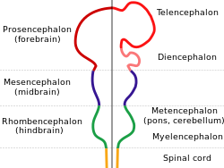
In the anatomy of the brain of vertebrates, the forebrain or prosencephalon is the rostral (forward-most) portion of the brain. The forebrain controls body temperature, reproductive functions, eating, sleeping, and the display of emotions.

<span class="mw-page-title-main">Midbrain</span> Forward-most portion of the brainstem

The midbrain or mesencephalon is the rostral-most portion of the brainstem connecting the diencephalon and cerebrum with the pons. It consists of the cerebral peduncles, tegmentum, and tectum.

<span class="mw-page-title-main">Piriform cortex</span> Region of the brain

The piriform cortex, or pyriform cortex, is a region in the brain, part of the rhinencephalon situated in the cerebrum. The function of the piriform cortex relates to the sense of smell.

<span class="mw-page-title-main">Middle cerebral artery</span> Paired artery that supplies blood to the cerebrum

The middle cerebral artery (MCA) is one of the three major paired cerebral arteries that supply blood to the cerebrum. The MCA arises from the internal carotid artery and continues into the lateral sulcus where it then branches and projects to many parts of the lateral cerebral cortex. It also supplies blood to the anterior temporal lobes and the insular cortices.

<span class="mw-page-title-main">Lobes of the brain</span> Parts of the cerebrum

The lobes of the brain are the major identifiable zones of the human cerebral cortex, and they comprise the surface of each hemisphere of the cerebrum. The two hemispheres are roughly symmetrical in structure, and are connected by the corpus callosum. They traditionally have been divided into four lobes, but are today considered as having six lobes each. The lobes are large areas that are anatomically distinguishable, and are also functionally distinct to some degree. Each lobe of the brain has numerous ridges, or gyri, and furrows, the sulci that constitute further subzones of the cortex. The expression "lobes of the brain" usually refers only to those of the cerebrum, not to the distinct areas of the cerebellum.

<span class="mw-page-title-main">Olfactory tubercle</span> Area at the bottom of the forebrain

The olfactory tubercle (OT), also known as the tuberculum olfactorium, is a multi-sensory processing center that is contained within the olfactory cortex and ventral striatum and plays a role in reward cognition. The OT has also been shown to play a role in locomotor and attentional behaviors, particularly in relation to social and sensory responsiveness, and it may be necessary for behavioral flexibility. The OT is interconnected with numerous brain regions, especially the sensory, arousal, and reward centers, thus making it a potentially critical interface between processing of sensory information and the subsequent behavioral responses.

<span class="mw-page-title-main">EMX1</span> Protein-coding gene in the species Homo sapiens

Homeobox protein EMX1 is a protein that in humans is encoded by the EMX1 gene. The transcribed EMX1 gene is a member of the EMX family of transcription factors. The EMX1 gene, along with its family members, are expressed in the developing cerebrum. EMX1 plays a role in specification of positional identity, the proliferation of neural stem cells, differentiation of layer-specific neuronal phenotypes and commitment to a neuronal or glial cell fate.

<span class="mw-page-title-main">Ganglionic eminence</span>

The ganglionic eminence (GE) is a transitory structure in the development of the nervous system that guides cell and axon migration. It is present in the embryonic and fetal stages of neural development found between the thalamus and caudate nucleus.

<span class="mw-page-title-main">Pallium (neuroanatomy)</span> Layers of grey and white matter that cover the upper surface of the cerebrum in vertebrates

In neuroanatomy, pallium refers to the layers of grey and white matter that cover the upper surface of the cerebrum in vertebrates. The non-pallial part of the telencephalon builds the subpallium. In basal vertebrates, the pallium is a relatively simple three-layered structure, encompassing 3–4 histogenetically distinct domains, plus the olfactory bulb.

The neuroanatomy of memory encompasses a wide variety of anatomical structures in the brain.

<span class="mw-page-title-main">Avian brain</span> Brain of birds

The avian brain is the central organ of the nervous system in birds. Birds possess large, complex brains, which process, integrate, and coordinate information received from the environment and make decisions on how to respond with the rest of the body. Like in all chordates, the avian brain is contained within the skull bones of the head.

References

  1. "BrainInfo". braininfo.rprc.washington.edu.
  2. Arnould-Taylor, William (1998). A Textbook of Anatomy and Physiology. Nelson Thornes. p. 52. ISBN   9780748736348 . Retrieved 27 January 2015.
  3. Angevine, J.; Cotman, C. (1981). Principles of Neuroanatomy. NY: Oxford University Press. ISBN   9780195028850 . Retrieved 25 January 2015.
  4. 1 2 Rosdahl, Caroline; Kowalski, Mary (2008). Textbook of Basic Nursing (9th ed.). Lippincott Williams & Wilkins. p. 189. ISBN   9780781765213 . Retrieved 28 January 2015.
  5. de Lussanet, M.H.E.; Osse, J.W.M. (2012). "An ancestral axial twist explains the contralateral forebain and the optic chiasm in vertebrates". Animal Biology. 62 (2): 193–216. arXiv: 1003.1872 . doi:10.1163/157075611X617102. S2CID   7399128.
  6. Ebbesson, Sven O. E.; Ito, Hironobu (1980). "Bilateral retinal projections in the black piranah (Serrasalmus niger)". Cell Tissue Res. 213 (3): 483–495. doi:10.1007/BF00237893. PMID   7448850. S2CID   2406618.
  7. Gilbert, Scott F. (2014). Developmental biology (10th ed.). Sunderland, Mass.: Sinauer. ISBN   978-0-87893-978-7.
  8. Kandel, Eric R., ed. (2006). Principles of neural science (5th ed.). Appleton and Lange: McGraw-Hill. ISBN   978-0-07-139011-8.
  9. 1 2 3 4 Romer, Alfred Sherwood; Parsons, Thomas S. (1977). The Vertebrate Body. Philadelphia, PA: Holt-Saunders International. pp. 536–543. ISBN   0-03-910284-X.
  10. T.L. Brink (2008). "Unit 4: The Nervous System.". Psychology: A Student Friendly Approach (PDF). p. 62.
  11. Jarvis ED, Güntürkün O, Bruce L, et al. (2005). "Avian brains and a new understanding of vertebrate brain evolution". Nat. Rev. Neurosci. 6 (2): 151–9. doi:10.1038/nrn1606. PMC   2507884 . PMID   15685220.
  12. Emery NJ (2006-01-29). "Cognitive ornithology: the evolution of avian intelligence". Philos. Trans. R. Soc. Lond. B Biol. Sci. 361 (1465): 23–43. doi:10.1098/rstb.2005.1736. PMC   1626540 . PMID   16553307.